Dry eye syndrome

Last updated date: 07-Apr-2023

Originally Written in English

Dry eye syndrome

Overview

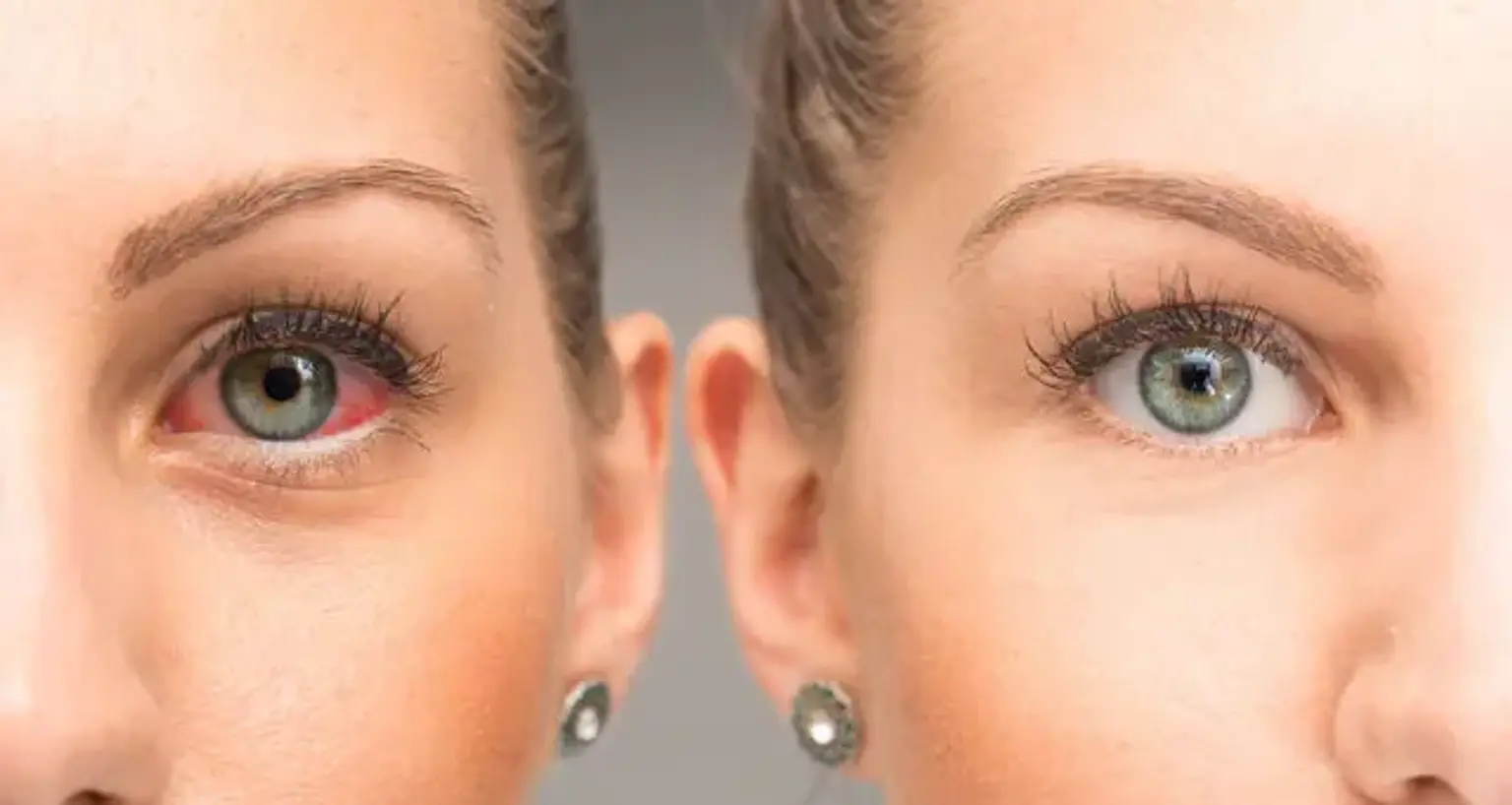
The condition of having dry eyes is known as dry eye syndrome (DES), also known as keratoconjunctivitis sicca (KCS). Irritation, redness, discharge, and easily tired eyes are some of the other symptoms. Blurred vision is also possible. Symptoms range from moderate and intermittent to severe and ongoing. Untreated instances may result in corneal scarring.

Dry eyes occur when the eye fails to produce enough tears or when the tears evaporate too rapidly. Contact lens usage, meibomian gland dysfunction, pregnancy, Sjögren syndrome, vitamin A deficiency, omega-3 fatty acid deficiency, LASIK surgery, and certain drugs including eye antihistamines, blood pressure medication, hormone replacement therapy, and antidepressants can all cause this. 

Chronic conjunctivitis, such as that caused by cigarette smoke or infection, can also cause the illness. The symptoms are used to make the diagnosis; however, a variety of additional tests may be employed.
Treatment is determined on the underlying cause. Artificial tears are typically used as the initial line of defense. Tear evaporation may be reduced by wearing wrap-around glasses that fit tight to the face. Certain drugs may be discontinued or changed. In certain circumstances, ciclosporin or steroid eye drops may be administered. Lacrimal plugs, which block tears from draining from the surface of the eye, are another alternative. Wearing contact lenses is sometimes impossible due to dry eye condition.Dry eye syndrome is a rather frequent eye condition.

It affects 5–34 percent of the population, depending on the demographic studied. It affects up to 70% of adults over the age of 65. It affects around 17% of the population in China.


How common is Dry eye syndrome?

 Dry eye

Dry eye disease is more frequent in women than in males, and its incidence rises with age. The incidence of dry eye illness varies depending on the diagnostic criteria used, and has been reported in population-based research to vary between 5 and 50 %.

It is more frequent in Asian populations than in whites, while regional, climatic, and environmental differences may also play a role. The most frequent kind of dry eye disease is evaporative dry eye. There may be a mismatch between dry eye signs and symptoms, with signs being more common and varied than symptoms. 


How Dry eye syndrome develops?

Dry eye syndrome development

Dry eyes have typically been divided into two types: aqueous deficient and evaporative. These categories, however, are not mutually exclusive, and many people have a mix of these dry eye disease processes.

  • Aqueous tear deficit is defined by insufficient tear production, with the most common causes being Sjogren Syndrome (primary or secondary), lacrimal gland illnesses such as blockage, or systemic medicines that impact tear production.
  • Evaporative dry eye is characterized by increased tear film evaporation and is most commonly caused by malfunction of the meibomian gland. Meibomian glands border the eyelid edges and release oils that form the lipid layer of the tear film, reducing tear evaporation. Inadequate secretion owing to atrophy, gland dropout, or blocking of gland orifices can all induce meibomian gland dysfunction.

Other main reasons of excessive tear evaporation include insufficient blinking (low rate, partial lid closure), lid aperture abnormalities, and environmental variables (low humidity, high airflow).

Hyperosmolarity of the tear film is a defining feature of dry eye illness, and it can directly or indirectly harm the ocular surface by inducing inflammation. Hyperosmolarity of the tear film causes a cascade of signaling events that release inflammatory mediators and damage to the ocular surface, which may further diminish tear film stability, leading to disease self-perpetuation in a 'vicious loop.' Aside from hyperosmolarity, other causes such as ocular surface inflammation produced by illnesses such as allergic eye disease, topical preservative toxicity, or xerophthalmia may trigger this pathologic cycle.


Dry eye syndrome causes

Dry eye syndrome causes

There are several different etiologies that might contribute to the development of dry eye disease, and many instances are complex. Local ocular variables, systemic disorders, and iatrogenic causes such as drugs or operations are examples of these. A list of some of these possible etiologies is provided below.

Potential causes and/or factors associated with dry eye disease include:

  • Systemic medications such as antihistamines, antihypertensives, anxiolytics/benzodiazepines, diuretics, systemic hormones, non-steroidal anti-inflammatory drugs, systemic or inhaled corticosteroids, anticholinergic medications, isotretinoin (causes meibomian gland atrophy), and antidepressants.
  • Topical medications such as glaucoma drops or preservative toxicity from eye drops that contain preservatives
  • Skin diseases on or around the eyelids such as rosacea or eczema
  • Meibomian gland dysfunction is a common co-morbidity with thickening and erythema of the eyelids and inadequate or altered secretions of meibomian glands.  
  • Ophthalmic surgery, including refractive surgery, cataract surgery, keratoplasty, and lid surgery.
  • Chemical or thermal burns that scar the conjunctiva.
  • Ocular allergies.
  • Computer or device usage as this may lead to decreased blinking when looking at the screen.
  • Excess or insufficient dosages of vitamins, particularly vitamin A deficiency which can lead to xerophthalmia and the appearance of Bitot spots on the conjunctiva in severe cases.
  • Decreased sensation in the cornea from long-term contact lens wear, herpes virus infections, or other causes of a neurotrophic cornea.
  • Graft-versus-host disease
  • Systemic diseases including Sjogren syndrome and other autoimmune or connective tissue disorders such as rheumatoid arthritis and lupus, and thyroid disease.
  • Environmental factors such as exposure to irritants like chemical fumes, cigarette smoke, pollution, or low humidity.


Signs and symptoms of Dry eye syndrome

symptoms of Dry eye

Dry eye disease may lead to any of the following symptoms:

  • Stinging, burning, or a feeling of pressure in the eyes. 
  • A sandy, gritty, or foreign body sensation.
  • Epiphora, or tearing, is a symptom that is often counterintuitive. This is due to dryness leading to pain or irritation that results in intermittent excess tearing, or epiphora.
  • Pain is a broad term, and sharp and dull pain can be described, which may be localized to some part of the eye, behind the eye, or even around the orbit.
  • Redness is a common complaint that is frequently exacerbated by the rebound effect of vasoconstrictors included in many over-the-counter eye treatments intended to diminish redness. Vasoconstrictors may reduce redness temporarily by constricting the vessels of the episclera, but they might have a rebound effect and cause greater redness once the drops wear off in a relatively short amount of time.
  • Blurry vision, particularly intermittent blurry vision, is a common complaint and may also be described as glare or haloes around lights at night.
  • A sensation of heavy eyelids or difficulty opening the eyes. 
  • Dryness is a common problem for contact lenses wearers, and irritation may make contact lenses uncomfortable or even impossible to wear.
  • Tired eyes.  Closing the eyes may provide relief to some individuals with dry eyes.


Evaluation of those with Dry eye syndrome

Dry eye syndrome

There is no one gold standard sign or symptom that can be used to diagnose dry eye disease. It is suggested to evaluate both symptoms and signs of dry eye disease since signs might exist without symptoms and vice versa.

1. Symptoms and signs:

A spoken history enables for the elicitation of dry eye symptoms that is not scripted. Furthermore, various questionnaires have been designed to check for dry eye disease symptoms. The use of a validated questionnaire as a screening tool enables for reliable measurement of symptoms as well as monitoring for progression and response to treatments. There are several questionnaires available, including the Ocular Surface Disease Index (OSDI), Dry Eye Questionnaire (DEQ-5), Symptoms Analysis in Dry Eye (SANDE), and others, that may be useful in assessing dry eye symptoms. Many questionnaires also ask about subjective visual function or problems that may be caused by dry eye.


2. Tear Stability:

Tear Film Break-up Time: The time elapsed between a full blink and the first break in the tear film. This is usually done in the clinic using a slit lamp microscope after administering sodium fluorescein dye to make the tear film more visible. A cut-off time of less than 10 seconds is frequently associated with dry eye disease. Alternatively, without the use of fluorescein, a non-invasive tear breakup time can be determined using apparatus that assesses the reflections of patterns or rings from the tear film, or by utilizing interferometry to evaluate for the appearance of lipid layer discontinuities after a blink.


3. Tear Volume: 

Tear meniscus assessment: The inferior tear film meniscus height is measured at the slit-lamp to examine the tear meniscus. This approach is simple to use; however, it has low intervisit reproducibility. Although instruments for more objective assessment of the tear film meniscus have been developed, they are not yet readily available in most clinics.

Schirmer test: A Schirmer paper strip is folded at the notch and the shorter end is hooked over the lateral lid edge to minimize corneal irritation while the patient relaxes with his or her eyes closed. The Schirmer I test is conducted without the use of topical anesthetic to evaluate basic and reflex tearing with less than 5 to 10mm (depending on the cut-off used) of wetness after 5 minutes, which is indicative of aqueous insufficiency. Alternatively, a topical anesthetic can be applied and residual fluid wiped from the inferior fornix prior to measuring basic secretion, with less than 5 to 10mm of wetness considered diagnostic for aqueous insufficiency.

Phenol red test: A cotton thread colored with phenol red is hooked over the temporal eyelid into the sulcus for 15 seconds as the patient sleeps with closed eyes, similar to Schirmer testing. When wet, the thread becomes red, with clinical cut-off values ranging from less than 10 to 20mm.


4. Ocular Surface Assessment:

Fluorescein staining: Fluorescein staining is used to examine corneal damage. A little amount of fluorescein is injected into the tear film, with optimal viewing occurring 1 to 3 minutes later. More than 5 spots of staining are considered a positive result, and many grading methods, including the Oxford grading scale, are utilized.

Lissamine green staining: Lissamine can be used to assess conjunctival and lid margin damage, as well as corneal injury to a lesser extent. More than 9 positions is a good performance. A positive result for lid wiper epitheliopathy, or staining of the lid edge, is 2 mm or more staining in length and/or higher than 25% in sagittal width.

Conjunctival redness: Conjunctival redness or hyperemia, is not particular to dry eye disease since it can be caused by any stimulus that causes conjunctivitis, including infective, allergy, chemical, or mechanical etiologies. Slit-lamp examination is commonly used to determine grading, while various equipment with automatic grading or digital photography can also be utilized.


5. Tear Film Assays:

Film of tears Osmolarity: Dry eye illness is characterized by elevated osmolarity and greater variability of osmolarity of tears. Osmolarity levels often rise as disease severity increases. Various cutoff values have been reported, with 308 mOsm/L being used as a threshold to diagnosis mild/moderate disease and 316 mOsm/L being used to diagnose more severe disease.

Matrix Metalloproteinases: These proteases are detected in the tears of dry eye patients. A point-of-care test can be used to assess matrix metalloproteinase-9 (MMP-9) levels.  


6. Eyelid Evaluation:

Blepharitis: Examining the eyelids is an important component of determining whether or not there are any variables leading to dry eye syndrome. The examination involves a look for anterior blepharitis and Demodex blepharitis, both of which are common comorbidities of dry eye illness.

Lid wiper epitheliopathy: refers to the region of the conjunctiva at the lid border that touches the ocular surface to disperse tears. Lid wiper epitheliopathy, or staining of the lid wiper with fluorescein or lissamine green, may be more prevalent in dry eye patients, owing to greater friction between the lid and the ocular surface.

Meibomian gland structural evaluation: Meibography can be used to assess the structure of the meibomian gland. While the contour of meibomian glands may be observed at the slit lamp or with a penlight by transilluminating the everted eyelid, better visibility is gained when meibography is performed utilizing infrared imaging devices. External blockages of orifices can be detected by inspecting the meibomian gland orifices along the eyelid border. Meibomian gland function may be evaluated by assessing meibum quantity, quality, and expressibility. Applying digital pressure along the eyelid edge with a clear meibum readily expressed from the natural eyelid is used to test expressibility. Meibum is turbid or viscous and difficult to produce when the meibomian glands are dysfunctional.

Blinking and closing of the eyelids: Dry eye illness can be caused by insufficient blinking and nocturnal lagophthalmos. The blink can be evaluated with or without the use of a microscope or video capturing equipment. Lagophthalmos can be measured by softly closing the patient's eyes and checking for incomplete closure.


7. Evaluation for systemic disease:

Many systemic disorders, including primary Sjogren syndrome and secondary Sjogren syndrome induced by other conditions such as rheumatoid arthritis, lupus, progressive systemic sclerosis, and dermatomyositis, can cause dry eye disease. Other systemic disorders related with dry eye disease include Parkinson's disease, androgen insufficiency, thyroid disease, and diabetes. If an underlying problem is suspected, an evaluation for systemic disease causing secondary dry eye may be necessary.

A system evaluation is recommended to test for underlying systemic illnesses. Sjogren syndrome may also affect the salivary glands, causing dry mouth and predisposing to periodontal disease, as well as other mucous membranes such as the vaginal, stomach, and respiratory mucosae.

Sjogren syndrome (antibodies to Ro/SS-A or La/SS-B), rheumatoid factor, and antinuclear antibodies are all tested for in the lab. A referral to a rheumatologist may be necessary, and some instances of Sjogren syndrome may necessitate a salivary gland biopsy performed by an oral surgeon.


Is Dry eye syndrome curable?

Dry eye syndrome

The treatment of dry eye syndrome is done in a step-by-step manner that varies depending on the severity of the condition. Initial treatments include education about the condition, environmental modification (eliminating direct high airflow/fans, reducing screen time, and using a humidifier), identification and elimination of offending topical and systemic agents, topical ocular lubricants, and lid hygiene (warm compresses and lid scrubs), and oral essential fatty acids.

Preservative-free ocular lubricants, reversible punctal occlusion (punctal plugs), night-time ointment or moisture goggles, device-assisted heating and/or expression of the meibomian glands, intense pulsed light therapy, topical anti-inflammatory medications (corticosteroids, cyclosporine, lifitegrast), and oral antibiotics are the next treatment options (macrolide or tetracycline).

Serum eye drops, oral or topical secretagogues, therapeutic contact lenses, amniotic membrane grafting, surgical punctal occlusion, and tarsorrhaphy are other therapy possibilities. 


Prognosis of Dry eye syndrome

Prognosis of Dry eye syndrome

Dry eye disease is frequently seen as chronic, with periods of exacerbation caused by intermittent contributing factors. Post-surgical dry eye (such as after cataract surgery or refractive surgery) frequently improves over time, which may be due to corneal nerve regeneration or a reduction in ocular inflammation.


Complications of untreated Dry eye syndrome

Dry eye syndrome complications

Complications from dry eye disease range from mild to severe.  Mild to moderate dry eye disease causes symptoms detailed above including ocular irritation and/or visual disturbances.  More severe disease can result in corneal complications including infectious keratitis, ulceration, and scarring which may cause subsequent loss of vision.  While causation has not been established, there are several non-ocular associations with dry eye disease including depression, sleep and mood disorders, dyslipidemia, and migraine headaches.


Conclusion

Dry eye

Dry eye is a multifactorial ocular surface condition defined by a loss of tear film homeostasis and followed by ocular symptoms, with etiologic roles played by tear film instability and hyperosmolarity, ocular surface inflammation and injury, and neurosensory abnormalities.

There are several different etiologies that might contribute to the development of dry eye disease, and many instances are complex. Local ocular factors, systemic disorders, and iatrogenic causes such as drugs or operations are examples of these.

A stinging, burning, or scratchy feeling in your eyes is one of the signs and symptoms of DES, which generally affects both eyes. Mucus that is stringy in or around your eyes, Light sensitivity, Redness in the eyes, A feeling that something is in your eyes, Difficulty wearing contact lenses, Difficulty driving at night, Watery eyes are the body's reaction to the discomfort of dry eyes. Blurred vision or eye strain

Treatment is determined on the underlying cause. Artificial tears are typically used as the initial line of defense. Tear evaporation may be reduced by wearing wrap-around glasses that fit tight to the face. Certain drugs may be discontinued or changed. In certain circumstances, ciclosporin or steroid eye drops may be administered. Lacrimal plugs, which block tears from draining from the surface of the eye, are another alternative. Wearing contact lenses is sometimes impossible due to dry eye condition.