Ear Disorders

Last updated date: 15-May-2023

Originally Written in English

Ear Disorders

Overview

The auditory system allows us to hear and assists us in maintaining our balance and body position. When anything goes wrong with the auditory system, you might have a wide range of symptoms. People of all ages are affected by Ear disorders, which can be chronic or transitory.


Ear Anatomy

Ear Anatomy

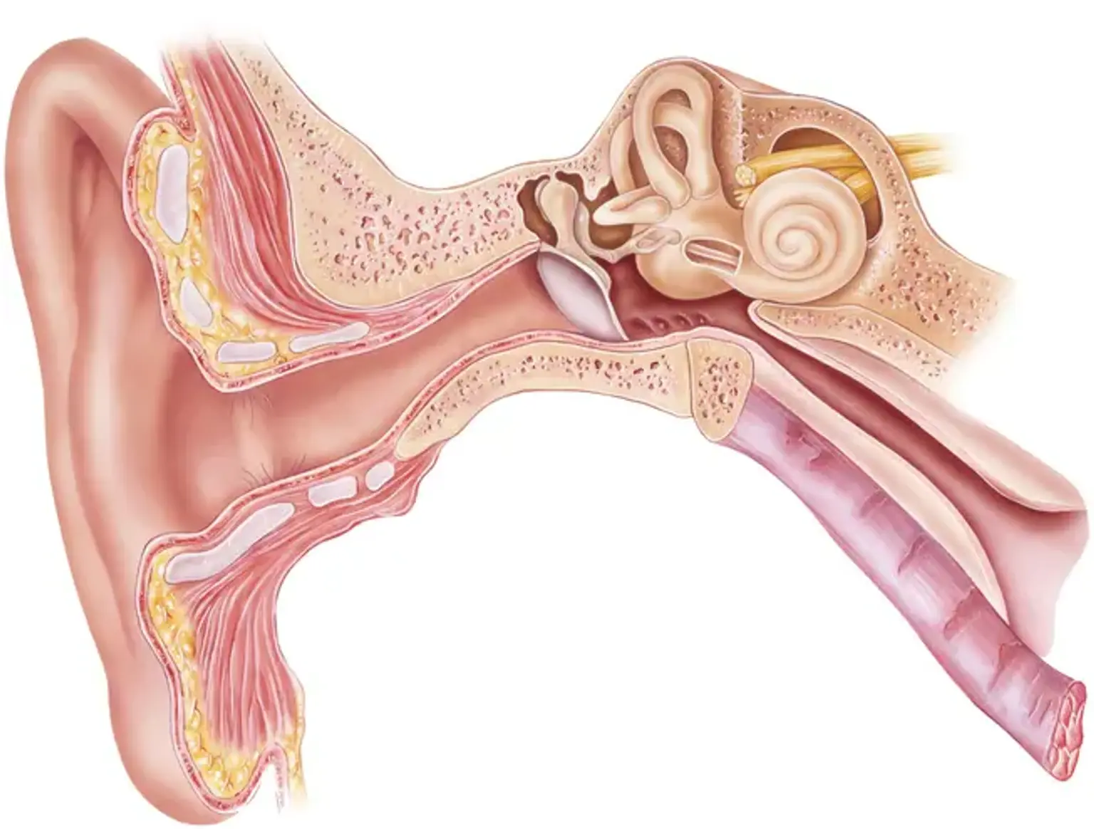
The relevant anatomy centers on the petrous bone and the inner ear. The ear is divided into 3 sections:

  • External, 
  • Middle, and 
  • Inner

The external ear is made up of three parts: the auricle, the external ear canal, and the tympanic membrane. The tympanic membrane divides the external ear from the middle ear components. The middle ear is an air-filled area that contains the three hearing bones: the malleus, incus, and stapes. The cochlear-vestibular apparatus and its accompanying nerves comprise the inner ear, which is fully encased in bone.

The cochlear-vestibular apparatus is a complicated structure with an exquisite spatial alignment. This structure is contained in a network of meandering tunnels and linking areas since it is totally covered in bone. The labyrinth is named from the tunnels' maze-like arrangement. The bony labyrinth is the bone that surrounds it.

The cochlea is a snail-shaped chamber that contains the Corti organ. It is in charge of converting mechanical vibrations into electrical impulses and transmitting them to the brain via the cochlear nerve.

The vestibular system is made up of a huge chamber (the vestibule) into which three semicircular canals emerge. Two sensors (the utricle and the saccule) detect linear acceleration within the vestibule, whereas the semicircular canals detect rotational motions in three planes of rotation.

The cochlea and vestibular system are connected in the center by a dual-chambered hydraulic system. Endolymph and perilymph are the fluids that bathe these hydraulic chambers. 

The mechanism may be imagined as a floating water balloon in a pool. The water within the balloon is the endolymph in this comparison, and the balloon itself is the membrane labyrinth that holds the endolymph. The perilymph in the pool water nourishes the fragile nerve tissues of the membranous labyrinth. The pool's walls symbolize the boundaries of the bony labyrinth area, and the earth that surrounds the pool represents the bone that encases the labyrinthine region.

The endolymphatic sac is a reservoir pouch located on the petrous bone's posterior surface, against the posterior fossa dura. It drains into the endolymphatic space of the cochlea through the vestibular duct.

Endolymphatic flow has been compared to a "lake-river-pond" model. The endolymph travels from the endolymphatic fluid space (the lake) to the endolymphatic sac through the vestibular aqueduct (the river). Endolymphatic hydrops will develop if there is an impediment.


How We Hear?

Hear

The outer ear is where hearing begins (external auditory canal). When someone makes a sound outside, the sound waves, or vibrations, enter the outer ear and travel to the eardrum (tympanic membrane). The eardrum vibrates, and the vibrations are transmitted to the ossicles, which are three small bones in the middle ear (malleus, incus, stapes). The ossicles amplify sound and send it to the inner ear and the fluid-filled hearing organ (cochlea)

Sound waves are transformed into electrical impulses when they reach the inner ear. The auditory nerve provides information to the brain, which interprets them as sound and meaning. The inner ear is also in charge of maintaining equilibrium.


Otalgia (ear pain)

Otalgia

Otalgia (ear pain) is classified into two types: primary and secondary otalgia. Primary otalgia is ear discomfort caused by disease in the inner, middle, or external ear. Secondary or referred otalgia is ear discomfort caused by disease outside the ear. As a result of intricate embryologic development, the ear is innervated by a complex neural network. This neuronal network is shared by the ear and other organs, which leads to a plethora of potential reasons of referred ear pain.

Cranial nerves V (trigeminal), VII (facial), IX (glossopharyngeal), X (vagus), all innervate the ear. 

  • The auricle is innervated by cranial nerves V, VII, X
  • The ear canal is innervated by cranial nerves V, VII, and X.
  • The tympanic membrane is innervated by cranial nerves VII, IX, and X.  
  • The middle ear is innervated by cranial nerves V, VII, and IX.  

Cranial nerves V, VII, IX, X, C2, and C3 also innervate organs outside of the ear, leading to numerous potential causes of referred ear pain. 

  • The trigeminal nerve (cranial nerve V) innervates the face, sinuses, palate, and teeth. The temporomandibular joint is innervated by the auriculotemporal branch of cranial nerve V. (TMJ). This branch is most frequently associated with temporomandibular joint disorders. Secondary causes of otalgia include dental and TMJ disease.
  • The anterior two-thirds of the tongue, as well as the sublingual and submandibular salivary glands, are innervated by cranial nerve VII (facial). It also innervates the face expressive muscles.
  • The glossopharyngeal nerve (cranial nerve IX) innervates the posterior portion of the tongue, the carotid body, and the oropharynx.
  • The sinuses, thyroid gland, throat, and larynx are all innervated by the vagus nerve (cranial nerve X). The vagus nerve's superior laryngeal branch innervates the voice chords. It also innervates distant organs including the heart, lungs, and sections of the digestive tract.


Otalgia symptoms 

Otalgia symptoms

A thorough history and physical examination are required to diagnose otalgia. Both main and secondary reasons must be considered by the doctor. The following events should be included in your history:

Red flags associated with otalgia include:

  • Dysphagia, odynophagia, dysphonia, or hemoptysis
  • Loss of vision or black spots
  • Unintended weight loss


Key features on history include:

  • Shorter time-frames suggest more benign causes.  Longer time-frames suggest a secondary cause. 
  • Ear pain lasting over four weeks is more suspicious for malignancy, especially if in the presence of risk factors and normal otoscopy. 
  • Ear fullness rather than ear pain may be more associated with cholesteatomas.
  • Sharp, lancinating pain is more indicative of neuralgia or neuropathy.   
  • Malignancy tends to cause unilateral symptoms. 
  • Ear pain exacerbated by swallowing is suggestive of glossopharyngeal neuralgia. 


The following associated symptoms could indicate the following referred origins:

  • Sinus congestion - chronic rhinosinusitis
  • Toothaches - dental pathology
  • Hoarseness - vocal cord condition
  • Heartburn - gastroesophageal reflux
  • Chest pain - coronary artery disease
  • Shortness of breath- lung disease
  • Upper back pain - cervical disc disease or myofascial pain 


Physical examination should include the following: 

  • Examine the ears for evidence of infection or other major etiologies. It may also reveal vesicular lesions, which are associated with herpes zoster oticus infection.
  • A nasal examination is used to detect irritated nasal mucosa or nasal polyps. It might indicate chronic rhinosinusitis.
  • Examine the oral cavity for dental caries, loose fillings, aphthous ulcers, abnormal growths, or abscesses. An extended styloid process, which can occur in Eagle syndrome, can also be detected by intra-oral palpation. 
  • Examine the temporomandibular joint for discomfort in the temporalis, lateral/medial pterygoid, or masseter muscles. It might uncover trigger points or TMJ problems.
  • Head check to rule out parotid or other salivary gland disorders. It may show malignancies of the salivary glands or sialadenitis. Temporal arteritis can be identified by tenderness along the temporal artery.
  • Neck check to rule out lymphadenopathy or thyroid gland disease. It might detect thyroiditis or lymph node cancer. 
  • Cervical spine exam to detect cervical spine pathology and concomitant musculoskeletal exam abnormalities. Myofascial discomfort or cervical degenerative disc degeneration may be revealed.
  • Full cranial nerve examination to detect cranial nerve neuropathies. It may indicate trigeminal neuralgia, glossopharyngeal neuralgia, geniculate neuralgia, sphenopalatine neuralgia, occipital neuralgia, vagal neuralgia, or Ramsay Hunt syndrome.


Otalgia diagnosis

Otalgia diagnosis

A thorough history and physical examination are the initial steps in assessing otalgia. The evaluation should rule out any red flags or risk factors for a severe diagnosis. If a problem is discovered, a head and neck CT and MRI, as well as panendoscopy, including nasolaryngoscopy and direct vision of the upper aerodigestive tract, might be requested.

The next step should be to evaluate primary otalgia. A detailed history and physical examination are seldom required for primary otalgia evaluation. The most prevalent cause of primary otalgia is acute otitis media. Pneumo-otoscopy will demonstrate tympanic membrane opacification, swelling, and immobility.

Another prevalent cause of otalgia is Eustachian tube dysfunction. Tympanometry can detect anomalies such as negative pressure in the middle ear.

If the ear exam is normal and there is no clear reason of the otalgia, the next step is to do a thorough investigation for secondary causes. The requirement for lab or imaging investigations should be guided by clinical evaluation. Secondary or referred otalgia is frequently caused by dental and temporomandibular joint problems. An orthopantogram is a quick and straightforward approach to get a panoramic image of the lower jaw and teeth.

Imaging investigations are not always necessary to diagnose temporomandibular joint dysfunction. Petrous apicitis can be diagnosed with CT imaging of the temporal bone. CT and MRI imaging can also be used to diagnose malignant otitis externa. To assess cranial neuropathy, an MRI of the cranial nerves might be ordered. A complete blood count (CBC) can aid in the detection of infection.


Otalgia Treatment 

The treatment of otalgia is determined on the diagnosis. This section will go through the key points. Most cases of primary otalgia are caused by infections and are treated with antibiotics, whereas mechanical otalgia is treated with decongestants, nasal steroids, or myringotomy. Secondary causes encompass a wide range of diagnosis. The requirement for early referral and steroid therapy for temporal arteritis, an uncommon cause of secondary otalgia, is one significant therapeutic aspect.

Otherwise, treatment focuses on the underlying medical issue, such as cancer, dental cavities, temporomandibular joint dysfunction, coronary artery disease, or gastric reflux. Patients with an unremarkable clinical examination and no red flags or risk indicators for severe illness might be treated conservatively with analgesics for 4 weeks before being re-evaluated.


Acute Otitis Media

Acute Otitis Media

Acute otitis media (AOM), defined as the first three weeks of a process in which the middle ear exhibits signs and symptoms of acute inflammation, is the most prevalent illness requiring medical treatment in children under the age of five.


Otitis Media symptoms

Although the course of AOM differs with age, certain consistent characteristics emerge over the otitis-prone years, including the following:

  • Neonates: Irritability, feeding difficulties 
  • Older children: This age group begins to demonstrate a consistent presence of fever and otalgia, or ear tugging
  • Older children and adults: Hearing loss becomes a recurring symptom of AOM and otitis media with effusion (OME); ear stuffiness is seen prior to the presence of middle ear fluid.

Adults with external otitis media, tooth abscess, or pain transferred from the temporomandibular joint experience otalgia without hearing loss or fever. As the dental occlusion changes, orthodontic equipment can cause referred pain.


Otalgia Diagnosis

Pneumatic otoscopy is the gold standard for diagnosing acute and chronic otitis media. Examination of AOM patients may reveal the following findings:

  • Signs of inflammation in the tympanic membrane
  • Bulging in the posterior quadrants of the tympanic membrane may bulge; scalded appearance of the superficial epithelial layer
  • Perforated tympanic membrane (most frequently in posterior or inferior quadrants)
  • Presence of an opaque serumlike exudate oozing through the entire tympanic membrane
  • Pain with/without pulsation of the otorrhea
  • Fever


Imaging studies

In most cases of simple AOM, radiologic investigations are not required. However, CT scanning may be required to assess whether or not a problem has developed. For suspected intracranial problems, an MRI may be more suitable.


Procedures

Tympanocentesis is the process of aspirating the contents of the middle ear cleft using a needle and collecting that material for diagnostic testing. Tympanocentesis is recommended for the following AOM patients:

  • Neonates less than 6 weeks old (and therefore are more likely to have an unusual or more invasive pathogen)
  • Immunocompromised or immunosuppressed patients
  • Patients in whom appropriate antibiotic therapy has failed and indications of local or systemic sepsis persist.
  • Patients with a complication that need a culture for proper treatment


Otalgia Management

  • Pharmacotherapy

Because antibiotics are the only drugs with proven effectiveness in the treatment of AOM, they are the first-line therapy of choice. The antibiotic chosen should cover the majority of the prevalent bacterial diseases and be tailored to the child's allergy, tolerance, past antibiotic exposure, cost, and community resistance levels. The duration of treatment may also be a factor in antibiotic selection.

  • Surgery

Surgical management of AOM can be divided into the following 3 related procedures:

  • Tympanocentesis
  • Myringotomy
  • Myringotomy with insertion of a ventilating tube


Swimmer's ear (Otitis Externa)

Swimmer's ear

Otitis externa (OE) is an infection or inflammation of the external auditory canal (EAC), or both. This illness affects people of all ages.


Otitis Externa Symptoms

Pain on palpation of the tragus (anterior to the ear canal) or application of traction to the pinna is the most common physical finding of OE. The following signs and symptoms may also be present in patients:

  • Otalgia - Ranges from mild to severe, typically progressing over 1-2 days
  • Hearing loss
  • Ear fullness or pressure
  • Erythema, edema, and narrowing of the EAC
  • Tinnitus
  • Fever (occasionally)
  • Itching (especially in fungal OE or chronic OE)
  • Severe deep pain - Immunocompromised patients may have necrotizing (malignant) OE
  • Discharge - Initially, clear; quickly becomes purulent and foul-smelling
  • Cellulitis of the face or neck or 
  • History of exposure to or activities in water (frequently) (eg, swimming, surfing, kayaking)
  • History of preceding ear trauma (usually) (eg, forceful ear cleaning, use of cotton swabs, or water in the ear canal)


Otitis externa Diagnosis

The history and physical examination of the patient, including otoscopy, typically offer enough information for the doctor to determine the diagnosis of OE. A diabetic or immunocompromised patient with acute ear discomfort should have necrotizing OE excluded by an otolaryngologist.

  • Laboratory testing

Laboratory tests are not usually required, although they may be useful if the patient is immunocompromised, if the standard therapeutic procedures are inadequate, or if a fungal cause is suspected. The following tests may be administered:

  1. Gram stain and culture of any discharge from the auditory canal
  2. Blood glucose level
  3. Urine dipstick


  • Imaging studies

Most OE instances do not need imaging investigations. However, radiologic examination may be useful if an invasive infection, such as necrotizing (malignant) OE, is suspected or if mastoiditis is suspected.

Imaging modalities may include the following:

  1. High-resolution computed tomography (CT) - Preferred; better depicts bony erosion
  2. Radionucleotide bone scanning
  3. Magnetic resonance imaging (MRI) - Not used as often as the other modalities; may be considered secondarily or if soft-tissue extension is the predominant concern


Otitis externa Management

Most persons with OE are treated empirically. Primary treatment involves the following:

  • Pain management
  • Removal of debris from the EAC
  • Administration of topical medications to control edema and infection
  • Avoidance of contributing factors


Presbycusis

Presbycusis

Presbycusis is the term used to describe bilateral age-related hearing loss. Presbycusis literally means "old hearing" or "older hearing." It appears at the age of 60 and advances slowly; however, there is evidence that some stresses might accelerate the pace of decline. Audiometry can be used to confirm the diagnosis.

The inability to understand high-frequency components of speech is a defining feature of presbycusis. There is no cure, although hearing aids that magnify sounds can help to alleviate symptoms. Presbycusis affects various components of the auditory system anatomically. It is mostly caused by alterations in hair cells, the stria vascularis, and afferent spiral ganglion neurons with age.

Sound, in the form of air vibration, is caught by the funnel-shaped external ear and delivered to the tympanic membrane during normal hearing. The tympanic membrane vibrates at a specified frequency and amplitude as a result. The malleus, incus, and stapes are three tiny bones in the middle ear that help to amplify this movement. The signal is then conveyed to the cochlea as vibrations through the fluid of the inner ear. Hair cells in the cochlea convert the information represented in vibrations into a neuronal signal that goes to the auditory brain through the cochlear nerve.


Presbycusis Treatment 

There is no treatment for presbycusis. Hearing aids are the mainstay of therapy and have been proved to improve quality of life and communication dramatically. Hearing aids are not without limits. They do not restore normal hearing; rather, they magnify noises. Devices can be highly costly and are sometimes not covered by the patient's insurance. While smaller hearing aids may be more comfortable and inconspicuous, senior individuals' diminished dexterity may make these devices less practical. Importantly, hearing aid management does not end after the devices are installed.

It requires tremendous effort and experience to learn to wear hearing aids and adjust to both physical discomfort and cognitive adjustment. For ongoing auditory rehabilitation, a coordinated, multidisciplinary approach involving the primary care physician and an audiologist is advised. Many patients require encouragement since hearing aids are painful, ugly, and humiliating.

Hearing aids are recommended at specific levels of hearing loss. Patients with severe bilateral hearing loss who have not improved with hearing aids may be candidates for cochlear implants. For patients to be considered candidates, specific criteria must be met, which frequently include a preset level of impairment in word identification.


Meniere Disease

Meniere disease

Meniere disease, commonly known as idiopathic endolymphatic hydrops, is an inner ear ailment. Endolymphatic hydrops is a disorder characterized by elevated hydraulic pressure inside the endolymphatic system of the inner ear. Excess pressure in the endolymph can result in a triad of symptoms:

  1. Fluctuating hearing loss
  2. occasional episodic vertigo (usually a spinning sensation, sometimes violent),
  3. Tinnitus or ringing in the ears (usually low-tone roaring), and 
  4. Aural fullness (eg, pressure, discomfort, fullness sensation in the ears).

Endolymphatic hydrops is sometimes used interchangeably with Ménière disease and Ménière syndrome, both of which are thought to be caused by increased pressure inside the endolymphatic system. Ménière illness, on the other hand, is idiopathic by definition, although Ménière syndrome might emerge as a result of different mechanisms interfering with normal endolymph production or resorption (eg, endocrine abnormalities, trauma, electrolyte imbalance, autoimmune dysfunction, medications, parasitic infections, hyperlipidemia).


Pathophysiology of Ménière disease

The precise pathogenesis of Ménière disease is debatable. The fundamental process is thought to be membranous labyrinth deformation caused by endolymph buildup. Some authors have questioned whether endolymphatic hydrops is a symptom of illness or a cause. A examination of temporal bones discovered that while all individuals with Ménière's disease had hydrops in at least one ear, hydrops was also observed in people who showed no indications of the condition.

The fluids that fill the chambers of the inner ear, known as endolymph and perilymph, are divided by thin membranes that house the neuronal machinery of hearing and balance. Pressure fluctuations stress these nerve-rich membranes, resulting in hearing loss, tinnitus, vertigo, imbalance, and an ear pressure feeling.

Hydrops attacks are most likely triggered by an increase in endolymphatic pressure, which produces a rupture in the membrane that divides the perilymph (potassium-poor extracellular fluid) from the endolymph (potassium-rich intracellular fluid). The resulting chemical combination bathes the vestibular nerve receptors, causing depolarization and temporary loss of function. An acute vestibular imbalance is caused by a rapid shift in the rate of vestibular nerve activity (ie, vertigo).


Meniere disease diagnosis 

All patients with Meniere illness must have an audiometric assessment. The condition is characterized by fluctuating low frequency unilateral sensorineural hearing loss. Hearing loss can spread to all frequencies. Tinnitus is a frequent and ipsilateral condition.

To rule out retrocochlear disease, all patients with one-sided hearing loss should have magnetic resonance imaging (MRI). A BERA (brainstem evoked response audiometry) is adequate in some countries. Imaging is not required in the acute situation, although it may be performed a few weeks following the beginning of symptoms. High-resolution MRI imaging of the afflicted organs may reveal endolymphatic hydrops. More study is being conducted to see whether this is clinically useful.


Meniere disease Management

There are several therapy options for Meniere illness, with significant variation between nations. None of the available treatments cure the condition. Because many therapies have a major impact on the functioning of surrounding structures, non-invasive techniques with the fewest potential side effects should be used first, followed by more intrusive measures.

  1. Sodium restriction diet: Low-level data shows that limiting salt consumption may aid in the prevention of Meniere's episodes.
  2. Betahistine: There is much dispute in the medical profession over the usage of betahistine. The usage of betahistine varies significantly between research. Oral betahistine is commonly used to begin medical therapy in various medical institutes.
  3. Intratympanic steroid injections may lessen the frequency of vertigo attacks in Meniere's disease patients.
  4. Gentamycin intratympanic injections: Gentamycin has a severe ablative effect on vestibular cells. Sensorineural hearing loss is one of the side effects caused by toxicity to cochlear cells. 
  5. Surgery with vestibular nerve section or labyrinthectomy: Nerve section is a therapeutic alternative for individuals who have failed conservative therapy approaches and when surgical treatments have failed. Labyrinthectomy results in total hearing loss on the afflicted side. 


Conclusion 

Ear difficulties can be caused by issues in the ear, neck, sinuses, or head. Ear infections are more common among infants and toddlers. Ear disorders, if not treated properly, can cause long-term and devastating difficulties such as infection, hearing loss, vertigo (dizziness), and facial paralysis or weakness.