Echocardiography
A human heart is susceptible to a wide range of health conditions and malfunctions. It includes aneurysms, heart failure, cardiomyopathy, and congenital cardiac disease, among others. Some of these disorders are minor and temporary, while others are chronic and life-threatening. Such severe conditions usually require accurate and effective diagnosis as this also translates to a successful recovery.
There are several diagnostic tests and procedures to check and assess cardiac disorders. Echocardiography is one of the common types of test procedures that enable medical providers to diagnose various diseases effectively. It utilizes sound waves (ultrasound) to generate live pictures of the heart, making it easier to identify any underlying issue.
What is Echocardiography?
In general, echocardiography, also known as echocardiogram or echo, is a diagnostic imaging procedure used to evaluate the structure and functions of the heart. Furthermore, it enables the doctor to visualize the beating and blood-pumping function of the heart.
With the use of ultrasound (high-frequency sound waves), images of the heart chambers and valves are generated. This thus allows the physician to check for heart problems, including;
- Chamber enlargement
- Damaged cardiac tissue
- Blood clots within the heart
- Stiffening of the cardiac muscle
- Poorly functioning or damaged heart valves
- Fluid in the heart
Why Echocardiography is performed
While there are various significant ways of diagnosing and testing heart diseases and malfunctions, doctors can sometimes recommend an echocardiography procedure. Some of the reasons why echocardiography is often performed include;
- To assess the general functionality of the heart.
- To check the presence of various forms of cardiac diseases. It can include congenital heart conditions, heart valve disease, infective endocarditis, pericardial disease, myocardial disorder, and cardiac masses.
- To follow up the progress of heart valve disease with time.
- To determine the effectiveness of surgical treatment or medications.
- To diagnose congenital heart abnormalities in the fetus while still in the womb (fetal echocardiogram)
- To assess if the heart disorders are the triggers of various symptoms, including chest pain and shortness of breath.
Types of Echocardiography
There are several types of echocardiography procedures that doctors use to diagnose cardiac disorders. The type of echocardiogram to use depends on the heart condition and the information the doctor requires.
Overall, the commontypes of echocardiogram include;
- Transthoracic echocardiography
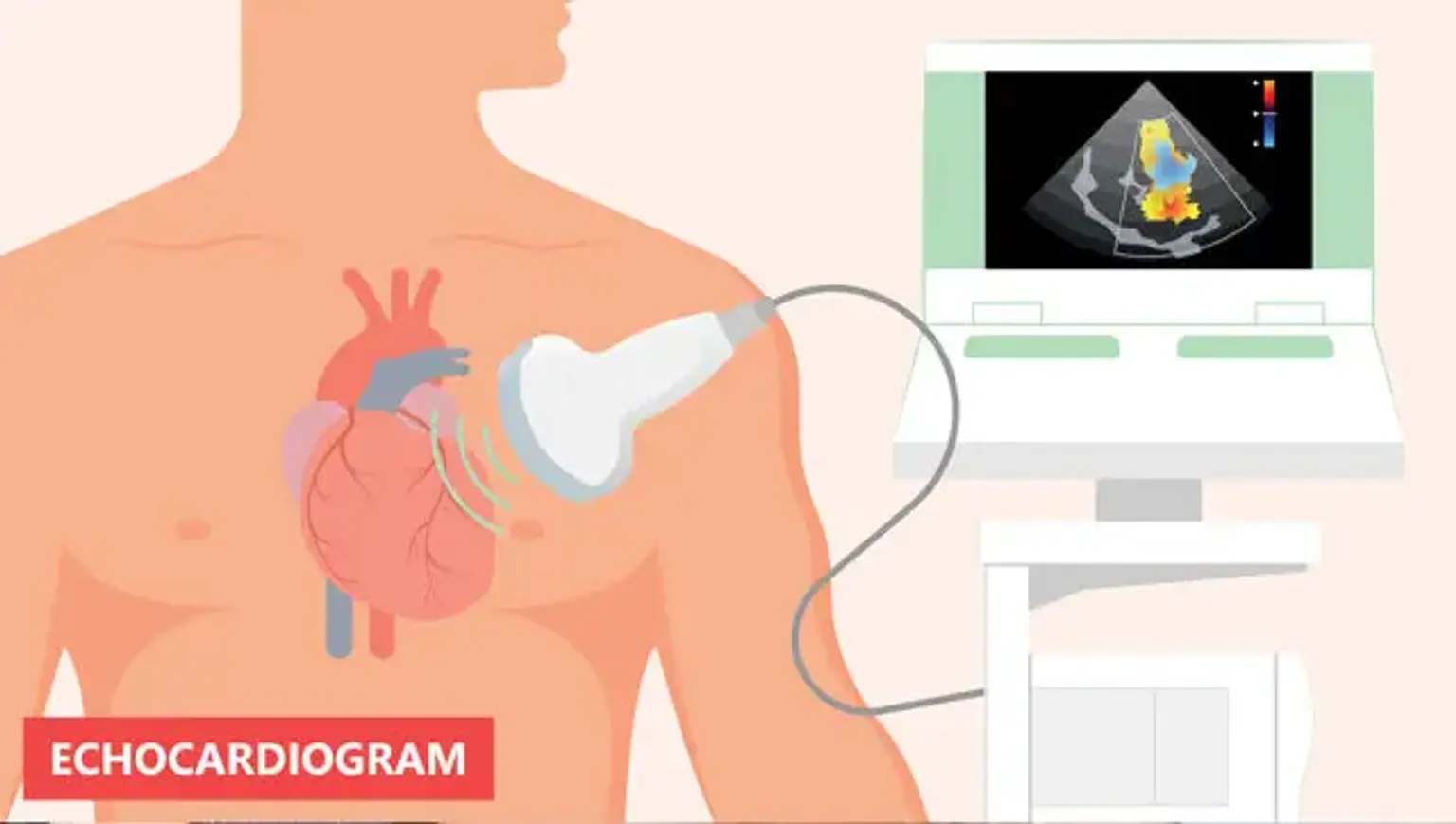
This is the most common echocardiogram type that diagnoses a range of cardiac disorders. With this technique, the sonographer or physician spreads gel to the mechanism (transducer). He or she then presses the transducer tightly against the skin, pointing the ultrasound beam via the chest to the heart.
This transducer will record the echoes or reflections of the sound wave from the heart. Using computer technology, the recorded echoes will be translated into moving pictures on the display screen.
- Transesophageal echocardiography
Sometimes, the doctor needs more accurate pictures or finds it hard to obtain a clear image of the heart using a regular echocardiogram. In such cases, they can suggest a transesophageal echocardiogram.
During this procedure, they will numb the throat and administer medications to keep you calm and relaxed. They will then direct a small flexible tube with a transducer down the throat and towards the tube that connects the mouth to the stomach or esophagus.
This transducer will record the echoes of the sound wave within the heart. A computer will then transform the echoes into detailed and accurate live pictures of the heart viewed on the monitor.
- Doppler echocardiogram
The sound waves usually cause changes to the pitch as they deflect off the blood cells that move via the blood vessels and heart. Such changes known as Doppler signals enable the doctor to test the direction and blood flow speed within the heart.
The Doppler methods are widely applied in transesophageal and transthoracic echocardiograms. Doctors can also use these methods to monitor blood flow issues and blood pressure within the arteries of the heart. The traditional ultrasound approach may not detect this. The blood flow displayed on the monitor is normally colored to help the doctor identify any issues.
- Stress echocardiography
Certain heart problems only arise during physical activities. This is especially those conditions that involve the arteries supplying blood into the heart muscle or coronary arteries. Therefore, the doctor can suggest a stress echocardiogram to evaluate coronary artery disorders. However, the echocardiography technique doesn’t provide details on the blockages within the arteries of the heart.
With a stress echocardiogram, the ultrasound pictures of the heart are recorded before and shortly after walking on a treadmill or riding a stationary bike. For individuals who can’t exercise, they can receive an injection of drugs. This helps stimulate the heart pump as vigorously as if they are exercising.
- Three-dimensional (3-D) echocardiography
A 3-D echocardiogram utilizes either transesophageal or transthoracic echocardiography to produce a 3-D view of the heart. This includes numerous pictures from various angles. This technique is also used before the surgery of the heart valve. Furthermore, it helps detect cardiac defects among children.
What to Expect During Echocardiography Procedure
Echocardiogram procedures can take about an hour or less and are normally performed in the physician’s office or hospital. During the transthoracic echocardiogram process, you should expect the following;
- Undressing from your waist up and lying on the examination bed or table
- Attaching sticky patches or electrodes to the body to make the detection easy and help conduct electrical currents in the heart.
- Applying gel on the transducer that increases the conductivity of sound waves
- Moving or passing the transducer to and from the chest to capture pictures of the heart's sound wave echoes. You might hear a pulsing whiz that is an ultrasound capturing the blood moving through the heart.
- If necessary, the doctor can ask you to breathe in a particular manner or rollover on the left-hand side.
In case you are undergoing a transesophageal echocardiogram, you should expect the following;
- Numbing of the throat using gel or spray
- Administration of a sedative to keep you calm and relaxed.
- Insertion of a tube containing the transducer into the throat and directing it via the esophagus. It’s then placed in a strategic location to take pictures of the heart.
Recovery and Results
People usually return to normal day-to-day activities immediately after echocardiography. If the results are good, then further diagnosis is not necessary. However, if they are concerning, the physician can refer you to a professional cardiologist for additional tests.
The echocardiogram results and information can indicate the following;
Changes in the size of the heart: High blood pressure, damaged or weakened heart valves, or any other disorders may cause the heart's chambers to expand. This can also make the heart's walls abnormally thickened.
Heart muscle damage: Echocardiogram allows the doctor to evaluate if all heart wall areas normally contribute to the beating activity of the heart. Parts of the heart wall that shift weakly might have been weakened or injured during a heart attack. It can also be because it’s getting too little amount of oxygen.
The pumping power: The measures derived from an echocardiogram test comprise the percentage of blood pumped out of the full ventricle for every pulse (ejection fraction). It also includes the amount of blood pumped through the heart within a minute (cardiac output). If the heart fails to pump sufficient blood to satisfy the needs of the body, it can result in heart failure symptoms.
Defects of the heart: Echocardiogram test can indicate issues and defects of the chambers of the heart. It also evaluates irregular connections between the main blood vessels and the heart and severe heart abnormalities present during birth.
Heart valve issues: An echocardiogram test enables the doctor to check whether the heart valves are large enough to have enough blood supply. It also helps check if it closes completely to avoid blood leakage.
Risks of Echocardiography
There are no complications associated with the normal transthoracic echocardiography technique. However, you can experience some pain due to the transducer being pressed tightly against the chest. Such firmness is needed to deliver the best-detailed pictures of the heart.
If you undergo a transesophageal echocardiogram, you are likely to have a sore throat that can last for several hours. In rare cases, the tube inside the throat can scratch. During the procedure, to screen for any respiratory issues that occur due to sedation drugs, the doctor will monitor the oxygen intake.
When undergoing stress, echocardiogram tests, exercise or medication can temporarily trigger an abnormal heartbeat. However, the echocardiogram itself doesn’t result in any complications. Besides, severe complications, including heart attacks, are uncommon.
Conclusion
Echocardiography is a non-invasive technique that doctors use to assess the heart, including the structure, functions, and underlying disorders. It involves the use of a transducer that transmits high-frequency sound waves which echo the heart structures. These echoes will be transformed into moving images that doctor views through the monitor. Based on the information the doctor requires, you might have to undergo a particular type of echocardiogram.
CloudHospital specializes in performing echocardiography test procedures to diagnose various heart diseases and disorders. It works with several skilled and experienced experts who effectively conduct the procedure and use the result to develop a treatment plan.