Emphysema
Overview
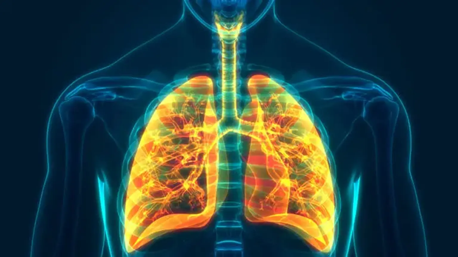
Emphysema, also known as pulmonary emphysema, is a condition of the lower respiratory tract characterized by air-filled spaces in the lung that can vary in size and can be quite big. The voids are created by the disintegration of alveolar walls and replace the spongy lung parenchyma. This lowers the total alveolar area accessible for gas exchange, resulting in a decrease in blood oxygen delivery.
What is emphysema?
Pulmonary emphysema is described as a pathological, permanent dilation of the distal airways (respiratory bronchioles, alveolar ducts, and alveolar sacs) caused by airway wall disintegration without fibrotic alterations. Emphysema kills vital ventilatory units and disrupts gas exchange.
Functionally, emphysema creates an obstructive ventilatory deficit, as indicated by spirometry, and is part of the chronic obstructive pulmonary disease (COPD) group of illnesses. Advanced emphysema is a common and significant cause of chronic respiratory failure as well as sudden decompensation, which can be fatal. Because of the severe morbidity and mortality associated with this illness, detecting and treating it in its early stages is critical.
Emphysema is distinguished from interstitial pneumonia by the lack of pulmonary interstitium fibrosis. Nonetheless, in many individuals with established emphysema, combined pulmonary fibrosis and emphysema (CPFE) has been described as a discrete clinical entity. Repeated emphysema exacerbations and subsequent decompensation induce a fast loss in lung function that is more than predicted with age.
COPD is the third most common cause of death in the United States and the fourth most common cause of mortality worldwide. According to World Health Organization (WHO) projections, it will be the third leading cause of death globally by 2020. COPD sufferers have chronic bronchitis and emphysema. Despite being defined as distinct entities, the majority of COPD patients exhibit characteristics of both. COPD frequently coexists with comorbidities, which impair the disease's progression.
Emphysema is a pathological condition that affects the air spaces distal to the terminal bronchiole. It is distinguished by abnormal permanent expansion of lung air gaps with degradation of their walls and lack of elasticity, as well as deterioration of lung parenchyma.
Epidemiology
Emphysema, or the larger term COPD, which also encompasses chronic bronchitis, is a common ailment that affects almost 250 million people worldwide. This condition affects around 14 million individuals in the United States of America (USA). COPD is becoming more common as a result of a societal shift toward the use of cigarettes in the United States.
The incidence is also higher in places with higher levels of pollution. Pneumoconiosis in coal miners is another cause of emphysema, even in nonsmokers. Males have a somewhat greater frequency than females, and it is connected to the level of smoking.
Its prevalence is gradually growing, owing mostly to a rise in cigarette smoking and environmental pollutants. Another factor is a reduction in mortality from other causes, such as cardiovascular and infectious illnesses. Genetic variables also play a key influence in predicting whether or not patients will experience airflow restriction. The severity of emphysema in coal miner pneumoconiosis is much greater, regardless of smoking status.
Emphysema causes
Despite the fact that several academics throughout the world have hypothesized on various factors, cigarette smoking is the most major acknowledged cause of emphysema. Emphysema affects around 10 to 15% of cigarette smokers throughout the course of their lives, depending on the severity and length of smoking.
Genetic factors, such as alpha-1 antitrypsin (AAT) deficiency, an autosomal recessive illness, are a cause of emphysema, particularly in children, and mostly affect the lower lobes of the lungs. Emphysema from cigarette smoking, on the other hand, is more frequent in the fifth decade and above, affecting mostly the upper lobes of the lung.
Emphysema commonly develops after 20 pack-years of smoking. Environmental exposure to specific chemicals, passive smoking, occupational drug exposure, and recurring lung infections have all been linked to emphysema. In the past, one of the most common causes of emphysema in underdeveloped nations was exposure to smoke from the customary use of a wood burner for cooking. There have been stories of young people developing emphysema after using marijuana.
Pathophysiology
Emphysema is the degeneration of the ventilatory functional respiratory unit, which includes the alveolar walls, respiratory bronchioles, alveolar ducts, and alveolar sacs. This results in persistent dilatation, which reduces the surface area of the ventilatory units. An unbroken alveolar wall, interstitium, and capillaries with appropriate blood flow are required for ventilation exchange.
Emphysema disrupts gas exchange, resulting in acute hypoxia and hypercapnia and, ultimately, respiratory failure. Emphysema is divided into three types: centriacinar, panacinar, and paraseptal. Paraseptal emphysema can occur in smokers as well as in those who are deficient in alpha-1 antitrypsin (AAT). Centriacinar is frequent among smokers, while panacinar is common in those with AAT deficiency.
Inflammatory mediators such as macrophages, T lymphocytes, and neutrophils are triggered by smoke and other substances, producing cytokines. Proteinases are also produced, damaging the lung parenchymal connective tissue and ventilatory units, resulting in severe hypoxia and hypercarbia in advanced instances.
Excessive mucus production is another complication. It is worth emphasizing that AAT is an anti-proteinase that inhibits lung deterioration by counteracting the proteinase, and emphysema arises owing to the loss of this protection due to hereditary deficiency.
Centrilobular emphysema
Centrilobular emphysema is a chronic lung condition that worsens with time. It is thought to be a kind of chronic obstructive pulmonary disease (COPD). The higher lobes of the lungs are primarily affected by centrilobular emphysema. It is distinguished by damage to your respiratory airways.
Bullous emphysema
Bullous emphysema is distinguished by damaged alveoli that distend to generate unusually wide air gaps, particularly in the topmost parts of the lungs. This syndrome can appear in otherwise healthy young people.
Symptoms of emphysema
A typical emphysema patient is in their fifth decade or older, has a long smoking history, and presents with a persistent, productive cough and progressive shortness of breath. The symptoms of severe COPD grow over time, interfering with daily activities. They may also lose weight unintentionally and seem cachexic.
Patients decompensate and typically have a cough with copious amounts of phlegm, significant wheezing, and proceed to hypercarbia with concomitant hypoxic respiratory failure in acute exacerbations. Patients often exhibit 'purse lip breathing,' are tachypneic, and engage their auxiliary muscles of respiration. Auscultation often demonstrates decreased air intake.
Cor pulmonale is a consequence of long-term emphysema that can induce symptoms and indications of right heart failure. These individuals are referred to as 'pink puffers' because they are plethoric and have a hyperexpanded (barrel-shaped) chest. In the early stages, they are not cyanotic.
Patients with decompensated chronic bronchitis, on the other hand, are referred to as "blue bloaters" because they are cyanotic. Hemoptysis is uncommon in COPD, and if it occurs, more severe disorders such as malignancy should be considered in an appropriate clinical context.
It is worth noting that there is a disease known as asthma COPD overlap syndrome (ACOS) in which both of these disorders coexist. It is critical to have a history in order to discern between the two, as asthma often has triggers for aggravation and may have seasonal changes. Upper airway illnesses such as chronic rhinosinusitis can coexist, thus a thorough history should be taken.
AAT insufficiency is a genetically transmitted disorder, hence family history is critical. Cigarette smoking is often a cultural trait that runs through families. Similarly, information regarding ill contacts would be useful, as viral infections are the most prevalent cause of COPD exacerbation. A vaccination history, including the administration of the yearly influenza vaccine, is also required.
The person's smoking history is significant, with an emphasis on the age at which they began smoking and the overall number of pack years. If the person has stopped smoking, it is critical to know how long it has been since he or she last smoked. It is critical to have a history of environmental and occupational exposure, as well as a family history of chronic respiratory diseases and COPD.
The physical examination may be normal in the early stages of the illness. Emphysema patients are commonly referred to as "pink puffers," which means cachectic and non-cyanotic. Expiration via pursed lips raises airway pressure and avoids airway collapse during respiration, and the employment of respiratory accessory muscles suggests advanced illness. COPD is not characterized by digit clubbing. Many more comorbidities might exist. Current smokers may have a smoke odor as well as nicotine stains on their hands and fingernails.
Percussion may be normal in the early stages of the condition. The remainder of the test may include extended expiration or wheezes on forced exhale, as well as increasing resonance, which indicates hyperinflation as the airway blockage worsens. Auscultation detects distant breath sounds, wheezes, crackles at the lung bases, and/or distant heart sounds.
Diagnosis
Following a thorough history and physical examination, the next step in the diagnosis of COPD is a full pulmonary function test (PFT). Spirometry, bronchodilator reversibility testing, lung volumes, carbon monoxide diffusion capacity (DLCO) measurement, and flow volume loop are all part of the PFT (FVL). The inclusion of arterial blood gas (ABG) to the PFT is useful in many circumstances.
Spirometry often reveals a decrease in both forced vital capacity (FVC) and forced expiratory volume at first second (FEV1). FEV1 drops more proportionally than FVC declines, and the FEV1/FVC ratio is less than 0.7. However, this 'obstructive' ventilatory defect is not pathognomonic for COPD, and spirometry may occasionally indicate a'restrictive' defect as well, depending on other underlying illnesses.
Bronchial hyperreactivity would be indicated by a significant improvement in FEV1 or FVC (defined as greater than 200 mL or 12%, respectively). Based on the FEV1, obstruction is classified as minor, moderate, or severe. The lung volumes may reveal signs of air trapping, hyperinflation, or both. In COPD, the DLCO is frequently decreased, indicating difficulty with gas exchange. It should be noted that DLCO may be lowered in different disorders such as heart failure, pulmonary hypertension, pulmonary vascular diseases, and so on.
The Global Initiative for Chronic Obstructive Lung Diseases (GOLD) classifies the degree of blockage in individuals with a FEV1/FVC ratio less than 0.7 as follows.
- Mild when FEV1 greater than or equal to 80 of predicted
- Moderate when FEV1 greater than or equal to 50 and less than 80% of predicted
- Severe when FEV1 greater than or equal to 30 and less than 50% of predicted
- Very severe when FEV1 less than 30% of predicted
Radiology is important in the diagnosis of emphysema. An X-ray of the chest would reveal hyperinflation and a flattened diaphragm. A chest computed tomogram (CT) is more specific and confirming for emphysema. Radiological imaging of pulmonary hypertension show a prominent or dilated pulmonary artery. In the diagnosis of emphysema, MRI of the chest has no established advantage over standard CT scan of the chest.
Genetic testing for AAT deficiency is required in young individuals with emphysema. Coexisting asthma or pulmonary fibrosis would necessitate further testing, such as a blood eosinophil count, immunoglobulin E levels, an autoimmune and vasculitis screen, allergy tests, and so on. An echocardiography would be used to determine secondary pulmonary hypertension.
Exercise oximetry (also known as the 6-minute walking test) is a simple method for determining chronic hypoxic respiratory failure that is required before beginning long-term oxygen treatment (LTOT)
Treatment for emphysema
Treatment includes both pharmaceutical and nonpharmacological interventions. Smoking cessation, as well as pollution avoidance, are critical to preventing additional lung damage. Annual influenza and pneumococcal immunizations are required in these patients unless contraindicated. Exacerbations can be avoided by avoiding ill contacts. In these individuals, adequate diet and, preferably, the aid of a nutritionist might be beneficial.
Patients who have an oxygen saturation of less than 88 percent or a PaO2 of less than 55 mmHg at rest or with exertion require long-term oxygen treatment. The benefits of pulmonary rehabilitation in increasing lung function and exercise capacity have been demonstrated. Monitoring lung function at regular intervals is useful for detecting rapid deterioration.
Pharmacological treatment is the cornerstone of COPD management. Albuterol, levalbuterol, anticholinergics such as ipratropium, inhaled corticosteroids (ICS), long-acting beta agonists (LABA), and long-acting muscarinic antagonists are examples of inhaled bronchodilators (LAMA). Though a combination of ICS and LABA was formerly used more frequently, with new findings of rising pneumonia in these patients, several physicians have successfully adopted a more 'nonsteroidal' strategy with the combination of LABA and LAMA.
LABA has been recommended for COPD without the addition of ICS or LAMA, despite evidence of higher fatalities when used in asthma. In severe situations, triple treatment with an ICS, LABA, and LAMA combination is required. For patients who have trouble utilizing inhalers, most of these drugs are available in nebulized form.
Roflumilast, a phosphodiesterase inhibitor, has been shown to minimize exacerbations in refractory individuals. Many additional medications, such as theophylline and long-term antibiotics such as azithromycin, might be attempted. Long-term oral corticosteroids may be the sole choice in a few individuals, despite the fact that they are not frequently advised.
Systemic corticosteroids, in addition to inhaled medicines, are primarily beneficial in COPD exacerbations. Methylprednisolone, hydrocortisone, and dexamethasone are commonly used around the world. Prednisone used orally is just as effective as steroids given intravenously. Antibiotics and, in rare cases, antivirals are utilized in acute exacerbations.
Patients with alpha-1 anti-trypsin deficiency would be candidates for the enzyme's "augmentation treatment." Lung volume reduction surgery may be an option for certain people with bullous emphysema. In appropriate individuals with "end-stage" illness, a lung transplant may be a possibility.
Supportive Therapy
Supportive therapy comprises oxygen therapy and ventilatory support, pulmonary rehabilitation, and palliative care.
In stable patients, routine supplementary oxygen has little effect on quality of life or clinical results. Continuous long-term, i.e., more than 15 hours of supplemental oxygen, is advised in patients with COPD who have a PaO2 less than 55 mmHg (or oxygen saturation less than 88 percent) or a PaO2 less than 59 mmHg in the event of cor pulmonale. Oxygen treatment has been demonstrated to improve the survival of these individuals with severe resting hypoxemia. Intermittent oxygen will aid people who desaturate during exercise. The objective is to keep the oxygen saturation over 90%.
Ventilation-perfusion mismatch (V/Q mismatch) is a prominent cause of hypoxemia in COPD, especially in low V/Q regions. The purpose of hypoxic vasoconstriction of the pulmonary arteries is to increase total gas exchange efficiency. Supplemental oxygen can successfully reach the alveoli in these lungs, preventing vasoconstriction and so increasing perfusion and improving gas exchange, resulting in hypoxemia improvement.
In patients with acute respiratory failure, noninvasive positive pressure ventilation (NPPV) has been shown to reduce morbidity and death. It should be tried as the initial mode of ventilation in patients with COPD exacerbation and respiratory failure who have no absolute contraindications since it improves gas exchange, minimizes hospitalization length, decreases labor of breathing, improves VQ matching, and increases survival. If NPPV fails to function in a COPD patient in a hospital environment, the patient should be intubated and placed on a ventilator.
Although palliative care is available from the time a person is diagnosed with COPD, it is primarily recommended for GOLD stage D patients. It is an addition to the patient's current treatment regimen. The objective is to give the highest possible quality of life. It not only aids in the assessment and management of symptoms, but it also assists patients in understanding their condition and promotes discussions about the patient's objective of care, advance care, and end-of-life care plans.
Advance care planning include communication between patients, their families, and their physicians, and it assists patients in developing treatment choices. A key component of palliative care is reassuring patients with a clear plan for dealing with dyspnea in late illness as well as the management of despair and anxiety.
These can be treated with low-dose opioids, lifestyle changes, and relaxation methods, in that order. Because most patients underestimate the condition, it is critical to check for transition time and address advanced care as early as possible in the disease. It is also critical to find out where the patients wish to spend their final days (for example, at home or in the hospital) and to offer them with as much comfort as possible.
Differential Diagnosis
Because the symptoms of COPD are vague, the differential diagnosis is fairly wide. Bronchial asthma also has an obstructive ventilatory problem. Fixed obstruction can also occur in long-term asthma without accompanying COPD. Many chronic respiratory disorders, such as pulmonary fibrosis, bronchiectasis, heart failure, pulmonary hypertension, and pulmonary vascular diseases, have many symptoms in common.
Chronic cough can be caused by infectious illnesses such as pneumonia, TB, and fungal infections, as well as inflammatory disorders such as interstitial pneumonia. Chronic aspiration should be considered a treatment option in older people and those suffering from neurological illnesses. In suitable clinical conditions, nonpulmonary disorders such as severe acid reflux disease and upper airway diseases such as chronic rhinosinusitis might be included in the differential diagnosis.
Prognosis
Emphysema is defined as the persistent breakdown of the alveolar walls. Early detection and treatment, on the other hand, can avoid the rapid decrease of lung functioning. The most important element in preventing additional lung damage is quitting smoking.
A bad prognosis is associated with inadequate nutrition, the presence of bronchial hyperreactivity, male gender, bacterial colonization, a concomitant immunocompromised condition, cardiac or other respiratory disorders. Many doctors utilize the BODE index (Body mass index, Level of Obstruction, Dyspnea, and Exercise capacity) to predict COPD mortality.
Chronic hypercarbia is an alarming indicator of long-term sickness and is connected with a bad prognosis. Pulmonary function falls more than predicted for age with each exacerbation. The presence of emphysema on a CT chest is a sign of a bad prognosis. However, smoking cessation and long-term oxygen treatment (LTOT) have been shown to lower COPD mortality.
The presence of additional disorders worsens the prognosis in COPD patients. Patients having characteristics of both asthma and COPD, for example, have a worse quality of life and a greater death rate. Patients with emphysema who have elevated serum alpha-1 antitrypsin levels are also more likely to die. Comorbid disorders that are often observed include metabolic syndrome, cardiovascular disease, hypertension, and bronchiectasis.
A widely used tool for prognosis prediction is the BODE index which takes into account the following:
- BMI
- FEV (obstruction)
- Dyspnea
Complications
COPD patients can develop life-threatening decompensated acute respiratory failure. Home oxygen would be required even in compensated individuals with persistent respiratory failure. Continued smoking in these people would put them at danger of developing cancer. COPD consequences include decreased exercise capacity, cachexia, cor pulmonale, and recurring chest infections. Depression, anxiety, and irritability are more common in these individuals, and the treating practitioner should address them appropriately.
Conclusion
Lung emphysema is a kind of chronic obstructive pulmonary disease described as a pathological, permanent dilatation of distal airways (respiratory bronchioles, alveolar ducts, and alveolar sacs) caused by airway wall degradation without fibrotic alteration. Emphysema disrupts gas exchange, resulting in restrictive ventilation.
An interprofessional team including a pulmonologist, internist, pharmacist, dietician, social worker, respiratory therapist, primary care provider, and thoracic surgeon manages emphysema. There is no treatment for emphysema, and the underlying cause must be addressed. This includes informing the patient about the dangers of smoking and obtaining the yearly influenza vaccination.
Most patients' prognoses are bleak, and their quality of life is marred by frequent exacerbations and remissions of severe respiratory distress. Some patients may experience recurring pneumothoraces, which must be handled by a thoracic surgeon. There are several types of lung volume reduction surgeries available, but they are high risk, have major consequences, and need lengthy intensive care.
Pharmacists must examine medications for interactions and emphasize the need of compliance to patients and their families. Nurses should monitor and educate patients while also informing the team of any changes in their condition.