What is Endometrial Hyperplasia?
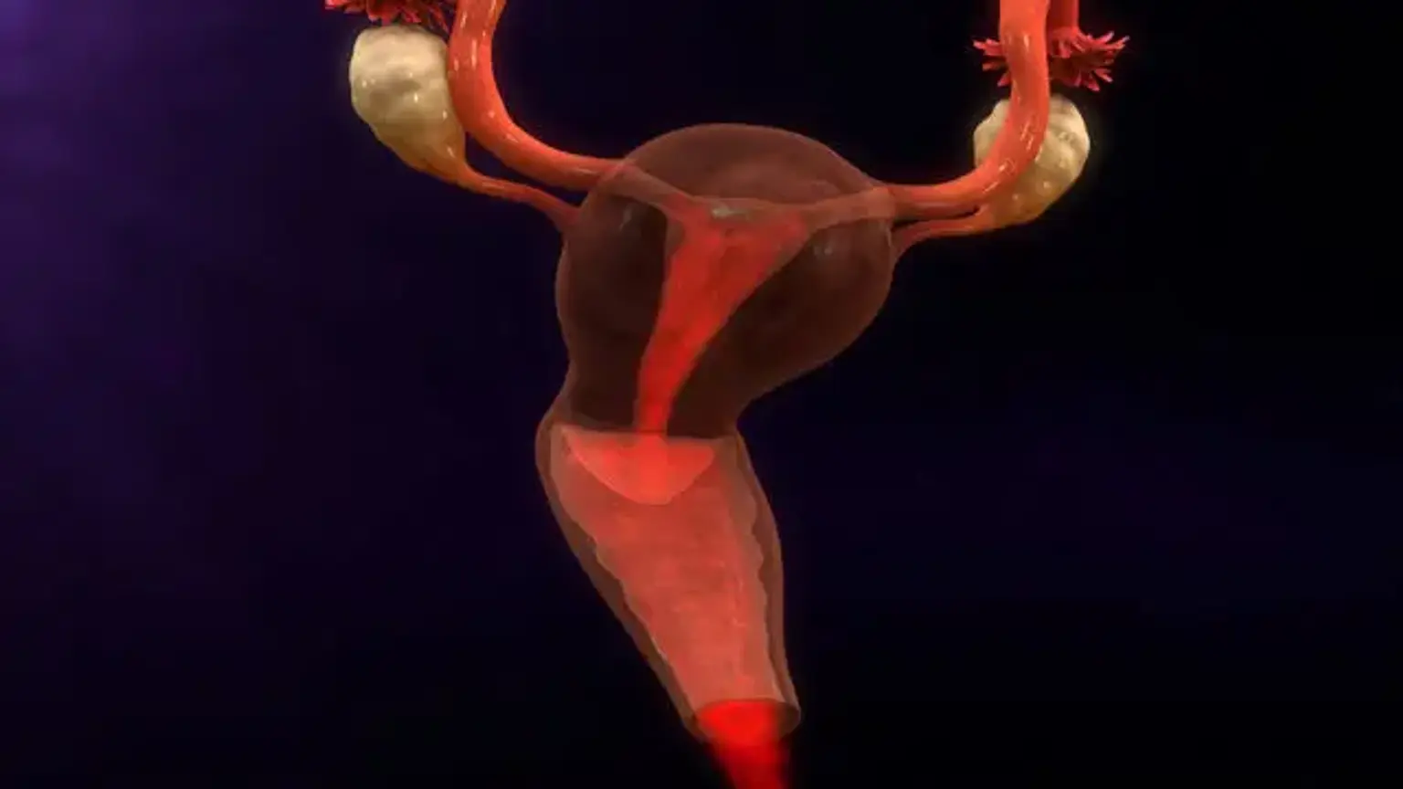
Endometrial hyperplasia is a medical condition in which the lining of the uterus (the endometrium) becomes abnormally thick. This thickening occurs due to an overgrowth of cells in the endometrial lining, usually as a response to prolonged exposure to estrogen without adequate progesterone to balance its effects. The endometrial lining, which normally thickens and sheds each month in response to hormonal changes, becomes excessive in this condition.
While endometrial hyperplasia can occur in women of all ages, it is most common in women who are in their reproductive years or approaching menopause, especially when there is an imbalance in their hormone levels. The condition may be classified as simple or complex, with or without atypical cells. The presence of atypical cells increases the risk that the hyperplasia may progress to endometrial cancer.
Endometrial hyperplasia can be precancerous in some cases, but it is not cancer itself. However, if left untreated, certain types of hyperplasia, especially those with atypical cells, may develop into endometrial cancer, which makes early detection and treatment crucial.
Importance of Treatment
The treatment of endometrial hyperplasia is crucial because, if left unmanaged, the abnormal cell growth in the uterine lining can become more severe and potentially lead to endometrial cancer. For this reason, early diagnosis and appropriate intervention are key to reducing the risk of progression.
Endometrial hyperplasia presents with symptoms like abnormal uterine bleeding, which includes heavy or irregular periods, spotting between periods, or postmenopausal bleeding. Such symptoms can be alarming, and they may lead women to seek medical advice early on. Treatment is vital not only to alleviate these symptoms but also to prevent the condition from progressing to something more serious.
The appropriate treatment also depends on the type and severity of the hyperplasia, as well as a woman’s individual health and reproductive goals. Hormonal therapies can be effective for women who wish to preserve fertility, while surgical treatments, such as a hysterectomy, may be necessary in more severe cases.
By addressing endometrial hyperplasia early, most women can avoid complications and improve their overall reproductive health.
Global Prevalence and Recognition
Endometrial hyperplasia is a prevalent condition worldwide, though its exact prevalence can vary based on factors like age, geographical location, and the presence of underlying health conditions like obesity or polycystic ovary syndrome (PCOS). It is particularly common in women who are peri-menopausal or postmenopausal, where hormone imbalances—especially elevated levels of estrogen—are more frequent.
Recent studies show that endometrial hyperplasia affects approximately 4-5% of women of reproductive age. In postmenopausal women, the prevalence increases, especially among those who are not on hormone replacement therapy (HRT). In fact, it is one of the most commonly diagnosed gynecological conditions associated with abnormal uterine bleeding in postmenopausal women.
Globally, the recognition of endometrial hyperplasia has led to better screening and early diagnosis, improving the chances of effective treatment. However, despite being widely acknowledged in medical practice, there are significant challenges in its awareness among the general public. As women become more informed about the condition, early intervention will be more likely, leading to better outcomes.
For many women, the thought of endometrial hyperplasia can be concerning, as they might confuse it with cancer. Understanding that it is often a treatable condition is essential for reducing unnecessary anxiety. With advances in medical technology and treatments, women today have more options than ever to address this condition effectively.
Hormonal Imbalance and Endometrial Hyperplasia
Hormonal imbalance is the primary cause of endometrial hyperplasia. Normally, the menstrual cycle is regulated by a balance of estrogen and progesterone. Estrogen is responsible for stimulating the growth of the endometrial lining, while progesterone counters this effect by stabilizing the lining and preparing it for menstruation.
In women with endometrial hyperplasia, estrogen is typically present in excess or is not balanced adequately by progesterone. This imbalance causes the endometrial lining to continue growing beyond its normal thickness, eventually leading to hyperplasia. This is why endometrial hyperplasia is most commonly seen in women who experience estrogen dominance, particularly those who:
Have irregular periods (e.g., in polycystic ovary syndrome or PCOS),
Are going through perimenopause,
Are obese (as fat tissue produces additional estrogen),
Use certain types of estrogen-only hormone replacement therapy (HRT).
Other factors, such as diabetes, hypertension, and certain genetic conditions, can also increase the risk of hormonal imbalance, further elevating the likelihood of developing endometrial hyperplasia.
It is important to note that while hormonal imbalance is the leading cause, unopposed estrogen therapy (where estrogen is taken without progesterone) is a significant contributing factor. This is why women who take estrogen-only therapies, such as those used for menopause or birth control, are often given progesterone as well to reduce the risk of endometrial hyperplasia.
Hormonal Therapy Overview
Hormonal therapy is a first-line treatment for endometrial hyperplasia. The most common approach is the use of progestin, a synthetic form of the hormone progesterone. Progestin works by balancing estrogen levels, which helps to shrink the overgrown endometrial tissue. This therapy is typically recommended for women who wish to preserve fertility or avoid surgery.
Progestin can be administered in various ways:
Oral tablets (taken daily),
Intrauterine device (IUD) releasing progestin,
Injectable progestin (every 3 months).
Hormonal therapy is particularly effective for non-atypical hyperplasia. It can also help regulate menstrual cycles and reduce heavy bleeding. However, it requires ongoing monitoring to assess its effectiveness, and side effects such as mood swings or irregular bleeding may occur.
Hysterectomy: A Definitive Treatment
For severe cases of endometrial hyperplasia, especially when there is atypical hyperplasia or the risk of cancer, a hysterectomy (removal of the uterus) may be necessary. This is considered the most definitive treatment, eliminating the risk of recurrence.
A hysterectomy is typically recommended when other treatments have failed, or if the woman is no longer interested in having children. The procedure can be done in several ways: through the abdomen or vaginally, depending on the patient’s condition and surgeon’s recommendation.
While a hysterectomy is highly effective, it is a major surgery with risks such as bleeding and infection. It also results in the permanent loss of fertility and may induce menopause if the ovaries are removed. Recovery can take several weeks, but it provides a long-term solution for managing hyperplasia and preventing cancer.
D&C (Dilatation and Curettage)
For more severe or recurring endometrial hyperplasia, a D&C (Dilatation and Curettage) may be necessary. This procedure involves scraping or suctioning the excess tissue from the uterus. It is both a diagnostic and therapeutic tool, especially when biopsy results show abnormal tissue.
The D&C is performed under anesthesia and can provide immediate relief from symptoms like abnormal bleeding. However, it does not always offer a permanent solution, especially for women with recurrent hyperplasia or atypical hyperplasia. For persistent cases, further treatments such as a hysterectomy might be considered.
Ablation Therapy
Endometrial ablation is a procedure that destroys the uterine lining to prevent further abnormal growth. This treatment is an option for women who no longer wish to bear children and have not responded to other therapies. It works by using heat, cold, or lasers to destroy the endometrium.
Endometrial ablation can significantly reduce heavy bleeding and prevent the recurrence of hyperplasia. However, it is not a cure for the underlying hormonal imbalance and does not completely eliminate the risk of cancer. Women should consider this option carefully, as it makes future pregnancies impossible.
Role of Endometrial Biopsy
An endometrial biopsy is the gold standard for diagnosing endometrial hyperplasia. It involves removing a small tissue sample from the uterine lining to examine under a microscope. This procedure helps determine if the abnormal thickening is caused by hyperplasia or if there are signs of precancerous or cancerous cells.
The biopsy is usually done in the office with local anesthesia. Though mildly uncomfortable, it is quick and provides essential information about the type of hyperplasia, guiding treatment decisions. It is especially important if there are concerns about atypical hyperplasia or endometrial cancer.
Hormonal Therapy for Endometrial Hyperplasia
Hormonal therapy is a cornerstone of treatment for endometrial hyperplasia. Progestin, a synthetic form of progesterone, is often prescribed to counteract the effects of excess estrogen. Progestin can be given in several ways:
Oral pills,
Intrauterine device (IUD),
Injections.
Hormonal therapy is effective for non-atypical hyperplasia and can reduce symptoms like heavy bleeding. It's especially beneficial for women who wish to preserve fertility, as it can prevent the need for surgery. However, it may cause side effects such as changes in menstrual cycles or mood swings. Regular follow-up is necessary to assess treatment effectiveness.
Hysterectomy: The Last Resort
For women with severe endometrial hyperplasia or those at high risk of progression to cancer, a hysterectomy may be the most definitive treatment. A hysterectomy involves removing the uterus and, sometimes, the ovaries. This is typically considered for women who no longer wish to bear children or when other treatments have failed.
Although a hysterectomy eliminates the risk of recurrence, it is a major surgery with significant recovery time. Women who undergo this procedure may experience changes in hormone levels, especially if the ovaries are removed, leading to menopause. Despite the risks, a hysterectomy offers the best long-term solution for managing endometrial hyperplasia, particularly in cases with atypical hyperplasia.
Managing Side Effects of Treatment
After treatment for endometrial hyperplasia, patients may experience side effects, depending on the type of therapy. For those undergoing hormonal therapy, side effects may include irregular bleeding, mood swings, weight gain, and headaches. These symptoms are often temporary and improve as the body adjusts to the hormonal changes.
For women who undergo a D&C, post-procedure bleeding and cramping are common but usually subside within a few days to weeks. Endometrial ablation may cause some discomfort during recovery, and light bleeding can last for a few weeks.
For hysterectomy patients, recovery is more involved. The initial weeks may involve pain management and light bleeding, with full recovery taking about 6-8 weeks. Emotional recovery can also be significant, especially if the procedure results in menopause.
Regular follow-ups are essential to monitor for any recurrence of symptoms or complications, ensuring the treatment remains effective.
Lifestyle Changes to Support Healing
Post-treatment lifestyle changes can play a crucial role in recovery and long-term health. Maintaining a healthy weight, staying physically active, and managing stress are important steps. Excess weight can contribute to estrogen dominance, potentially increasing the risk of recurrence. A balanced diet rich in fruits, vegetables, and whole grains can support overall health and hormone balance.
For women who have undergone surgery, avoiding strenuous activity and lifting heavy objects is essential during the initial recovery phase. Once cleared by a healthcare provider, resuming light exercises like walking can help with overall well-being and reduce the risk of complications.
Monitoring for Recurrence
Even after successful treatment, there’s always a risk of recurrence, especially for women with atypical hyperplasia. Regular monitoring is necessary, including:
Routine pelvic exams,
Ultrasounds to check the thickness of the endometrial lining,
Follow-up biopsies if abnormal bleeding recurs.
For those on hormonal therapy, follow-up visits are crucial to adjust medications and ensure the treatment is still effective. For women who have undergone ablation or a hysterectomy, routine gynecological evaluations can ensure the condition does not return or develop into cancer.
Early detection of recurrence is vital to managing the condition before it escalates.
Emotional and Psychological Support
Dealing with endometrial hyperplasia and its treatment can take an emotional toll, particularly for women undergoing major procedures like a hysterectomy or ablation. The prospect of infertility, hormonal changes, or cancer can cause anxiety, depression, or feelings of loss.
Seeking emotional and psychological support is critical. Joining support groups, talking to a therapist, or seeking counseling can help women cope with the emotional impact of their treatment journey. Additionally, discussing concerns with a healthcare provider can offer reassurance and help develop a care plan that includes mental health support.
Self-care practices such as meditation, relaxation techniques, and journaling can also help in managing emotional distress. It's important for women to know that they are not alone, and support is available to help them through the recovery process.
Can Endometrial Hyperplasia Be Prevented?
While endometrial hyperplasia cannot always be prevented, certain lifestyle changes and medical interventions can reduce the risk of developing the condition. For women at risk—especially those with irregular menstrual cycles, obesity, or polycystic ovary syndrome (PCOS)—managing weight and maintaining hormonal balance can help.
Progestin therapy, especially for women on estrogen-only hormone replacement therapy, can also help prevent the overgrowth of the endometrial lining. Regular gynecological check-ups are important to catch early signs of hyperplasia, especially for women who have risk factors like obesity, diabetes, or a family history of uterine cancer.
Maintaining a healthy lifestyle, avoiding excessive estrogen exposure, and following medical advice can reduce the likelihood of developing endometrial hyperplasia and its complications.
Advancements in Treatment
Medical advancements continue to improve the treatment and management of endometrial hyperplasia. New forms of progestin therapy and IUDs are becoming more effective and easier to use. Researchers are also exploring targeted therapies that focus on hormonal regulation to prevent the growth of abnormal endometrial tissue without relying on traditional methods like hysterectomy or ablation.
Additionally, minimally invasive procedures, such as laparoscopic surgery and robot-assisted surgeries, are making treatments like hysterectomy safer and less traumatic for patients. These techniques often result in shorter recovery times, fewer complications, and better cosmetic outcomes.
In the future, the development of more advanced genetic testing and biomarker identification may help identify women at higher risk for developing hyperplasia or endometrial cancer, leading to earlier, more personalized treatments.
The Importance of Early Detection
Early detection of endometrial hyperplasia plays a crucial role in preventing the progression to endometrial cancer. Women who experience abnormal uterine bleeding or other symptoms of hyperplasia should seek medical attention promptly. A simple endometrial biopsy or ultrasound can help diagnose the condition early, allowing for more effective treatments that prevent serious complications.
Additionally, women with risk factors for endometrial hyperplasia should undergo regular screenings and health assessments. This can include monitoring for hormonal imbalances, maintaining a healthy weight, and treating conditions like PCOS or diabetes, which increase the risk of developing hyperplasia.
By staying vigilant about reproductive health and engaging in regular medical evaluations, women can take proactive steps toward reducing their risk of severe complications.
Conclusion
Endometrial hyperplasia is a treatable condition, and with early diagnosis and appropriate intervention, most women can manage it effectively. Whether through hormonal therapy, D&C, ablation, or hysterectomy, various treatment options cater to different patient needs. Women diagnosed with hyperplasia can benefit from personalized care plans that consider fertility goals, overall health, and the severity of the condition.
While the condition may pose risks, particularly when left untreated, the outlook is generally positive for women who follow their doctor's recommendations. With ongoing medical advancements and a focus on early detection, endometrial hyperplasia can often be managed without leading to more severe complications like endometrial cancer.
For women experiencing symptoms, seeking medical care early and discussing treatment options openly with healthcare providers can help ensure the best possible outcome and long-term reproductive health.