Endoscopic laser treatment
Last updated date: 13-Mar-2023
Originally Written in English
Endoscopic Laser Treatment
Overview
Laser research towards medical applications began shortly after its invention in 1960. Even the earliest research revealed that this unique technology had a high therapeutic potential.
Since those early studies, low-power lasers have been employed in diagnostic and therapeutic applications in a wide range of medical fields. However, due to uncertainty about the mechanism of action by which therapy is achieved and a lack of consensus protocols for certain applications due to the large number of parameters (e.g., frequency, energy, treatment timing, positioning) that can be selected, this modality of therapy remains highly controversial.
Using a tiny fiber led to the tumor tissue, a laser may be focused to a specific location of the brain. The laser transfers energy, which warms up the tissue surrounding the laser fiber's tip. High temperatures can induce irreversible tissue damage.
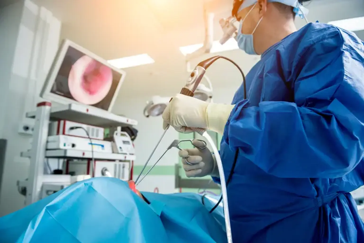
Advantages of Endoscopic Laser Surgery
- Endoscopic laser surgery helps patients with head and neck cancers to avoid chemoradiation, which is typically protracted and associated with a number of treatment-related consequences.
- There is no post-operative swelling, which may make breathing and, in some disorders, eating more difficult.
- Less trauma and a faster healing time following surgery.
Types of Endoscopic lasers
- Nd:YAG laser: The Nd:YAG laser was invented in the mid-1980s. After positioning the fluoroscopically guided probe, 1,000-1,850 J of energy was injected into the disk in continuous 1 second 20 W pulses and 1 second pauses, producing tissue ablation within the disk's nucleus. The vaporized products (water and carbon particles) were able to exit through the spinal needle. The FDA has now approved a 1.06 Nd: YAG laser for PLDD.
- KTP laser: Early research revealed that KTP lasers were just as successful as Nd:YAG lasers for discectomy. KTP lasers are currently intended to deliver 1,250 J of energy. KTP was proven to be safe and helpful, and the FDA licensed it for PLDD. Manufacturers have now improved on this technology by creating side firing probes that allow laser energy to be targeted to specific regions, reducing the danger of harm to nearby structures.
- Ho:YAG laser: A Ho:YAG laser with mid-infrared wavelengths is utilized in a pulsed manner to ablate the disk's nucleus. At 10 Hz, the laser delivers 1.6 J of energy per pulse with a pulse width of 250 microseconds. In comparison to prolonged exposure, treating the disk with a pulsed laser is anticipated to cause no temperature rise in adjacent tissue, reducing any adverse effects on normal tissue. The Ho:YAG laser is FDA approved and considered safe and effective.
Preparing for Endoscopic Laser Treatment
- Preoperative Testing
A history and physical examination will be required within two weeks of your scheduled operation. If your insurance company requires this to be done by your primary care physician, please make your own appointment and request that your primary care physician send the findings to our office as soon as possible. Regardless of insurance, you will need to make an appointment with the neurosurgery department to have the following explained to you:
-
- Review of outside history and physical.
- Instructions on eating, drinking and taking necessary medications before surgery such anticoagulants.
- A discussion about anesthesia and what will happen before the surgery with a nurse or doctor.
If applicable, the following will happen at this appointment as well:
-
- Blood tests.
- Chest x-ray.
- EKG.
- MRI or CT scan.
- Consent Forms
Before asking you to sign three consent forms, your surgeon will explain the advantages and dangers of surgery and answer any concerns you may have. When possible, this will happen during your clinic visit.
- Medications
Before and after your operation, you will continue to take the majority of your usual prescriptions. Your doctor may also advise you to use extra drugs. They may consist of the following:
-
- Dexamethasone (Decadron®) is a steroid that helps prevent brain swelling. The tumor or surgery might both induce edema. This drug will be taken before, during, and after surgery.
- If you have been prescribed steroids, you will also be given a stomach protecting agent since they might cause stomach distress and irritation.
- Some individuals are additionally given anti-seizure drugs. Brain inflammation that might lead to seizures can arise depending on the location of the tumor or the type of operation.
- When you are discharged from the hospital following surgery, you will be given instructions on how to take these drugs at home.
Take no aspirin or anti-inflammatory medications (e.g., Advil®) for at least two weeks before your operation. Nonsteroidal anti-inflammatory medicines (NSAIDs) can be identified by your pharmacist (NSAIDS). Please with your doctor before discontinuing aspirin or blood thinners such as warfarin (Coumadin®). If you have any questions regarding your drugs or believe they are causing difficulties, please contact your neurosurgeon's office directly.
- MRI or CT Scans
A special MRI or CT scan of your brain may be performed a few days before surgery. This is known as a WAND MRI or CT scan. Small, circular stickers (fiducials) will be applied around your head before the scan. Please do not remove these stickers before surgery, but do not be concerned if one or two fall off. This implies you should not wash your hair once the fiducials have been inserted. This enables the surgeon to employ a navigation device in the operating room, which helps him remove your tumor by delivering pictures of your brain.
- Admission
On the day of operation, you are normally admitted. An IV will be inserted into your arm. After you have fallen unconscious, further IVs and catheters will be inserted. This will be discussed in further detail at the preoperative session.
- Surgical Experience
Your surgeon will be assisted by one or more others. There are also anesthesiologists and nurses in the operation room. The length of the surgery depends on the size and location of the tumor. Your family should wait in the OR waiting area. This allows the operating room team to keep your family updated during the process, and your surgeon to speak with your family once the treatment is finished.
Surgical Applications of Endoscopic Laser Surgery
1. Lesional epilepsy
MRI-guided laser thermal ablation for epilepsy is gaining popularity as a treatment option for a wide range of epileptogenic foci, including hypothalamic hamartomas, cortical dysplasias, cortical malformations, and the amygdalohippocampal complex. The placement of the Visualase laser probe by the robotic stereotactic assistance, ROSA, with MR-guided thermal ablation for medically resistant epilepsy has proven effective.
Multiple entrypoint trajectories and repeated ablations following probe pullback might be used to tailor the ablation region in bigger epileptogenic foci. The surgery can be conducted while the patient is awake, but it is commonly done when the patient is under general anesthesia.
Only one study with a limited number of patients has been reported, which showed good outcomes and a low complication rate. The ablation of the amygdalohippocampal complex was shown to produce curative effects similar to those with open surgical procedures but in a less invasive manner; however, the shortcoming of this study is its small number of patients.
2. Hypothalamic hamartomas
Hypothalamic hamartomas are an uncommon form of nonneoplastic lesion seen on the third ventricle's floor. Gelastic seizures, early puberty, hormone abnormalities, cognitive impairment, behavioral disorders, and emotional difficulties are more likely in kids. Indications for more extensive treatment options occur as a result of intolerance to or ineffectiveness of conservative therapy, which is most commonly caused by antiseizure drug adverse effects.
Surgical excision and Gamma Knife treatment are alternatives, however they come with a significant risk of damaging nearby eloquent brain areas. As a result, laser ablation is an appealing option for such patients. Laser ablation may be stereotactically administered with great accuracy to the location where the hamartoma joins to the brain tissue during a single surgery, separating the aberrant firing neurons from the normal tissue.
Prior to ablation, a biopsy can be collected using the same probe. The main risk of this treatment is hypopituitarism and severe memory loss caused by fornix column, mammillary body, or mammillothalamic tract injury. As a result, when performing ablations, surgeons may leave certain aberrant hamartoma connections to spare memory-related structures. If the initial operation is ineffective, a slightly modified trajectory might be used for subsequent ablation.
In half of the instances, the short-term memory deficit is transient. Treatment effects might appear within three months, generally within the first two weeks after the operation. Additional ablation is often undertaken only after a follow-up evaluation if it is judged necessary.
Because hypothalamic hamartomas are formed of relatively avascular tissue that warms up quicker than surrounding normal brain tissue, they may reach the threshold of permanent damage before surrounding structures. Furthermore, when ablation is repeated, the amount of energy deposited in the hamartoma increases. With a mean follow-up of 9 months and no surgical complications, neurologic impairments, or neuroendocrine changes, 12 of 14 (86 percent) hypothalamic hamartoma patients reported seizure freedom following LITT.
In general, MR-guided laser thermal ablation has great potential as a less invasive therapy option for brain epileptogenic foci. However, its longterm effectiveness is uncertain. As more institutions undertake clinical studies of this new technology and begin to utilize it, evidence will become available.
3. Brain metastases
Cerebral metastases have been effectively treated using laser therapy. This method might be used for lesions with diameters less than 3 cm, even those with elongated geometries, and especially for radioresistant lesions. Initially, tumor volume increases following the surgery, but with time, tumor volume reduces and, in most cases, disappears completely. In 7 patients with 15 metastatic brain tumors, no recurrences were seen inside thermal ablation zones 30 months following laser thermal therapy.
Despite the fact that small case series have shown satisfactory control, two incidences of recurrence following LITT have been reported: metastatic lung adenocarcinoma 11 months after LITT and metastatic breast adenocarcinoma 6 months after LITT. Larger trials with different histological tumor types are needed to discover the sorts of tumors and patient sub - populations that may benefit from laser thermal ablation. Simultaneous photodynamic treatment with interstitial MRI-guided thermal ablation is a potentially helpful idea, however there has been little clinical evidence published.
4. Ependymomas
Ependymomas are a form of brain tumor that is moderately radiosensitive but chemoresistant, and they usually advance if total resection is not possible. Depending on the tumor grade, the typical treatment for ependymomas is surgery followed by radiation. Many studies discussed their preliminary experience with MRI-guided LITT for recurrent intracranial ependymomas in 9 ablations for 5 lesions.
They described the prospect of long-term tumor management; nonetheless, severe ablation is essential since an incompletely ablated ependymoma will ultimately reappear. Another possible restriction of LITT in intraventricular or paraventricular lesions is the presence of cerebrospinal fluid around the ablation site, which presumably functions as a heat sink and prevents thermal energy from spreading.
One study concluded that new thermal‑damage estimate software based on MR‑thermal imaging may increase the efficacy of LITT for recurrent ependymomas.
5. Gliomas
MRI guided LITT might be used in individuals with glioblastoma multiforme (GBM) who would normally only get a biopsy. Deep-seated or difficult-to-access lesions, elderly age, and comorbidities that preclude open surgical procedures due to potentially high morbidity and mortality risks are all indications. A minimally invasive method like this one enables cytoreduction by breaking the blood-brain barrier in the peritumoral regions and therefore promoting medication distribution to any residual tumor cells, thereby increasing the efficacy of adjunct therapy choices.
LITT can be administered after treatment with other minimally invasive options, such as Gamma Knife or CyberKnife, or it can be saved for future use. Furthermore, thermal cytoreduction allows for the use of low subsequent radiation doses. Thus, LITT is used to expand treatment options for patients with GBM who have exhausted all other treatment options. Future clinical trials are required due to the lack of information about the survival benefit of LITT for GBM patients.
6. Radiation necrosis
Radiation necrosis develops months to years after intracranial radiotherapy and is difficult to distinguish on standard MRI from tumor recurrence. Radiation necrosis can cause an increased rim of enhancement and brain edema on MR imaging, as well as headache, nausea, and somnolence. When there is symptomatic steroidrefractory radiation necrosis or the diagnosis is ambiguous, biopsy and resection may be considered. LITT may be a viable therapy option for radiation necrosis, according to many case studies. The "Laser Ablation After Stereotactic Radiosurgery" trial was completed in 2016, but study results are still pending. In support of LITT for radiation necrosis, only case reports and a small case series of cerebral edema resolution in postradiosurgery metastases following LITT are currently available.
What Happens After Surgery?
You will most likely spend the night in the Neuro-Critical Care Unit (NCCU). You will have IVs, a heart monitor, and a catheter in your bladder. A loose-fitting mask can be put over your mouth to provide oxygen. You will also be wearing a cause dressing for a day or two. When you are discharged from the NCCU, you will be sent to one of the neurosurgical nursing units. A three or four-day hospital stay is usual. The day following surgery, an MRI or CT scan will be conducted.
You should not feel any discomfort following surgery. Acetaminophen (Tylenol®) is commonly used to treat mild pain. If you experience any discomfort or headaches, notify your nurse so that your prescriptions may be altered if required.
It's anticipated that you will be up and about the day after surgery, eating and going for brief walks. The IVs will be withdrawn from your arm after you are eating and drinking properly. The nurses will record everything you eat and drink, as well as how much you pee. For a few days, you may be advised to drink less than normal.
You are welcome to bring some comfortable clothing (such as a bathrobe) to wear after surgery.
Please contact your surgeon's office once you have been released from the hospital to schedule a postoperative appointment.
What are the Risks of Brain Surgery?
As with any operation, there is a risk of bleeding, infection, or anesthetic responses. Other acute side effects that may arise following surgery include:
- Aphasia (difficulty speaking).
- Brain swelling.
- Confusion or delirium.
- Dizziness.
- Headaches.
- Movement or balance problems.
The biggest long-term risks after brain surgery include:
- Behaviour changes.
- Brain damage.
- Difficulty walking.
- Memory loss.
- Problems with speech.
- Weakness in your arms or legs.
When to Call Your Doctor?
It is usual to have side effects and even feel worse following brain surgery before feeling better. However, there are specific abnormalities that should be reported to your surgeon or other healthcare provider:
- Peeing difficulties (urinating). Control issues with urine or bowel motions.
- Having difficulty remaining awake or waking up.
- Fever, nausea, or vomiting.
- Severe disorientation, significant changes in mood or behavior, or hallucinations.
- Difficulties with vision, hearing, or speech.
- Walking difficulties or weakness in the legs or arms.
- A stiff neck or headaches that are more severe than usual.
- You have no sensation, numbness, or pins and needles in your arms, legs, or face.
- Fainting.
- Seizure.
Conclusion
Endoscopic laser therapy involves inserting a small fiber into the tumor tissue and directing a laser to a specific area in the brain. The laser transmits energy, which causes the tissue around the laser fiber's tip to warm up. High temperatures have the potential to cause irreversible tissue damage.
Endoscopic laser surgery allows patients with head and neck tumors to avoid chemoradiation, which is often prolonged and associated with a number of treatment-related side effects, and allows for faster recovery time after surgery.