Endoscopic mucosal resection

    Last updated date: 13-Mar-2023

    Originally Written in English

    Endoscopic Mucosal Resection

    Endoscopic Mucosal Resection

    Overview

    Endoscopic mucosal resection of the gastrointestinal tract (EMR) is a surgery used to remove precancerous, early-stage cancer or other abnormal tissues (lesions) from the digestive tract.

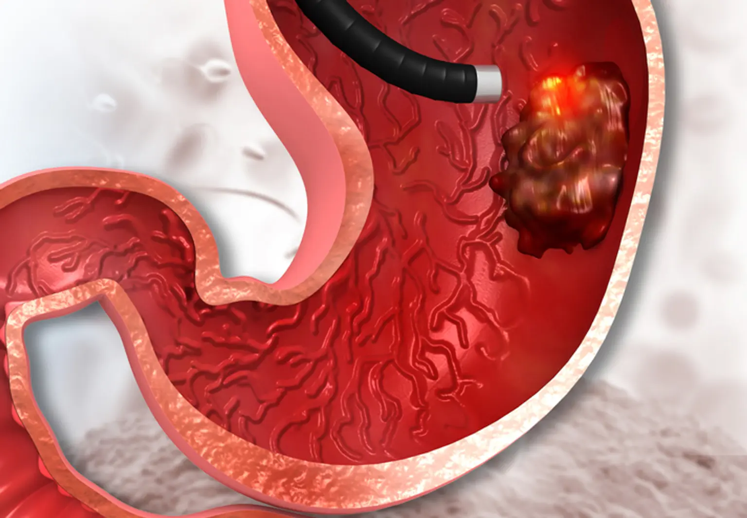
    Endoscopic mucosal resection is carried out using a long, thin tube fitted with a light, video camera, and other devices. The tube (endoscope) is inserted down your throat during upper digestive tract EMR to reach an abnormality in your esophagus, stomach, or upper section of the small intestine (duodenum).

    EMR is mostly used for treatment, but it is also utilized to gather tissues for diagnosis. If cancer is found, EMR can assist establish whether it has spread to areas beneath the digestive tract lining.

    Risks of the endoscopic mucosal resection include: bleeding (This most common complication often can be detected and corrected during the procedure.), narrowing of the esophagus, or puncture (perforation).


    What is Endoscopic Mucosal Resection (EMR)?

    Endoscopic Mucosal Resection

    Endoscopic mucosal resection (EMR) is a minimally invasive surgery that uses an endoscope — a flexible, tube-like equipment — to remove gastrointestinal (GI) cancer and precancerous lesions. These tumors are removed using an electrical snare, which is a tiny wire passed through the endoscope and formed into a loop at the end that tightens around the tumor. When an electrical current is sent via the wire, the tumor is separated and the wound is cauterized.

    A gastroenterologist, a specialist who specializes in treating problems of the GI tract, performs EMR. The endoscope allows the doctor to view within the body in great detail. It can also aid in the detection and removal of small growths such as polyps from the GI tract. The doctor will put the endoscope through the mouth for upper GI tumors or through the anus for lower GI tumors, depending on where the tumor is situated in the GI tract.


    Advantages & Benefits of Endoscopic Mucosal Resection (EMR)

    Benefits of Endoscopic Mucosal Resection

    The following are some of the benefits of EMR for the treatment of esophageal and gastric cancer:

    • The organ itself (esophagus, stomach) is kept in place, allowing the patient's quality of life to be preserved.
    • As long as the disease has not progressed via the lymph nodes to other regions of the body, EMR is considered a cure for early small esophageal and gastric cancer. Larger lesions may need more specialized techniques, such as endoscopic submucosal dissection.
    • Patients who have successfully undergone EMR for esophageal and gastric cancer have a low risk of recurrence (the cancer rarely comes back).


    Indications for Endoscopic Mucosal Resection

    Indications for Endoscopic Mucosal Resection

    In the absence of lymph node or distant metastases, endoscopic resection of superficial premalignant and well-to-moderately differentiated malignant GI lesions may be considered for final therapy. They also play an important part in the early GI cancer staging algorithm by producing a bigger resection specimen than traditional forceps biopsy, allowing accurate T staging and demonstrating the existence of lymphovascular involvement. In addition, pathologic evaluation of resected specimens frequently results in major changes in patient care.

    Esophageal lesions

    1. Squamous cell carcinoma of esophagus

    EMR is reserved for lesions less than 2 cm in size, confined to the esophageal mucosa (equivalent to stage T1a), and occupying less than one-third of the esophageal circumference. Endoscopic Submucosal Dissection (ESD)can be used to treat larger lesions. Disease-specific survival rates can reach 95%, and complication rates are modest. However, there was no change in survival after a 4-year follow-up. EMR and ESD can be used to stage lesions that lack architectural or EUS signs that indicate profound submucosal invasion.


    2. Adenocarcinoma of esophagus

    ESD can be used to treat early esophagogastric junction cancer (EGJ). Several case series have evaluated the function of ESD in non-Barrett adenocarcinoma of the EGJ in the past. En-bloc resection rates were 100%, with curative resection rates ranging from 68% to 79%.

    Non-curative resection patients were generally handled surgically. Patients with curative ESD resections had no local recurrences or metastatic cancer in trials with typical follow-up lengths of 15-30 months; in studies with longer follow-up periods, patients with curative resections had a 5-year disease-specific survival rate of 100 %.

    3. Barrett esophagus with dysplasia and early adenocarcinoma

    EMR should be employed in Barrett esophagus (BE) segments with mucosal abnormalities such as nodularity, ulceration, or a flat but irregular mucosal shape. This allows for more accurate lesion staging as well as the possibility of therapeutic benefit if a lesion is totally removed. EMR's stated benefits in this situation include accurate pathologic staging, the possibility of treatment, and a good safety profile.

    EMR is regarded final therapy for cancer if the lesion is well-to-moderately differentiated, restricted to the mucosal layer, 2 cm or less in size, and has no lymphovascular invasion.

    If an EMR specimen is a definitively resected Barrett cancer or shows only HGD, endoscopic ablative therapy of the remaining BE should be performed. If the EMR specimen demonstrates neoplasia at the deep margin, residual neoplasia should be assumed, and surgical or systemic therapy (chemotherapy, radiation, or a combination of the two) should be considered. 

    Gastric cancer

    The JSGE has developed recommendations for the use of EMR in the treatment of gastric cancer as an established alternative to surgery.

    Differentiated malignancies are well-to-moderately differentiated tubular and papillary adenocarcinomas, whereas undifferentiated cancers are signet-ring cell carcinomas and weakly differentiated adenocarcinomas. Endoscopic resection indications are defined as absolute, expanded, or out of indication based on the following criteria:

    • Depth of invasion.
    • Finding of ulceration.
    • Lesion size.
    • Classification as differentiated vs undifferentiated.

    Absolute indications for endoscopic resection include the following:

    • Macroscopically intramucosal differentiated carcinomas measuring less than 2 cm in diameter
    • Macroscopic type does not matter but no ulceration scar 


    Duodenal lesions

    Ampullary and periampullary adenomas, early carcinomas, and ampullary submucosal lesions have all been treated and staged using EMR and ESD. Nonampullary adenomas, neuroendocrine tumors, and submucosal lesions are other possible uses. According to the research, duodenal EMR and ESD may be linked with a high risk of complications, particularly in the treatment of submucosal lesions.


    Colorectal lesions

    EMR and ESD are often used to remove laterally spreading benign or malignant colon cancer, including as early-stage colorectal malignancies, flat adenomas, large superficial colorectal tumors, and rectal carcinoids. Additional indications include individuals who refuse surgical intervention or who have major comorbidities.


    When EMR is Contraindicated?

    When EMR Is Contraindicated?

    The existence of or a high index of suspicion for lymph node or distant metastases is a contraindication.

    Because of fibrosis at the biopsy site, a biopsy of the lesion prior to endoscopic resection (including ESD and EMR) may result in a false-positive result. Biopsies prior to referral for ESD/EMR should be avoided because the results are unlikely to change treatment strategies. If a biopsy is performed, reducing the time delay between the biopsy and the endoscopy may help decrease false-positive findings.

    The nonlifting sign, defined as a lesion's failure to elevate above the surrounding mucosa following submucosal injection underneath the lesion, has been proven as a highly accurate and specific predictor of colonic lesion submucosal invasion. ESD may commonly resect mucosal lesions with a nonlifting sign, as well as persistent or recurring lesions, for which EMR is often difficult and en-bloc resection is required.

    Because of the danger of bleeding, the presence of coagulopathy is a relative contraindication. Any contraindication to standard endoscopy (for example, significant cardiovascular comorbidities) also applies to ESD and EMR.


    How to Prepare For the Procedure?

    How To Prepare For the Procedure?

    1. You will be required to supply the following information before having endoscopic mucosal resection:
    1. Before the surgery, your doctor may advise you to stop taking certain drugs, such as those that alter blood coagulation or interact with sedatives.

    2. The day before the operation, you will be given written instructions. These instructions may differ based on the location of the lesion or lesions being removed. In general, the instructions will most likely include:
      • Fasting. When to begin fasting will be specified. You may be unable to eat, drink, chew gum, or smoke after midnight the night before the surgery. You may be requested to follow a clear liquid diet the day before your surgery.
      • Cleaning the colon. If the procedure requires you to empty your bowels and clear your colon, you will most likely need a liquid laxative or an over-the-counter enema kit.
      • After the risks and benefits have been described to you, you will sign an informed consent document giving your doctor permission to undertake the operation. Before you sign the document, ask your doctor about any questions you have regarding the operation.


    How is Endoscopic Mucosal Resection (EMR) Performed?

    Endoscopic Mucosal Resection Performe

    The EMR procedure for esophageal or gastric cancer includes the following.

    • During the surgery, the patient is given general anesthesia and sleeps.
    • The endoscope is inserted into the patient's mouth.
    • A precancerous or cancerous lesion is discovered.
    • The surgeon either injects a liquid into the submucosal layer beneath the lesion or puts a rubber band beneath the lesion to raise it and make it easier—and safer—to remove. The most frequent method for lifting a lesion is to use a rubber band. (The submucosa is the tissue layer immediately under the mucous membrane.)
    • The surgeon next uses the snare device to trap the lesion and cut it out. The endoscope is then used to grip and remove the lesion out of the patient's mouth.


    What Happens After Endoscopic Mucosal Resection?

    After Endoscopic Mucosal Resection

    1. You'll be kept in a recovery room until the majority of the sedative's effect has worn off. 
    2. You will be given written instructions on when you may begin eating and drinking and when you can resume normal activities.
    3. Within 24 hours of the treatment, relatively mild adverse effects may develop, including:
      • Sedative reactions. You may continue to feel drowsy and may suffer nausea and vomiting.
      • Sore throat. If the endoscope was directed down your esophagus, your throat may be painful.
      • Cramps or gas. If air was pushed into your digestive tract to make it more accessible, you may have gas, bloating, or cramping following the treatment.
    1. You will also be given written instructions on when to contact your doctor or seek emergency care following the surgery. The signs and symptoms listed below may suggest a significant consequence after endoscopic mucosal resection:
      • Fever.
      • Chills.
      • Vomiting.
      • Black stool.
      • Bright red blood in the stool.
      • Chest or abdominal pain.
      • Shortness of breath.
      • Fainting.


    Are There Any Risks?

    Are There Any Risks?

    A colonoscopy with EMR is a low-risk technique with few consequences. However, the risk of severe consequences increases when larger polyps are removed rather than smaller ones. However, before the operation, the doctor will discuss these risks with you. If a problem occurs, hospitalization is required, and surgery may be required. Please see your doctor if you are concerned about any of these risks. Prior to the treatment, you can also seek a formal face-to-face conversation with your doctor.

    You may feel bloated, nauseous, or gassy shortly after an EMR. When you cough or use the bathroom, you may notice small quantities of blood in the esophagus or rectum (depending on where the growths were removed). A painful throat is common after an EMR of the upper gastrointestinal tract.

    More severe complications can also occur, including:

    • Esophageal narrowing (strictures), which may need medical intervention to resolve.
    • Perforation or perforation in the gastrointestinal tract, which may necessitate surgery.
    • Persistent bleeding, which may end on its own or necessitate intervention.
    • Sedative-related complications, such as decreased respiration, heart rate, or blood pressure.

    Get medical attention right away if you experience:

    • Chest pain.
    • Difficulty breathing.
    • Worsening throat pain.
    • Difficulty swallowing that gets worse.
    • Vomit that is bloody or resembles coffee grounds.
    • Bowel movements that are bloody, black or tar-colored.
    • Fever.


    Post-Surgery Appointment

    Post-Surgery Appointment

    A follow-up consultation with the gastroenterologist will most likely be scheduled to discuss the results of your endoscopic mucosal resection and laboratory testing done on lesion samples. The following are some questions to ask your doctor:

    • Were you successful in removing all abnormal tissues?
    • What were the laboratory test results? Were any of the tissues malignant?
    • Is it necessary for me to consult a cancer expert (oncologist)?
    • Will I require extra treatments if the tissues are cancerous?
    • How will you keep an eye on my health? 


    Follow-up Exams

    Follow-up Exams

    A follow-up exam is usually performed three to 12 months following your treatment to ensure that the whole lesion was removed. Depending on the results, your doctor will recommend more tests.

    An exam will most likely include a visual inspection with an endoscope. Your doctor may tattoo the region where the lesion was removed so that when a follow-up endoscopy is performed, he or she can be sure the lesion was entirely eliminated.


    Who Can I Contact If I Have Any Questions?

    Please do not hesitate to speak to your doctor. If after the investigation you experience severe stomach pain or bleeding (more than an eggcup full), please attend your nearest acute Accident and Emergency Department taking with you the endoscopy report and photos. 


    Conclusion

    Endoscopic mucosal resection

    Endoscopic mucosal resection (EMR) is an operation in which a tiny, lighted tool with a camera is inserted into the digestive system to find and remove aberrant or malignant growths. An endoscope is a hollow tube through which a doctor transmits the surgical tools required to remove the tumor. To diagnose and treat malignancies of the esophagus, stomach, or upper intestine, the tube is put via your mouth and down your esophagus. The endoscope is introduced into the anus for colorectal cancer (lower intestine).

    EMRs are performed by doctors who have had endoscopic experience and training. The doctor is frequently a gastroenterologist, a specialist in detecting and treating digestive problems.

    An EMR is not the same as an endoscopic biopsy, which removes only a tiny quantity of abnormal tissue for testing. Doctors can use the resection process to completely remove growths, perhaps curing some malignancies without the need for more severe surgery. These might include precancerous growths, such as those seen in Barrett's esophagus, or early-stage gastrointestinal malignancies, such as stomach cancer.

    The growths are sent to a laboratory for testing and analysis, exactly like biopsies, although they frequently yield a more reliable and thorough diagnosis than smaller biopsy samples. Pathologists, or diagnostic specialists, perform tests on the specimens and analyze them under a microscope to identify the particular type of cancer and whether it is likely to spread, among other variables.

    Doctors in Japan, where stomach cancer is very common, developed the EMR as a potential therapy option for early-stage stomach cancer. Because screening tests are not standard in the United States, stomach cancers are often not detected until they are advanced. As a result, EMRs are seldom utilized, thus doctors who perform the surgery must have extensive knowledge and experience.