Introduction
Endoscopic neck surgery refers to a minimally invasive technique used to treat various conditions of the neck. With the aid of small incisions and specialized tools, surgeons perform operations that once required larger, more invasive cuts. This approach, which uses a camera for better visualization, has revolutionized neck surgeries by offering quicker recovery, reduced pain, and smaller scars.
Unlike traditional open surgery, where larger incisions are made, endoscopic procedures utilize tiny cuts, minimizing trauma to surrounding tissues. This has led to its increasing adoption globally, as patients seek more efficient and aesthetically pleasing alternatives.
How Endoscopic Neck Surgery Works
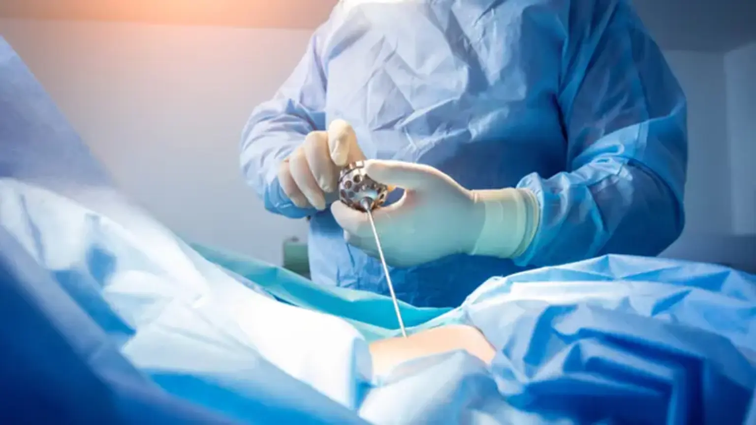
Endoscopic neck surgery involves the use of specialized instruments that are inserted through small incisions in the skin. A tiny camera, known as an endoscope, allows the surgeon to see the internal structures of the neck with high precision. Here’s how the procedure typically unfolds:
Incision: The surgeon makes small incisions, usually around 1-2 cm in length.
Insertion of Instruments: A long, thin tube with a camera is inserted through one of the incisions to provide live video footage.
Procedure: The surgeon uses specialized tools inserted through the other incisions to perform the surgery, whether it’s removing thyroid tissue, lifting the neck, or excising a tumor.
Completion: After the procedure, the incisions are closed with stitches or adhesive strips.
This approach minimizes blood loss and reduces post-operative pain, making the recovery process much quicker than traditional surgery.
Cosmetic Advantages: Endoscopic Neck Lift Surgery
Endoscopic neck lift surgery is a popular cosmetic procedure that targets sagging skin in the neck and jawline. Unlike traditional neck lift surgeries, which require larger incisions, endoscopic techniques use tiny cuts and specialized instruments to tighten and smooth the skin.
This procedure offers several benefits:
Less Invasive: Small incisions mean less disruption to the skin and tissues.
Minimal Scarring: The incisions are strategically placed to ensure scars are inconspicuous.
Faster Recovery: Patients can often return to work and other activities more quickly than after a traditional neck lift.
For those looking to enhance their appearance, the endoscopic neck lift provides a safer and less painful option with results that are just as effective as traditional methods.
Types of Endoscopic Neck Surgeries
There are several types of endoscopic neck surgeries, each designed for specific medical and cosmetic concerns:
Endoscopic Thyroidectomy: Used primarily for thyroid disorders, including thyroid cancer, hyperthyroidism, and benign growths. The procedure involves removing part or all of the thyroid gland with minimal scarring.
Endoscopic Neck Lift: A cosmetic procedure aimed at rejuvenating the neck's appearance by tightening sagging skin. This is a less invasive alternative to traditional facelifts, with a focus on improving the contour of the neck and jawline.
Endoscopic Surgery for Tumors: This technique is also used to remove benign or malignant tumors from the neck area, offering a less traumatic alternative to traditional neck surgeries.
Each of these procedures offers distinct advantages depending on the patient's needs, whether for medical treatment or cosmetic enhancement.
Recovery and Aftercare for Endoscopic Neck Surgery
One of the major advantages of endoscopic neck surgery is the quicker recovery time compared to traditional open surgeries. However, recovery still requires attention and care to ensure the best results.
Immediate Aftercare: After surgery, patients are typically monitored for a few hours before being discharged. Most can go home the same day.
Rest and Pain Management: Some soreness and discomfort are expected in the first few days, but most patients report that pain is minimal. Over-the-counter pain relievers are often enough to manage discomfort.
Follow-up Appointments: Regular follow-up visits to the surgeon are essential to monitor healing and address any concerns.
Return to Normal Activities: For cosmetic procedures, patients can often return to work within a week. For medical surgeries like thyroidectomies, full recovery may take several weeks, depending on the extent of the surgery.
Endoscopic surgery’s minimally invasive nature allows for a smoother, faster recovery with fewer restrictions, making it an appealing choice for many.
Endoscopic Thyroidectomy: A Safer Alternative to Traditional Surgery
Endoscopic thyroidectomy is a breakthrough technique in the removal of thyroid tissue, particularly for patients with thyroid cancer, benign growths, or other thyroid disorders. Traditionally, thyroid surgery involved a large incision in the neck, which not only left visible scars but also came with a longer recovery time and more risk of complications.
Endoscopic thyroidectomy offers several significant advantages:
Minimal Scarring: The small incisions made in the skin allow for less noticeable scarring, particularly when compared to traditional surgery.
Faster Recovery: Patients typically experience a faster recovery with fewer restrictions, often resuming normal activities within a week.
Reduced Risk of Complications: The procedure's precision reduces the likelihood of damaging surrounding structures, such as the parathyroid glands or recurrent laryngeal nerve, which can lead to issues with calcium regulation or voice changes.
For many patients, the ability to undergo thyroid surgery with less invasive methods, faster recovery, and fewer complications makes endoscopic thyroidectomy a preferred option.
Global Popularity and Availability of Endoscopic Neck Surgery
Endoscopic neck surgery is gaining popularity worldwide due to its benefits, including faster recovery times, minimal scarring, and reduced complication risks. As technology has advanced, this minimally invasive approach has become increasingly accessible in both developed and developing countries.
In regions like North America, Europe, and parts of Asia, many top hospitals and clinics now offer endoscopic procedures for both medical and cosmetic purposes. Its rise in demand is attributed to growing awareness and the desire for less invasive treatment options. Additionally, advancements in surgical tools and techniques continue to make these procedures safer and more efficient.
Who Is a Good Candidate for Endoscopic Neck Surgery?
Endoscopic neck surgery is suitable for a variety of patients, both for medical and cosmetic reasons. Ideal candidates include:
For Medical Conditions: Patients with thyroid disorders (like cancer or benign growths) who are in good overall health and seek a less invasive approach.
For Cosmetic Enhancements: Individuals seeking a neck lift or rejuvenation with minimal downtime.
General Requirements: Patients who have realistic expectations and are non-smokers, as smoking can slow down recovery.
It’s important to consult with a qualified surgeon who can assess each individual’s suitability based on their medical history, health condition, and goals for surgery.
Benefits of Endoscopic Neck Surgery
Endoscopic neck surgery offers several key advantages over traditional methods:
Minimized Scarring: Because the incisions are small, scarring is less noticeable, particularly for cosmetic procedures like neck lifts or thyroid surgeries.
Reduced Recovery Time: Patients generally recover much faster, with many able to return to normal activities within a few days to a week, compared to the weeks of recovery often needed after open surgery.
Lower Risk of Complications: Smaller incisions mean less trauma to surrounding tissues, which can result in lower risks of infection, nerve damage, and excessive bleeding.
Precision and Less Trauma: The camera and tools used in endoscopic surgery allow for highly precise removal of tissue, reducing damage to healthy structures in the neck.
Patients who choose endoscopic neck surgery typically experience less pain, shorter hospital stays, and a faster return to their daily routines.
Endoscopic Neck Surgery vs. Traditional Surgery
Endoscopic surgery is quickly becoming a preferred choice over traditional open surgery due to its less invasive nature. Key differences include:
Smaller Incisions: Traditional surgeries require larger cuts, resulting in longer recovery times and more visible scars.
Shorter Recovery Time: Patients typically recover in days rather than weeks, with less pain and fewer restrictions.
Reduced Risks: The risk of complications such as bleeding, scarring, and infection is lower with endoscopic procedures.
While traditional surgery is still necessary in some cases, the endoscopic approach is gaining traction due to its faster recovery and improved outcomes.
Technological Advancements in Endoscopic Neck Surgery
Recent advancements in technology have greatly enhanced the safety and precision of endoscopic neck surgery. Some key innovations include:
Robotic Assistance: Robots can assist in guiding instruments with extreme precision, improving outcomes in delicate surgeries like thyroidectomies.
High-Definition Imaging: Advanced cameras provide surgeons with clearer views of internal structures, minimizing errors and improving precision.
Smarter Tools: Newer instruments, like tissue-sealing devices, reduce the risk of bleeding and enhance safety.
These technological advancements continue to evolve, making endoscopic neck surgery safer, more efficient, and even more accessible to patients worldwide.
Preparing for Endoscopic Neck Surgery
Proper preparation is crucial to ensure the success of endoscopic neck surgery. Here’s what patients can expect:
Initial Consultation: During this visit, the surgeon will evaluate the patient's medical history, perform a physical examination, and discuss the best surgical options. Imaging studies, such as ultrasounds or CT scans, may be recommended.
Pre-Surgical Instructions: Patients may be advised to stop certain medications, such as blood thinners, and refrain from eating or drinking for several hours before surgery.
Post-Surgery Plan: Patients should arrange for someone to drive them home and help during the initial recovery phase.
Being well-prepared helps to minimize risks and ensures a smoother surgery and recovery process.
Cost of Endoscopic Neck Surgery
The cost of endoscopic neck surgery varies depending on the procedure, location, and the complexity of the surgery. Typically, cosmetic procedures like the endoscopic neck lift tend to be more expensive due to the specialized tools and expertise required.
Cosmetic Procedures: Endoscopic neck lifts can range from $5,000 to $15,000 or more, depending on the clinic and region.
Medical Procedures: Surgeries like endoscopic thyroidectomy can cost between $10,000 and $20,000, depending on factors such as the hospital, the complexity of the surgery, and insurance coverage.
While the initial cost may seem high, the advantages—such as quicker recovery and fewer complications—often lead to lower overall healthcare costs for patients.
Post-Surgical Care and Lifestyle Adjustments
After endoscopic neck surgery, patients need to follow specific aftercare instructions to ensure optimal healing:
Pain Management: Over-the-counter pain relievers usually suffice, but stronger medications may be prescribed for the first few days.
Avoid Strenuous Activity: Patients should avoid heavy lifting or vigorous exercises for at least 2-4 weeks after surgery.
Follow-up Appointments: Regular follow-up visits are essential for monitoring healing and addressing any concerns.
Patients should also avoid smoking, as it can slow down healing. Maintaining a healthy lifestyle, including a balanced diet and adequate hydration, can help speed up recovery.
Risks and Complications of Endoscopic Neck Surgery
While endoscopic neck surgery is generally considered safe, like all surgeries, it comes with potential risks and complications. These can vary depending on the specific procedure being performed and the individual patient.
Infection: As with any surgical procedure, there is a risk of infection. However, the smaller incisions used in endoscopic surgery tend to lower this risk.
Bleeding: Although rare, excessive bleeding can occur, especially in complex surgeries.
Nerve Damage: In some cases, nerves around the neck area may be injured, leading to temporary or permanent changes in sensation or movement.
Scarring: While scarring is minimized, it is still possible, especially if the patient has a tendency to form hypertrophic scars.
Surgeons take great care to minimize these risks through advanced techniques, careful planning, and post-operative care. Proper consultation and choosing an experienced surgeon can further reduce the chances of complications.
Frequently Asked Questions (FAQs)
Is Endoscopic Neck Surgery Safe? Yes, it is generally considered safe, with minimal risks compared to traditional open surgeries. However, as with any surgery, risks exist, so choosing an experienced surgeon is key.
How Long Does Recovery Take? Recovery time varies but is typically much shorter than traditional surgery. For cosmetic procedures, most patients return to work in a week. Medical surgeries may require a few weeks for full recovery.
Will There Be Visible Scarring? The small incisions used in endoscopic surgery result in minimal scarring, which is often barely noticeable.
How Much Pain Should I Expect? Pain is usually minimal and well-managed with medication. Most patients report discomfort, not severe pain, during the first few days post-surgery.
Long-Term Results of Endoscopic Neck Surgery
The long-term outcomes of endoscopic neck surgery are generally very positive. Most patients experience lasting improvements in both appearance and health. For cosmetic procedures like neck lifts, the results can last for several years, though aging and lifestyle factors may affect the longevity of the outcome.
Neck Lift: The tightening and rejuvenation of the neck can last 5–10 years, with gradual changes over time.
Thyroid Surgery: For medical conditions, such as thyroid removal, patients can expect a long-term resolution of symptoms like swelling or thyroid imbalances.
The minimally invasive nature of endoscopic surgery means that patients typically have more satisfactory and durable outcomes compared to traditional approaches.
Alternatives to Endoscopic Neck Surgery
While endoscopic neck surgery offers significant benefits, it’s not the only option available. Other alternatives may be more suitable depending on the patient’s condition and preferences:
Traditional Open Surgery: Still the preferred choice for more complex cases, especially if a large amount of tissue needs to be removed or if the surgeon requires direct access.
Non-Surgical Treatments: Procedures such as Botox injections, fillers, or laser treatments can help with mild cosmetic concerns like wrinkles or sagging but are not as effective for severe cases.
Radiofrequency Therapy: For non-invasive neck lifts, radiofrequency energy can tighten skin without surgery, though results are typically less dramatic.
Each option has its pros and cons, and a consultation with a skilled surgeon will help determine the most appropriate choice.
The Future of Endoscopic Neck Surgery
The future of endoscopic neck surgery looks promising, with ongoing advancements in technology and surgical techniques. Some potential developments include:
Improved Imaging Technology: Higher-definition cameras and real-time imaging allow for even more precise surgery with minimal risk.
Robotics and AI: Robotic assistance in surgeries can provide unprecedented accuracy, particularly in delicate neck surgeries.
Personalized Treatments: As understanding of genetics and individual anatomy improves, surgeons may offer more customized surgical plans that optimize results for each patient.
With these innovations, the scope and efficacy of endoscopic neck surgery will continue to grow, offering even better results with fewer risks.
Conclusion
Endoscopic neck surgery represents a major leap forward in surgical techniques, offering patients a safe, effective, and minimally invasive solution for both cosmetic and medical conditions of the neck. With reduced scarring, faster recovery, and fewer complications, it is an appealing option for many.
However, it’s essential to consult with a qualified surgeon who can assess your specific needs and help you understand the best approach for your situation. Whether you’re seeking relief from thyroid problems or aiming for a more youthful appearance, endoscopic neck surgery could provide the outcomes you’re looking for.
Remember, each patient’s journey is unique. With proper preparation, realistic expectations, and skilled surgical care, endoscopic neck surgery can offer significant benefits and improve your quality of life.