Endoscopic Sinus Surgery

Last updated date: 26-Apr-2023

Originally Written in English

Endoscopic Sinus Surgery

Overview

Since its inception, endoscopic sinus surgery (ESS) has been competing with technology; advancements in equipment provide a vast playground for the use of ESS beyond chronic rhinosinusitis (CRS). CRS, pituitary tumors, skull base deformities, sinonasal tumors, and complications of acute rhinosinusitis are among the most common indications. When addressing structures outside the sinuses, a progressive strategy targeting all sinuses delivers a favorable outcome in cases of CRS and a wide and safe channel.
 

Functional Endoscopic sinus surgery definition

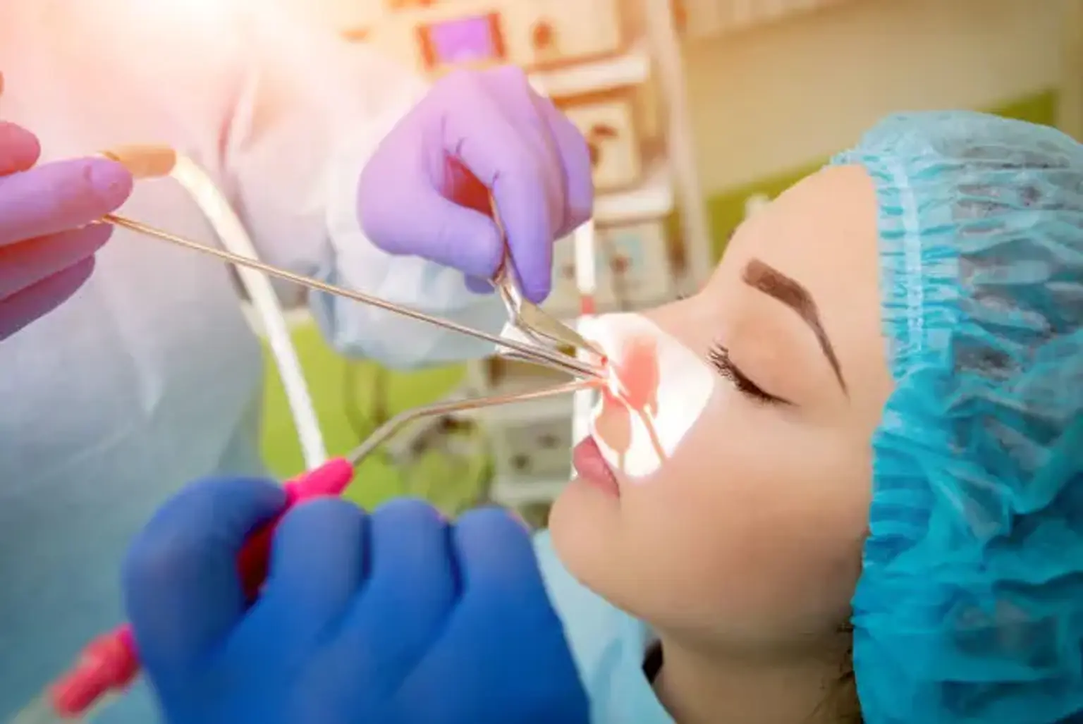
Functional Endoscopic Sinus Surgery (FESS) is a minimally invasive surgical treatment that uses an endoscope to open the sinus air cells and sinus ostia. The use of FESS as a sinus surgical approach is now generally acknowledged, and the word functional is intended to differentiate this form of endoscopic surgery from the more traditional non-endoscopic sinus operation.

Endoscopic examination of the sinuses was first performed in 1902. However, until the 1970s, ESS was not conducted on a regular basis during the majority of the past century. Sinus diseases were treated utilizing external techniques and a lamp. Since the 1970s, endoscopic sinus surgery procedures have been in a continual race with technological improvements, with new surgical instruments, imaging, simulation, and navigation.

In the treatment of sinusitis, the aims of functional endoscopic sinus surgery (FESS) are to increase sinus Ostia, restore appropriate sinus aeration, enhance mucociliary transport, and offer a better route for topical treatments. The concept of FESS may appear simple, but the anatomical variety and wide range of severity of illnesses treated in each FESS remain problems for the surgeon in every instance. Pre-operative preparation for sinus surgery is critical for achieving the best results and avoiding any problems.

Endoscopic sinus surgery is the gold standard for treating chronic rhinosinusitis because it targets sinus disease (CRS). With technology advancements, the frontiers of ESS are always growing. At this stage, the symptoms of ESS have outpaced those of rhinosinusitis. The adoption of this therapy cemented its place in the treatment of sinus malignancies and disorders that extend beyond the sinuses.


Anatomy and Physiology

Endoscopic Sinus Surgery Anatomy

Endoscopic sinus surgery requires a good grasp of nasal and paranasal anatomy. The structure of the paranasal sinuses varies significantly across persons and sides. As a result, prior to undergoing surgery, it is critical to review computed tomography and other radiologic investigations.

The nasal bones, upper lateral cartilages, and lower lateral cartilages make form the external nose. The septum divides the inside section of the nose into two nasal chambers.

A medial wall, which is the vertical septum, and a lateral wall exist in each nasal cavity. The ceiling of the nasal cavity is formed by the crista galli, cribriform plates, and the sphenoid body. The palatine process of the maxilla and the horizontal process of the palatine bone make up the floor.

The septum is a stiff structure made up of bony and cartilaginous components. The anterior section of the septum is formed by the septal cartilage. The ethmoid perpendicular plate is located postero-superiorly, the vomer is found postero-inferiorly, and the septum is formed by the crests of the maxillary and palatine bones. The septum is mucosa-covered and has a large blood supply that passes between the mucosa and the perichondrium.

The sphenopalatine artery, a branch of the maxillary artery, the anterior and posterior ethmoidal arteries deriving from the ophthalmic artery, the septal branch of the superior labial artery deriving from the facial artery, and the septal branch of the ascending and greater palatine arteries branching from the maxillary artery are the arteries that supply the septum. Little's region is a vascular area where numerous arteries connect to produce the Kiesselbach's plexus; it is a common site of anterior epistaxis.

The turbinates are bony outgrowths on the lateral nasal wall. Each side has three to four turbinates. The superior, intermediate, and, if present, supreme turbinate stem from the ethmoid bone and, as a result, can include conchas. The inferior turbinate is derived from a distinct bone. The turbinates are mucosa-covered and play an important role in the filtration, humidification, and control of the flow of breathed air.

A nasal channel, commonly known as the meatus, is located inferior to each concha. The inferior meatus is the channel via which the nasolacrimal duct empties via Hasner's valve. The middle meatus is the most complicated and serves as the main drainage path for the majority of paranasal sinuses (frontal, maxillary, and anterior ethmoid sinuses).

The uncinate process is a protrusion from the ethmoid that is linked to the lacrimal bone anteriorly, the inferior turbinate inferiorly, and has a two-dimensional aperture posteriorly, also known as the hiatus semilunaris, within the middle meatus. Superiorly, the uncinate can be attached to one of three structures: the lamina papyracea, the middle turbinate, or the ethmoid sinus roof.

The crescent-shaped region between the ethmoid bulla superiorly and the uncinate inferiorly is where secretions from the sinuses flow into the nasal cavity; this structure grows antero-superiorly into the ethmoidal infundibulum.

The ethmoid sinuses are bounded laterally by the lamina papyracea, superiorly by the fovea ethmoidalis, and medially by the nasal cavity. A basal lamella separates the ethmoid sinuses into anterior and posterior cells. The anterior cells drain into the middle meatus, whereas the posterior cells drain into the superior meatus's sphenoethmoidal recess. The agger nasi, or most anterior cells, are found near the superior attachment of the middle turbinate.

The biggest ethmoidal cell is the ethmoidal bulla, which is always present posterior to the semilunar hiatus. The posterior ethmoidal cells are found posterior to the basal lamella; they are bigger in size and fewer in number than the anterior cells. The ethmoid sinuses get blood supply from the anterior and posterior ethmoidal arteries, while the venous drainage is routed to the superior ophthalmic vein or pterygopalatine plexus.

The maxillary sinuses are placed superiorly, between the orbital floor and the alveolar processes of the maxilla, and inferiorly, between the orbital floor and the alveolar processes of the maxilla. The maxillary sinus ostium is found in the sinus's medial wall and usually opens into the posterior portion of the ethmoid infundibulum. It is also often placed on the superior portion of the medial wall; hence, it is critical not to enter the sinus superior to the ostium in order to avoid a breach of the medial orbital wall. Accessory maxillary ostium can be detected in up to 43% of instances and can be seen in either the anterior or posterior nasal fontanelle.

During FESS, it is critical to distinguish the maxillary ostium from the accessory ostia and to expand the genuine maxillary ostium. Ethmoidal cells can occasionally grow and spread laterally into the maxillary sinus wall; these cells are also referred to as Haller cells. The maxillary sinuses are supplied with blood by branches of the maxillary and face arteries. Venous return into the facial vein or the pterygoid plexus.

The sphenoid sinuses are paired cavities separated by a septum that are situated within the body of the sphenoid bone. The sphenoid ostium is positioned in the anterior wall of the sinus and drains into the superior meatus's sphenoethmoidal recess. The internal carotid arteries, cavernous sinuses, and the optic, vidian, maxillary, oculomotor, trochlear, and abducent nerves all surround the sphenoid sinus.

The sphenoid sinus allows endoscopic access to the skull base, pituitary gland, optic nerve, and many other tissues. Sphenoethmoidal cells, also known as Onodi cells, are occasionally present superolateral to the sphenoid and are strongly related to the optic nerve.

It is critical to detect the presence of these sphenoethmoidal cells and their interaction with the optic nerves, as well as optic nerve dehiscence and carotid dehiscence, in order to avoid potentially fatal consequences. The sphenopalatine artery supplies blood, and the maxillary vein drains venous blood.

The frontal sinuses are the most superior sinuses, located between the inner and outer frontal bone tables. A septum separates the two frontal sinuses. The frontal sinus ostium is placed in the middle of the sinus floor and drains into a frontal recess. Based on the superior attachment of the uncinate, the drainage of the frontal recess might run into the ethmoidal infundibulum or medial to it.

The supraorbital and supratrochlear arteries (branches of the ophthalmic artery) feed blood, and the superior ophthalmic vein drains blood.


Indications of Functional endoscopic sinus surgery

Indications of Functional endoscopic sinus surgery

Since the introduction of functional endoscopic sinus surgery, the indications for conducting this treatment have grown. Endoscope, camera, instruments, and navigation advancements have paved the way for an ever-expanding realm of endoscopic surgery to reach the skull base, optic nerve, cavernous sinus, pituitary, orbit, pterygopalatine fossa, and many other places and structures.

Chronic rhinosinusitis is the most prevalent and early cause of FESS. Rhinosinusitis is an inflammatory condition of the paranasal sinuses that is classified into many types based on the length of the inflammatory process:

  • Acute rhinosinusitis: less than four weeks
  • Subacute rhinosinusitis: between 4 to 12 weeks
  • Chronic rhinosinusitis: longer than 12 weeks

These disorders are among the most often seen by clinicians. Sinus sickness also places a huge financial strain on the healthcare system. In the United States, for example, chronic rhinosinusitis costs $8.3 billion each year on average.

Aside from the financial implications, CRS has a significant detrimental impact on patient's emotional and physical well-being. CRS should be diagnosed based on symptoms and objective findings on physical examination, such as anterior rhinoscopy, nasal endoscopy, or computed tomography scans.

CRS is initially treated with saline irrigation and/or topical intranasal steroids, according to clinical practice standards. When all other medical treatments have been exhausted, endoscopic sinus surgery is the next step. There is currently no broadly acknowledged consensus on what constitutes maximum medical care or when surgery should be performed. For many years, the significance of surgery in the treatment of CRS with or without polyposis has been researched, and FESS has demonstrated its importance in greatly enhancing the quality of life of people with CRS.

In addition to CRS, FESS is used to treat complex acute rhinosinusitis (ARS). Chandler's categorization is commonly used to classify extracranial and intracranial ARS problems. Pre-septal cellulitis, orbital cellulitis, subperiosteal abscess (SPA), orbital abscess (OA), and cavernous sinus thrombosis are the most severe. FESS is explored in cases of preseptal and orbital cellulitis when there is vision impairment or increased intraocular pressure, and when medical therapy does not improve the situation.

It is also essential in the treatment of SPA (particularly those bigger than 1 cm) and OA. The abscess is draining, the sinuses are being opened and drained to restore patency, and cultures are being obtained for targeted antibiotic therapy.

Endoscopic Sinus Surgery has also been utilized to access some orbital disorders trans-nasally while avoiding skin incisions due to the close relationship between the ethmoid sinuses and the orbit. Decompression of the orbit and optic canal in Graves disease or post-traumatic optic neuropathy, lesions of the extraconal medial orbital apex or space, benign sinonasal tumors invading the orbit medially, and medial orbital wall fractures are some of the indications for an endonasal approach to the orbit.

Mucoceles, invasive and non-invasive fungal sinusitis, silent sinus syndrome, pituitary tumors, cerebrospinal fluid leaks, benign and malignant sinonasal tumors, and ventral skull base lesions, petrous apex lesions, or pterygomaxillary fossa lesions are also treated with ESS. Malignancies in the nasal and paranasal passages, including those that extend into the anterior skull base, are treated with expanded sinus surgery.

In the field of rhinology, navigation-guided endoscopic sinus surgery has received a lot of attention. Based on imaging taken before to surgery, it advises the surgeon intraoperatively. In image-guided surgery, the tracking system aids in a more thorough and complete dissection of the sinuses, better imaging of tumor boundaries to acquire negative margins, and a decreased risk of complications.  

The indications for using the navigation system include: 

  • Revision sinus surgery
  • Distorted sinonasal anatomy
  • Benign or malignant sinonasal tumors
  • Repair of the cerebrospinal fluid leak; skull base defects or lesions
  • Pathologies close to the optic nerve, orbit, carotid artery, or skull base
  • Pathologies involving frontal, sphenoid, or ethmoid sinuses
  • Extensive polyposis


The surgical technique

Endoscopic Sinus surgical technique

Equipment

A television monitor, navigation system (if used), camera, sinus endoscopy tray with various curettes, downbiters, backbiters, elevators, ball-tip probes, through-cut instruments, Kerrison rongeurs, giraffe instruments, sinus forceps with different angulations, punch instruments, endoscopes and a powered debrider with straight and angled blades are required in the OR.


Personnel

A surgeon, usually an otolaryngologist, a scrub technician, a nurse, and an anesthesiologist are required in the operating room for Functional Endoscopic Sinus Surgery. A neurosurgeon is frequently present in the room with the otolaryngologist when a transsphenoidal technique for pituitary tumors or excision of tumors with intracranial extension is used.


Preparation

The patient is put on the operating table, which is angled toward the television display. The head of the bed is raised to place the patient in reverse Trendelenburg position. The endotracheal tube is placed in the left side corner of the patient's mouth.

The patient's eyes are covered with a transparent covering or only partially covered, leaving the medial area open to the surgeon, who will be looking for any swelling symptomatic of orbital hematoma on a frequent basis.

For decongestion, both nasal canals are initially filled with oxymetazoline-soaked cotton. After that, the patient is draped. If navigation is employed, imagery taken prior to surgery should be uploaded to the system, and the tracking system should be registered and confirmed to be correct.


Technique

Endoscopic Sinus Surgery Technique

A 0 or 30-degree scope is used to perform a complete nasal endoscopy. The lateral nasal wall at the uncinate and the axilla of the middle turbinate are then infiltrated with a 3 ml syringe and 27 gauge needle with 1 percent lidocaine and 1:100,000 epinephrine. Following that, cotton pledgetts soaked in oxymetazoline are put in the middle meatus

In the instance of septal deviation to one side, the side to be operated on initially is generally the side with the most illness or the side that is more open.

Excision of concha bullosa: 

A concha bullosa inside the middle turbinate may be found on occasion; excision of this cell is the first step in gaining greater access to the lateral nasal wall. A sharp sickle knife is used to make an incision in the anterior section of the middle turbinate, and the lateral half of the turbinate is removed.


Uncinectomy:

To reach the uncinate process, the central turbinate is gently medialized using a Freer elevator. Uncinectomy can be done retrogradely, using a ball tip probe used to identify and medialize the uncinate off the lamina payracea. To avoid harm to the medial orbital wall, the uncinate is sliced inferiorly using a backbiter. Blakesley forceps are used to grasp and remove the uncinate's free edge. The remaining uncinate process is removed using biting devices or a motorized debrider until the maxillary ostium is visible.

Another method of uncinectomy is to make an incision in the uncinate process using a sickle knife or the sharp edge of a Freer elevator.


Maxillary antrostomy: 

After removing the uncinate, the natural ostium of the maxillary sinus is visible. It has an oval form and is located at the bottom of the infundibulum. It is critical to distinguish between the native and accessory ostium. A ball-tip probe is used to confirm the entrance to the maxillary sinus, which is best viewed using a 30 or 45-degree scope. When the natural ostium is verified, it is widened with a through cutting instrument, punch forceps, and a powered debrider. To avoid harm to the orbit superiorly and the nasolacrimal duct anteriorly, the ostium is expanded posteriorly and inferiorly.


Ethmoidectomy:  

Ethmoidectomy can be done with either a 0 or a 30-degree scope. The ethmoid bulla is the first cell met in the ethmoid sinus. This big cell can be removed retrogradely from the retrobullar area by penetrating it medially and inferiorly with a powered debrider, curette, or punch forceps. The lamina papyracea has been identified, and it is suggested that its mucosa be preserved.

Dissection should be carried out posteriorly until the basal lamella is met and pierced. Posterior ethmoid cells encountered posterior to the basal lamella are dissected medially between the middle and superior turbinate and laterally between the lamina papyracea. Once the skull base is located posteriorly at the sphenoid's face, the ethmoid cells are dissected superiorly from posterior to anterior, with septations eliminated as the dissection progresses.

While dissecting the superior cells, a 45-degree scope can be employed. Prior to dissecting superiorly, it is critical to identify any dehiscence in the lamina payracea, the ethmoid artery, and the skull base with direct vision (or with navigation, if using); it is also important not to dissect medial to the superior attachment of the middle turbinate to avoid penetrating the fovea ethmoidalis.


Sphenoidotomy: 

Sphenoidotomy

Transnasally medial to the middle turbinate or transethmoidally lateral to the middle turbinate is where the sphenoid ostium can be found. The sphenoid ostium is located in the infero-medial region of the posterior ethmoid when detected transethmoidally. To improve vision of the spheno-ethmoidal recess and the sphenoid ostium, the inferior section of the superior turbinate is frequently resected. By gently moving a probe over the face of the sphenoid, the ostium is detected. When the probe reaches the ostium, it will glide inside it.

When the ostium has been located, it is expanded inferiorly with Kerrison tools or curettes. A motorized debrider can then be used to enlarge the sphenoidotomy. If sphenoethmoidal cells are present, the navigation system should be utilized to detect these cells, their septations, the skull base, and any adjacent tissues such as the optic nerve and carotid artery, as well as any structural dehiscence.


Frontal sinusotomy: 

To avoid bleeding from the frontal recess region impairing visibility while operating on more posterior and inferior cells, the frontal sinus is the final sinus to be treated. This sinus has complicated architecture as well, therefore navigation is essential while reaching the frontal recess. The posterior wall of agger nasi cells obstructs frontal sinus outflow. To remove the impediment, it is meticulously dissected.

The uncinate, as indicated in the anatomy section, can have a variety of superior attachments. Because the frontal sinus drains into the infundibulum when the uncinate process joins to the middle turbinate, the superior section of the uncinate should be excised to provide access to the frontal recess. After identifying the frontal recess using a registered frontal probe, curved curettes and frontal giraffe tools are used to remove septations, dissect frontal cells, and enlarge the frontal recess.

Sparing the mucosa at all stages reduces the likelihood of postoperative scarring and osteogenesis. To avoid turbinate instability, the vertical and horizontal attachments of the middle turbinate should be retained. In the event of instability, there is a substantial chance of turbinate lateralization, which can result in scarring and sinus drainage blockage.

To avoid lateralization, the anterior section of the turbinate can be excised, the turbinates can be sutured to the septum, or nasal packing can be inserted in the middle meatus. These solutions will assist in keeping the turbinate in the medial position.

Any remaining bone septations are removed at the end of the surgery, and hemostasis is achieved. As a middle meatus spacer, a dissolvable nasal pack can be used.


Endoscopic sinus surgery cost

There were also substantial increases in median operating times and expenses as the scope of surgery increased. Full ESS had a median total cost of $4,281, Intermediate was $3,716, and Anterior was $2,549 in 2016 USD.


Conclusion 

The most frequent treatment for CRS is functional endoscopic sinus surgery. Rhinologists may now dissect and open sinuses more aggressively thanks to the availability of fine equipment and high-resolution imaging. However, rhinologists should not rely just on imaging studies, navigation, and tools; they must also comprehend anatomy and be aware of all potential difficulties, both small and big. Following the FESS stages and finding the consistent landmarks at each stage is required for safe operation.