Endoscopic ultrasound
Overview
Endoscopic ultrasonography (EUS) allows your doctor to inspect the linings of your esophagus and stomach, as well as the walls of your upper and lower gastrointestinal tracts. The upper tract is made up of your esophagus, stomach, and duodenum, while the lower tract is made up of your colon and rectum. EUS is also used to examine organs close to the gastrointestinal tract, such as the lungs, liver, gallbladder, and pancreas.
Endoscopists are highly trained professionals who welcome queries about their qualifications, training, and experience. Your endoscopist will use an endoscope, which is a narrow, flexible tube with a small ultrasound probe integrated in. The endoscope will be sent via your mouth or anus to the location to be inspected by your doctor. Your doctor will then utilize ultrasonography to make visual pictures of the digestive tract by using sound waves.
What is an endoscopic ultrasound?
Endoscopic ultrasonography is a minimally invasive method that examines the GI system and beyond using a camera device (endoscope) and high frequency sound waves .
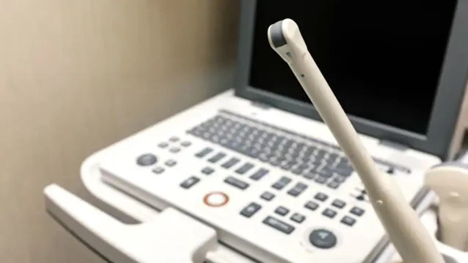
A standard endoscope is a thin, illuminated tube that is put through the mouth or anus to see the inside of your esophagus, stomach, or intestines. EUS makes use of an echoendoscope, which is an unique endoscope with a tiny ultrasound transducer on its tip. With the use of ultrasonography, the doctor can observe the GI system and adjacent organs.
Conventional EUS is divided into two types: radial scanners and sector scanners. There are also high-frequency ultrasonic probes that may be inserted through the biopsy channel of an endoscope. EUS has evolved as a key technique for the identification and staging of benign and malignant lesions of the gut wall and adjacent mediastinum, abdomen, and pelvic tissues.
It is also utilized as a diagnostic tool for evaluating upper gastrointestinal tract and rectosigmoid submucosal masses, detecting pancreatic endocrine cancers, and assessing vascular disease. Interventional applications, such as EUS-guided fine-needle aspiration (EUS-FNA) for acquiring tissue/fluid samples, pseudocyst drainage, and local treatment administration, are anticipated to improve the clinical value and cost-effectiveness of this imaging technique.
However, the most widespread use of EUS is in the diagnosis and staging of esophageal, gastric, rectal, and pancreaticobiliary cancer. EUS has been found to shift the clinical care approach of a large number of patients to a less expensive, dangerous, or intrusive method.
Why is EUS done?
By giving comprehensive pictures of your digestive tract, EUS gives your doctor with more information than conventional imaging procedures. EUS can be used by your doctor to detect disorders that cause stomach discomfort or unusual weight loss.
EUS is also used to analyze recognized abnormalities, such as lumps or lesions seen during a previous endoscopy or on x-ray examinations such as a computed tomography (CT) scan. EUS offers a thorough image of the lump or lesion, which can assist your doctor in determining the origin of the lump or lesion and in making treatment recommendations. When other tests are equivocal or contradicting, EUS can be used to identify disorders of the pancreas, bile duct, and gallbladder.
Your doctor can use EUS to detect the amount of spread of some malignancies of the digestive and respiratory systems. Your doctor can use EUS to properly determine the depth of the cancer and if it has spread to neighbouring lymph glands or important structures such as major blood arteries. In rare cases, EUS can be used to get a needle biopsy of a mass or lesion to assist your doctor in determining the best course of therapy.
EUS may be used by your doctor to assist identify a variety of digestive issues. EUS delivers precise photos of your interior structures to your doctor. It might be used by your doctor to:
- Detect small tumors in the pancreas
- Stage gastrointestinal cancers (determine how severe the cancer is)
- Detect stones in the biliary ducts
During EUS, your doctor may do fine-needle aspiration to perform a biopsy. He or she will insert a small needle through the endoscope to extract tissue samples, which will be sent to a lab for examination. Biopsies can be used to assess the existence of cancer or whether it has spread.
Endoscopic ultrasound for pancreas
One of the most helpful imaging investigations for identifying pancreatic cancer is endoscopic ultrasound (EUS). It is performed as an outpatient operation and offers precise pictures of the pancreas and adjacent organs such as the liver, blood arteries, and lymph nodes. EUS creates an image of the organs using high frequency soundwaves. When soundwaves bounce off inside organs, they transmit echoes to a computer, which produces a visual picture.
How should I prepare for EUS?
You should not eat or drink anything for at least six hours before an EUS of the upper gastrointestinal tract. Your doctor will advise you on when to begin fasting and if it is OK to continue taking your usual prescription drugs.
Prior to an EUS of the rectum or colon, your doctor will urge you to either ingest a colonic cleaning solution or follow a clear liquid diet supplemented with laxatives or enemas. If you do not properly follow your doctor's directions, the surgery may have to be postponed.
Most drugs can be taken as normal until the day of the EUS assessment. Inform your doctor about the drugs you're taking as well as any allergies you have. Before the operation, anticoagulant drugs (blood thinners such as warfarin or heparin) and clopidogrel may need to be changed. On the day of the EUS, insulin must also be changed. In general, aspirin and nonsteroidal anti-inflammatory drugs (ibuprofen, naproxen, etc.) can be taken safely before an EUS test. In advance, consult with your doctor about these suggestions.
Consult your doctor about the drugs you should take the morning of the EUS exam, and take only the necessary prescriptions with a little sip of water. If you have a latex allergy, you should notify your doctor before your test. Patients with latex allergies frequently require specialized equipment and may be unable to undergo a full EUS assessment.
Antibiotics are not often necessary before to or following EUS exams. However, if you are having specific EUS operations, such as draining a fluid collection or a cyst with EUS supervision, your doctor may prescribe antibiotics.
What Happens During an Endoscopic Ultrasound?
Under general anesthesia, EUS is performed in the operating or procedure room. It is normally performed as an outpatient operation. An endoscope (a thin, flexible tube) will be passed through the mouth and guided to the stomach and first section of the small intestine by the doctor. If the doctor wants to view the lower digestive tract, the endoscope may be sent through the rectum instead.
EUS can show places outside of the digestive system that conventional imaging procedures cannot. The probe can be placed extremely near to the "region of interest" within the body by the doctor. This allows the doctor to see clearly. A video screen displays images of the child's digestive tract and organs.
EUS is sometimes used just to see within the body to figure out what is going on. Occasionally, the doctor will diagnose or treat patients by slipping small surgical tools through the endoscope. If the EUS reveals stones in the bile or pancreatic channels, the doctor may opt to remove them. Endoscopic retrograde cholangiopancreatography is the medical term for this procedure (ERCP). Cincinnati Children's is one of the country's few pediatric medical institutions that provides ERCP.
EUS operations might take anything from 30 to 90 minutes. If ERCP is used, the procedure may take longer. Following that, the doctor will meet with the family to:
- Discuss findings and treatments that happened during the EUS
- Talk about next steps
- Answer questions
An EUS examination of the lower gastrointestinal tract may frequently be conducted safely and pleasantly without the use of drugs, but you will be given a sedative if the examination will be lengthy or if the doctor will be looking into the colon for a long period of time. Begin by reclining on your left side with your back facing the doctor. Most rectum EUS exams are completed in less than 45 minutes. If a needle biopsy of a lesion or cyst draining is performed during the EUS, the process will be lengthier and may take up to two hours.
Endoscopic Ultrasound Recovery
Most youngsters are able to eat and drink normally soon after waking up from anesthesia. Most people may return home the same day. If the EUS includes a test or treatment, the kid will be admitted to the hospital for the night. A liver biopsy or ERCP therapy are two examples.
The results of the EUS test are available three to seven days following the operation. A member of the care team will call the family to discuss the results. If you were given sedatives, you will be observed in the recovery area until the majority of the effects of the sedative medicine have worn off. Your throat may be a bit sore if you have an upper EUS. You may feel swollen as a result of the air and water injected during the test.
Unless otherwise directed, you will be permitted to eat after leaving the operation area. Your doctor can usually tell you the early findings of the surgery the same day, although the results of other tests, such as biopsies, might take several days.
Special indications of EUS
CONTRAST-ENHANCED EUS
Contrast agents are relatively recent ultrasonic adjuvant techniques. They are made up of gas-filled microbubbles encased by a phospholipid or albumin shell that is injected intravenously, and their uptake-washout characteristics through a specific lesion are caught by a specific color or the power Doppler mode of the ultrasound equipment.
As a result, utilizing contrast enhanced diffusion-EUS, it is feasible to distinguish between vascular-rich and hypovascular regions. With the advancement of harmonic imaging technologies (contrast enhanced harmonic-EUS), it was also feasible to get improved visualization of microcirculation and parenchymal perfusion, as well as better distinction of tissue enhancement for more accurate categorization.
EUS-ELASTOGRAPHY
Tissue elastic imaging is a technological method that enables for the computation and observation of tissue stiffness for non-invasive fibrosis assessment. The technique's operational parameters for identifying malignancy in pancreatic focal lesions provided a sensitivity of 93% and a specificity of 66%. A future review may improve its diagnostic accuracy and aid to obviate the requirement for tissue for diagnosis.
EUS-guided vascular interventions:
EUS has also been used to reduce GIT hemorrhage. To far, the majority of reports have been animal studies and limited case reports/series. New EUS-guided maneuvers for upper GIT variceal and non-variceal bleeding, pseudo-aneurysm control, coil application, and embolization operations, as well as the development of intrahepatic portosystemic shunts and endoprostheses, have been described. However, these studies are in their early phases, and further study is planned in the near future.
EUS-guided photodynamic therapy:
EUS-guided needle injection can also be used to deliver a photosensitizing medication, which causes targeted tissue necrosis when exposed to precise wavelengths of light. This therapy's practicality was evaluated in a healthy pig model, and more research is being conducted to determine its cost-effectiveness and biological adverse effects.
EUS-guided fiducial placement and brachytherapy:
A fiduciary marker, sometimes known as a fiducial, is a point of reference in external beam radiation treatment. Stereotactic body irradiation with gold fiducials is now accessible for the treatment of locally advanced pancreatic cancer. Similarly, interstitial brachytherapy using radioactive seeds has demonstrated some benefit in the local management of malignant pancreatic tumors.
These implants release constant gamma rays, which cause local ablation. The use of EUS-FNI to guide the placement of such implants allows for accurate targeting and prevents unnecessary laparotomy. According to research in this field, around 90% of patients were successfully placed.
Safety, Complications and Cost
The problem of diagnostic EUS safety and complications has been thoroughly evaluated, and the majority of the relevant literature indicated that diagnostic EUS treatments were safe, with exceptionally low associated consequences.
In terms of cost-effectiveness, studies have shown that incorporating EUS into a diagnostic algorithm is cost-effective, especially when combined with tiny needle guided procedures and when compared to other imaging modalities (e.g., CT and magnetic resonance imaging) and/or surgery. However, according to Schembre and Lin's assessment, which analyzed the cost of EUS processes, maintenance and repair of EUS equipment is extremely expensive and should be considered while operating an EUS unit.
Although difficulties can arise, they are uncommon when the EUS test is performed by specialists with particular training and expertise. Bleeding may occur at the site of a biopsy, although it is generally minor and seldom necessitates follow-up. You may get a mild sore throat for a day or two. Nonprescription anesthetic throat lozenges relieve sore throats.
Other possible but infrequent dangers of EUS include an allergic response to the sedatives used, aspiration of stomach contents into your lungs, infection, and problems from heart or lung illness. Perforation is a serious but infrequent complication of EUS. This is a rip in the gut lining that may require surgery to repair.
If a needle biopsy is performed during the EUS test, the risk of sequelae increases somewhat, including an increased risk of pancreatitis or infection. These hazards must be evaluated against the possible advantages of the surgery as well as the dangers of alternate treatments for the disease.
Conclusion
Endoscopic ultrasonography (EUS) is now widely recognized as a valuable tool in clinical practice. It evolved from solely diagnostic imaging to encompass tissue acquisition, which served as the foundation for therapeutic operations.
EUS is well established for subepithelial lesion differentiation, T-staging of luminal gastrointestinal and pancreaticobiliary malignancies, differentiation of benign pancreaticobiliary disorders, and diagnostic tissue acquisition, which can be accomplished via EUS-guided fine needle aspiration or EUS-guided fine needle biopsy using dedicated biopsy needles.