EUS-guided hepaticogastrostomy

Last updated date: 03-Mar-2023

Originally Written in English

EUS-guided hepaticogastrostomy

EUS-guided hepaticogastrostomy

Overview

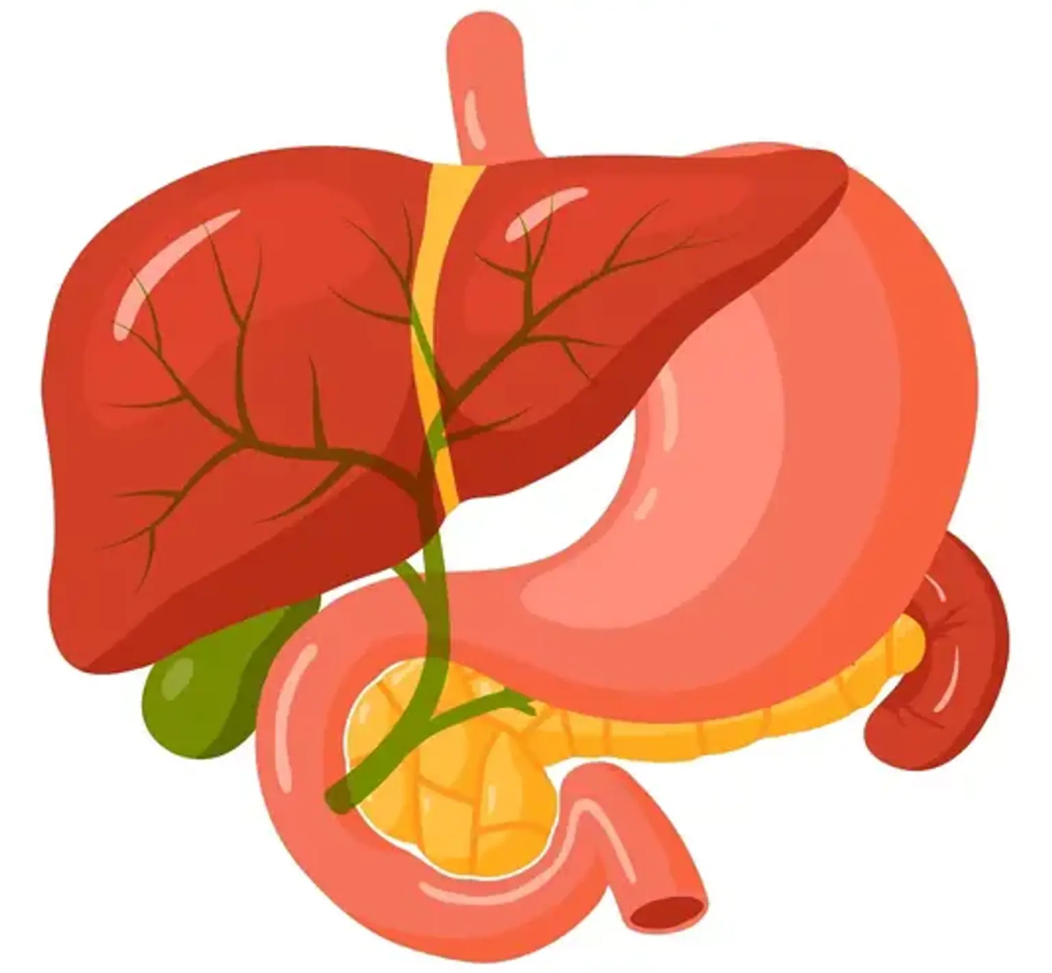
The liver produces and excretes bile. Bile travels through a network of tube-like structures known as bile ducts to the small intestine, where it aids in the digestion and absorption of food, and eventually out of the body via the digestive system. The liver, gallbladder, and pancreas are all connected to the small intestine through the common bile duct.

A biliary drain (also known as a biliary stent) is a thin, hollow, flexible tube with many tiny pores along one or both sides. When there is too much bile in the bile ducts, a biliary drain is employed. Bile can back up into the liver if the bile duct is blocked. Jaundice, a disorder in which the skin and whites of the eyes turn yellow, can result from this. A biliary drain aids bile passage from the liver into the gut when the bile duct is clogged. Depending on the kind of biliary tube, it may be attached to an external drainage bag.

Cholestasis is a condition in which the bile becomes clogged. Cholestasis can be caused by a number of conditions, including:

  • Hepatitis C (inflammation of the liver).
  • Cirrhosis (a slowly developing disease in which healthy liver tissue is replaced with scar tissue).
  • Biliary cholangitis (primary) (a liver disease caused by the destruction of the bile ducts in the liver).
  • Gallstones.
  • Tumors in the digestive system, such as the pancreas, gallbladder, or liver.
  • Infection.


What is Endoscopic Ultrasound (EUS)?

Endoscopic Ultrasound

The interior of your digestive tract is examined using endoscopic ultrasonography (EUS). It is capable of detecting pancreatic cancer, colon cancer, and cancer that has migrated to other regions of your body. Inflammatory bowel disease (IBD), pancreatitis, and other causes of stomach pain can also be detected with EUS.

Endoscopic ultrasonography (EUS) is a test that looks into your digestive tract. An EUS is performed by a gastroenterologist (a specialist who specializes in the digestive system).

An EUS test combines the following:

  • Endoscope, which is a long, thin, flexible tube with a camera and light at one end.
  • Ultrasound, a probe on the end of the endoscope that generates pictures of your organs, tissues, and blood arteries by emitting sound waves.


What is EUS-guided hepaticogastrostomy?

ERCP chart

The majority of individuals who require biliary drainage may be treated with endoscopic retrograde cholangiopancreatography (ERCP). However, ERCP can be difficult in individuals who have comorbidities such as malignant duodenal blockage or surgically changed anatomy, such as a Roux-en-Y anastomosis, which prevents the duodenoscope from being advanced into the ampulla of Vater.

Transhepatic or trans-duodenal techniques to biliary drainage have evolved as an alternate method of biliary drainage. Typically, both techniques can be used to conduct EUS-guided gallbladder drainage or choledochoduodenostomy, as well as EUS-guided hepaticogastrostomy (HGS). Because of its trans-gastric technique, EUS-HGS can be used in patients with malignant duodenal blockage.

When ERCP drainage fails, EUS-guided biliary drainage (BD) is an alternative for treating obstructive jaundice. These techniques are alternatives to surgery and percutaneous transhepatic BD, and they are made possible by the ongoing development and improvement of EUS scopes and accessories.

The most frequent treatment for obstructive jaundice is endoscopic biliary stenting. Selective cannulation of the main papilla fails in 3%-12% of instances, necessitating surgery or percutaneous biliary drainage (BD). Percutaneous BD methods (PTBD) have a high risk of complication such as hemorrhage or peritoneal bile leakage (20%-30%), and the morbidity and mortality of surgery for such palliative operations are 35%-50% and 10%-15%, respectively. A novel BD approach utilizing EUS- and EUS-guided bile duct puncture (common bile duct or left hepatic duct) is currently available. It is now feasible to produce biliodigestive anastomosis using EUS guidance and specific equipment.


What are the indications for EUS-guided hepaticogastrostomy?

ERCP Instrument

Endoscopic retrograde cholangiopancreatography (ERCP) is often used to treat malignant biliary blockage. The majority of individuals who require biliary drainage may be treated by ERCP. Despite the high success rate and low morbidity of BD acquired by ERCP, complications such as an ingrown stent tumor, tumor gut compression, periampullary diverticula, and anatomic variance might emerge.

As an alternative to bile duct drainage, percutaneous transhepatic biliary drainage (PTBD) has traditionally been explored. In patients with surgically changed anatomy, biliary drainage can also be accomplished via single-balloon enteroscopy (SBE) or double-balloon enteroscopy (DBE). However, while these operations have therapeutic benefits, they have drawbacks such as self-tube removal or aesthetic difficulties following PTBD, as well as lengthy procedures and the danger of perforation in SBE or DBE.

Endoscopic ultrasound-guided biliary drainage (EUS-BD) through transhepatic or trans-duodenal methods has recently emerged as a biliary drainage option. The first EUS-HGS surgeries were technically difficult and were linked with serious adverse outcomes such as stent migration. However, technical suggestions for EUS-HGS have matured with device and technology improvements.

This operation is used to clear blockages in the middle and lower bile ducts. The most common reason for EUS-HGS is pancreato-biliary cancer. The following are the indications for EUS-HGS:

  1. Failed EBD, including Vater inaccessibility induced by malignant duodenal blockage,
  2. Percutaneous transhepatic cholangiography drainage contraindications (PTCD)
  3. Obstruction of the middle or lower bile ducts


Case selection and special precautions

Precautions

The closeness to the left lobe of the liver is a benefit of the transhepatic route (via the liver). Careful examination of the patient's clinical state and cross-sectional imaging to rule out contraindications are necessary, as is dilatation of the left intrahepatic duct (IHD). The following conditions or circumstances should be regarded contraindications or necessitate particular precautions:


Ascites:

Ascites is a disorder in which fluid accumulates in your belly. Fluid accumulation in the belly can have an impact on your lungs, kidneys, and other organs. Ascites is characterized by stomach discomfort, swelling, nausea, vomiting, and other symptoms.

Because it raises the risk of biliary peritonitis and hematoma, the presence of ascites is regarded a relative contraindication for percutaneous transhepatic biliary drainage. Although experts have demonstrated that EUS-HGS may be conducted in the presence of some ascites, stent migration might occur, necessitating vigilance in such patients.

As a result, most authorities believe that having a lot of ascites is a contraindication for EUS-HGS because it can split the area between the liver and the stomach. To avoid difficulties, a percutaneous ascites drainage tube should be placed before doing EUS-HGS if the surgery is absolutely necessary.


Left lobe atrophy:

Before drainage, a thorough examination including cross-sectional imaging is required. Endoscopic drainage of the left IHD is not appropriate and should be avoided in the case of left hepatic lobe atrophy.


Tumor infiltration:

Because of the increased risk of tumor seeding and bleeding, tumor infiltration of the stomach wall at the puncture site is a contraindication. Because of the increased risk of stent migration, EUS-HGS should be avoided in patients with unresectable gastric cancer and low stomach volume.


Isolated right intrahepatic bile duct obstruction:

Despite the growing use of EUS-HGS for right IHD drainage, the duodenum is the preferred puncture location for isolated right IHD blockage.


How is EUS-guided hepaticogastrostomy performed?

EUS performed

The first critical step in this operation is to choose a puncture site. Bile aspiration can be conducted after the targeted bile duct section has been punctured. Following that, contrast is introduced, followed by wire passage. The puncture tract is dilated and a stent can be implanted once the guidewire has been carried through the needle channel and thoroughly into the appropriate bile duct. Many specialized tools have recently been created to increase success rates and reduce problems.


Liver segment selection:

It is critical that your surgeon understands the architecture of the liver and recognizes that liver segments 2 and 3 are often the puncture targets. Many parameters should be considered when selecting the target bile duct and liver segment, including stricture location, tumor or portal vein invasion, degree of segmental dilatation, and liver atrophy.

When the tip of the echoendoscope is positioned at the esophagogastric junction with the scope in the neutral position, the left lateral segment is generally visible. Although the scope position is straighter in segment 2 and guidewire manipulation is easier, the puncture site is generally in the esophagus, increasing the risk of mediastinitis or pneumomediastinum. As a result, puncturing segment 3 is favored.

To avoid puncturing the esophagus, a clip can be placed at the esophagogastric junction prior to the EUS test. To get EUS pictures of segment 3, the scope is frequently inserted into the stomach and angled upward toward the liver hilum.

The IHD component must also be chosen carefully. The intrahepatic duct is often bigger towards the liver hilum, making identification simpler; nonetheless, placing a completely covered self-expandable metallic stent (FCSEMS) in the hilar region should be avoided since the IHD branches should not be obstructed. When the bile duct width is greater than 5 mm and the hepatic part length is between 1-3 cm, the technical success rate is higher.


Scope position:

When doing EUS-HGS, the echoendoscope is often positioned in a short position with upward angulation toward the lesser curvature of the stomach. The scope position and needle orientation are critical factors for effective guidewire manipulation; hence, changing the scope position under fluoroscopy supervision is suggested before beginning the surgery. An oblique angle between the scope and the needle improves guidewire insertion success. However, when the scope is in an angulated position, the forward force during instrument insertion may be diminished, resulting in looping and displacement of the equipment.

Inserting a second guidewire close to the first wire is another strategy that has been recommended to increase the stability of the scope during the surgery. This approach assists in securing the scope to the tract, improving guidewire vision on EUS, and providing a rescue wire in the event of a technical failure.


Tract dilation:

The two primary forms of dilation devices are cautery and mechanical dilators. In previous research, a cautery-type dilator known as a needle-knife was utilized. When a needle-knife is put over an angulated scope, it is frequently in a tangential orientation, resulting in undesired incisions and an increased risk of problems.

The needle-knife cautery dilator has now been superseded with the coaxial electrocautery dilator. The electrocautery device is useful in individuals with fibrotic livers; nevertheless, greater bleeding is noted in such situations, prompting worries about "over-burning" effect.

The balloon dilator and the dilation catheter are the two most popular types of mechanical dilators. The danger of bile leakage during the surgery is a disadvantage of multistep tract dilatation. As a result, a novel dilation tool has been designed that enable tract construction, dilation, and stent implantation without the need for instrument swap. This "all-in-one" equipment consists of a 3-Fr tip balloon catheter, a 4-Fr tip balloon catheter paired with a stylet for tract dilatation, a metallic stent with a thin delivery system, or a metallic stent with electrocautery at the delivery system tip.


Stent selection:

Although straight plastic stents and completely covered self-expandable metallic stents (FCSEMS) have been employed in the past, plastic stents are known to have drawbacks such as stent occlusion and bile leakage, especially with large-tract dilatation. Meanwhile, FCSEMS has gained popularity since it allows for a wider drainage width through a short endoscopic route; nonetheless, it has limitations such as stent migration and probable occlusion of the IHD side branches.

Recently, a specialized plastic stent for interventional EUS has been created. To avoid migration, this newly designed plastic stent features a single pigtail with a tapered tip and four flanges. Furthermore, its tiny diameter may lessen the chance of IHD obstruction. Nonetheless, this form of stent is not generally accessible and requires stent substitution on a regular basis.

A specialized partly covered self-expandable metallic stent (PCSEMS) with an antimigration design has also been released. This stent has an uncovered piece for the IHD part to prevent minor branch occlusion, as well as a fully covered portion for the space between the liver and gastric wall to prevent bile leakage, and a flared end in the stomach side to prevent inward migration. Some specialists believe that a 6-mm FCSEMS is preferable to a large-bore metallic stent because it reduces the risk of IHD obstruction and biliary hyperplasia. Stent dysfunction and dislocation, on the other hand, have been documented in up to 50% of cases.


What is the outcome after EUS-guided hepaticogastrostomy?

Outcome after EUS

With improvements in equipment and processes, the technical success rate has risen to 90-100%. Recurrent biliary blockage might develop anywhere between 5.5 and 8.5 months following the operation. EUS-HGS is mostly used to diagnose malignant blockage. It has recently been utilized to treat anastomotic strictures in patients with surgically changed anatomy, as well as a portal for direct cholangioscopic-guided lithotripsy (stone break down) of IHD stones.

Lithotripsy via the hepaticogastrostomy tract is a safe technique that can be done 1-4 weeks following the original surgery. Although the technical success rate for benign diseases is comparable to that for malignant blockage, stent malfunction is common when metallic stents are utilized. As a result, they should be exchanged by plastic stents on a regular basis; nevertheless, the reintervention rate is significant due to stent malfunction. Long-term results under benign settings are yet unknown.


What are the possible complications of EUS-guided hepaticogastrostomy?

Complication of EUS

To date, 42 research including 1192 patients have been published on EUS-BD. The cumulative technical success rate (TSR), functional success rate (FSR), and adverse-event rate are respectively 94.71%, 91.66%, and 23.32%. Bleeding (4.03%), bile leakage (4.03%), pneumoperitoneum_ presence of air in the peritoneal cavity_ (3.02%), stent migration (2.68%), cholangitis (2.43%), abdominal discomfort (1.51%), and peritonitis (1.26%) were the most prevalent adverse events related with EUS-BD.


Vascular injury from needle puncture:

Injuries to the portal vein and hepatic artery can result in either immediate or delayed bleeding. There have been reports of bleeding from a hepatic artery pseudoaneurysm following EUS-HGS, with all patients successfully treated with trans-arterial procedures. If a vascular structure is mistakenly pierced, bleeding can be stopped by withdrawing the needle until the tip is in the liver parenchyma, then reintroducing the stylet to push back the trapped clot inside the needle and obstruct the puncture path.


Guidewire problems:

Shearing can occur during manipulation when the guidewire is shaved by the needle tip, especially if the angle between the guidewire and the needle is severe. To decrease this difficulty, gentle guidewire withdrawal, converting the needle to a catheter before guidewire manipulation, and utilizing a blunt needle tip with a sharp stylet have been advocated.

If shearing occurs, the liver impaction approach (forward guidewire insertion and mild retraction to retain the needle tip inside the liver parenchyma) has been documented. A steerable access needle device was recently designed to prevent wire shearing during manipulation.

Furthermore, during manipulation, coiling and inadvertent advancement of the guidewire toward the peripheral bile duct are common. To facilitate guidewire manipulation, the needle can be withdrawn and replaced with an ERCP cannula or coaxial dilator in certain cases.


Bile leak:

Bile leakage following EUS-HGS is not uncommon. The use of a needle knife, noncoaxial electrocautery, and the installation of a plastic stent are all factors related with bile leak. According to a retrospective study, the number of punctures (>1), procedure time (>20 min), distance to the liver parenchyma (2.50 cm), and presence of acute cholangitis were all significantly associated with bile peritonitis; however, only the short distance to the liver parenchyma (2.5 cm) remained significant in multivariate analysis. To avoid bile leak, a covered metal stent at the distal section between the liver and the stomach wall is advised. Thus, biliary peritonitis might be reduced by newly created equipment that decreases instrument interchange and process duration.


Stent migration:

One of the most serious problems related with mortality is stent migration. It might happen right away or a few days after the treatment. As a result, stent deployment technique is critical. Inadequate stent traction during deployment may result in a large gap between the liver and the outer stomach wall, resulting in stent entrapment between these two tissues, referred to as a "candy sign." As a result, adequate strain should be provided during stent opening to avoid this issue. In general, intra-scope channel stent placement is advised. Furthermore, a stent length of more than 3 cm on the stomach side is recommended.

If a stent is misplaced during deployment, rescue procedures can be undertaken while the wire remains in place and another stent is placed in tandem fashion. The use of foreign-body forceps to reposition the migrated metal hepaticogastrostomy stent and insert an additional stent, as well as EUS-guided penetration through the stomach hole and placement of another metallic stent, has been documented. If the bile leak persists, the biliary path from the HGS site may need to be converted to a trans-papillary or percutaneous route.

To prevent delayed stent migration, many preventive measures have been proposed, including using a longer stent or partially covered self-expandable metallic stent, inserting a double pigtail stent inside the metallic stent, using a plastic stent as an anchor to keep the metallic stent in place, and using a combination of uncovered and covered stents to lock the intrahepatic portion.


Conclusion

Acute hepatitis

With a high percentage of technical success and clinical effectiveness, EUS-guided hepaticogastrostomy is a valuable technique in the event of ERCP failure. The morbidity rate during biliary drainage (BD) is significant, necessitating a skilled staff. Despite evidence that EUS-BD is safe and successful, EUS-BD drainage remains one of the most technically difficult therapeutic EUS treatments. In cases with malignant duodenal involvement or changed architecture, hepaticogastrostomy is the preferred method.

The risk of adverse events with EUS-HGS is still significant, and such occurrences can occasionally be deadly, as with stent migration. Bile peritonitis may also arise after fistula dilatation. As a result, one-step stent placement with no device swaps is perfect. Additionally, advancements in stents, such as lumen apposing creation, are necessary to avoid stent migration. Finally, endoscopists should focus not only on technical achievement but also on preventing adverse events, in order to increase the therapeutic advantages of EUS-HGS in the patients who are chosen for it. It is recommended that EUS-guided hepaticogastrostomy be done only in carefully selected patients by experienced endoscopists trained in both EUS and ERCP, with well-trained surgical support.