Extracorporeal membrane oxygenation (ECMO)

Extracorporeal membrane oxygenation

Overview

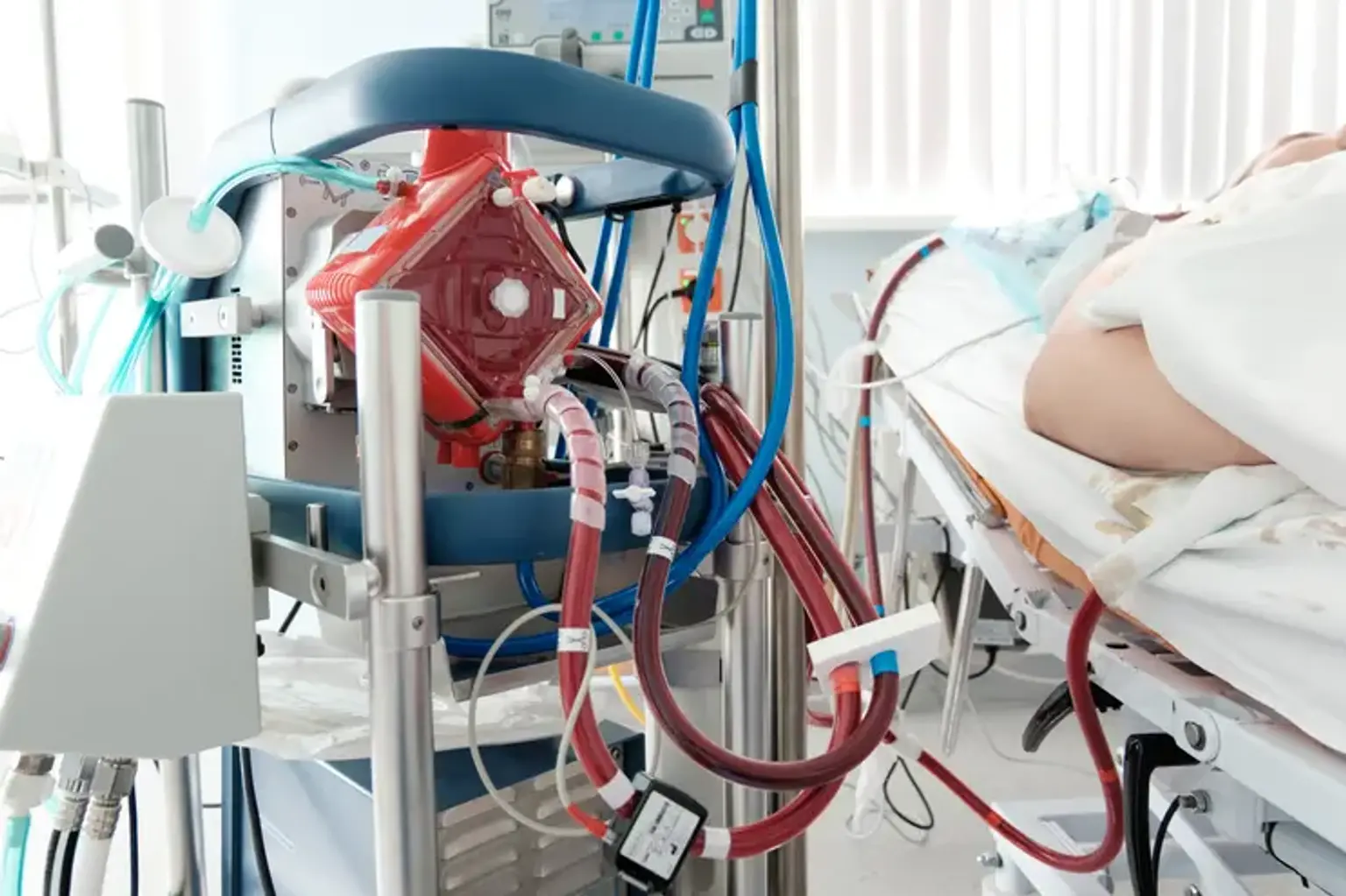
The heart and lungs are replaced by the Extracorporeal membrane oxygenation (ECMO) system. People who require ECMO assistance are cared for at a hospital's critical care unit (ICU). People are usually maintained by an ECMO machine for a few hours to days, but they may need it for a few weeks depending on how their disease improves. There are both similarities and variations in the use of ECMO in children and adults.


What is Extracorporeal membrane oxygenation (ECMO)?

Extracorporeal membrane oxygenation

Extracorporeal membrane oxygenation (ECMO), a life support system, is an invaluable tool for treating adults and children with life-threatening cardiac and pulmonary dysfunction that is resistant to conventional management or when cardiopulmonary resuscitation (CPR) measures return of spontaneous circulation (ROSC). The heart and lung functions are replaced by an ECMO system, which comprises of a pump and an oxygenator. The fundamental goal of ECMO is to replace the function of the heart and lungs, giving these organs ample time to heal.

ECMO has been used on 151,683 patients until 2020, according to the Extracorporeal Life Support Organization (ELSO) registry, including 45,205 newborns, 30,743 children, and 75,735 adults. ECMO began in 83 locations in 1990, and by 2020, the number has grown to 492 centers. Veno-venous ECMO (VV ECMO) supports the respiratory system, whereas veno-arterial ECMO (VA ECMO) supports the cardio-respiratory system.

ECMO is a form of supportive therapy rather than a disease-modifying treatment. Kolff and Berk reported oxygenation of the blood when it passed through the cellophane chambers of the artificial kidney in 1944. In 1953, Gibbon performed the first successful open-heart surgery using this notion of artificial oxygenation and perfusion. Prior to 1956, either a film or a bubble oxygenator was employed. Blood flows over numerous vertical discs in a film oxygenator, whereas oxygen bubbles through deoxygenated blood in a bubble oxygenator.

These devices' principal downsides were intravascular hemolysis, systemic inflammation, platelet destruction, and embolization. Clows and Basler created and utilized a prototype part of a membrane oxygenator suited for cardiopulmonary bypass surgery in 1956.

Rashkind used a bubble oxygenator for the first time on a baby with respiratory insufficiency in 1965. Dorson et al. published a paper in 1969 on the use of a membrane oxygenator for cardiopulmonary bypass in babies. Extracorporeal membrane oxygenation was first suggested by Baffes et al. in 1970 in neonates following heart surgery. The first application of ECMO for respiratory support in an adult patient with post-traumatic severe respiratory failure was described in 1972. In 1975, Bartlett et al. reported the first effective use of ECMO in infants suffering from acute respiratory distress.

From the 1980s until the early 2000s, ECMO circuits employed either silicone membrane or polypropylene hollow fiber oxygenators. However, due to plasma leakage in these units, a new generation of oxygenators based of polymethylpentene has been created (PMP). The most recent generation of oxygenators are simple to use, long-lasting, and enable improved gas exchange and less blood damage.

a study in 1994 failed to demonstrate the benefit of employing additional extracorporeal support in acute respiratory distress syndrome as compared to usual care with mechanical ventilation. The mechanical ventilation group had a 42 percent survival rate, whereas the low-flow VV ECMO group had a 33 percent survival rate.


Anatomy and Physiology

Anatomy and Physiology

Extracorporeal membrane oxygenation (ECMO) is a circuit in which blood is drained from the venous circulatory system through a catheter, pumped in a pump outside the body, and reinfused into the other venous or arterial vascular system for circulation in the body, depending on the ECMO circuit type.

Plastic cannulas are inserted into veins or arteries in the groins, neck, or chest. A catheter removes blood from the cannula via veins with high carbon dioxide (CO2) and low oxygen (O2) concentration. With the aid of a pump, deoxygenated blood retrieved from the venous catheter is delivered to the oxygenator. An oxygenator functions as an artificial lung, keeping the CO2 extraction and oxygenation flow rates constant.

The oxygenator's hollow threads carry air and oxygen. As blood flows through small fibers, oxygen escapes and replaces carbon dioxide in red blood cells (RBCs). CO2 is subsequently drawn into the fiber and removed in the exhaust gas. The catheter is used to return oxygenated blood to the patient.


What conditions is ECMO used to treat?

ECMO Treatment

ECMO can help infants and children with a variety of medical problems and conditions, including:

  • Meconium aspiration syndrome (MAS): A disorder in which meconium, a newborn's first feces, is ingested by the baby before or after delivery, causing lung issues.
  • permanent pulmonary hypertension of the newborn (PPHN): A condition characterized by unusually high blood pressure in the arteries that deliver blood to the lungs.
  • Congenital diaphragmatic hernia (CDH): A condition in which portions of the stomach and/or intestines protrude into the chest cavity through a hole in the diaphragm.
  • Respiratory distress syndrome (RDS) is a lung illness that mostly affects preterm newborns and makes it difficult for them to breathe on their own.
  • Pneumonia
  • Congenital heart conditions
  • Sepsis
  • End-stage cardiac or respiratory failure (as a bridge to transplant)


How does ECMO work?

ECMO Procedure

Surgery is required to connect a patient to an ECMO machine. The ECMO machine is connected to the patient via plastic tubes known as cannulas. The doctor inserts cannulas into big arteries and veins in the chest, neck, or legs after administering an anticoagulant, a medicine that stops blood clotting.

When the ECMO machine is linked, it pulls blood from the patient and sends it through cannulas into an artificial lung, which infuses the blood with oxygen and eliminates carbon dioxide. The ECMO machine then heats the treated blood to body temperature before returning it to the patient. When a patient's heart is unable to circulate blood on its own, a mechanical pump takes over and pumps blood through the patient's circulatory system.

Doctors continue to deliver sedatives and pain medicines after the treatment to make patients as comfortable and pain-free as possible. Routine chest X-rays will be performed by an ECMO team of doctors, nurses, and other health care workers, allowing clinicians to examine and follow the patient's lung and heart health. In addition, the team will do monthly blood tests to measure oxygen and carbon dioxide levels as well as look for any infections.


How does it feel to be on ECMO?

ECMO Experience

When a person is first linked to an ECMO machine, he or she is sedated and does not see the tubes being inserted into their veins and arteries. A person on ECMO is generally already hooked up to a breathing machine (ventilator) through a tube (endotracheal or ET tube) inserted into the mouth or nose and down into the windpipe.

The cannulas are not uncomfortable once linked to an ECMO machine. Patients on an ECMO machine may be given medications (sedatives or pain relievers) to make them comfortable. These medications may also cause them to fall asleep. While on an ECMO machine, some persons are awake and may converse and engage with others. In rare situations, patients using an ECMO machine can exercise to help them gain strength. However, certain motions might cause the ECMO tubes to get kinked, thus patients must be supported and closely monitored when moving.


After ECMO

After ECMO

Your child will require a ventilator to sustain oxygenation immediately after the cannulas and ECMO circuit are removed. Nitric oxide is a gas that is administered by the ventilator to relax blood vessels. The idea is to gradually wean your kid off the ventilator and drugs over time. A nurse will continue to closely monitor vital signs and indicators of discomfort, as well as complete any lab work that has been requested. While each kid is unique, be aware that your child's recovery after ECMO may take weeks or months rather than days.


Advice for parents when your child is on ECMO

ECMO Advice

Having an ECMO kid can be emotionally and even devastating for parents, guardians, and family members. The experience will most likely be packed with ups and downs. The advice below comes from other parents who have been in your situation.

  • First and foremost, take care of yourself. Every day, eat, sleep, and go outside. You will need to make medical decisions for your child, which you will only be able to do if you are rested and attentive.
  • Be an active participant in your child’s care. Most parents lack medical expertise and struggle to comprehend most of the medical language and information. Ask questions to help you comprehend what is going on and make decisions. The personnel at the hospital will make every effort to address all of your inquiries.
  • Use Hospital resources. There are numerous free services available at CHOP that may make your visit less stressful. These include sleep rooms (when available), a reference library, laundry, computers with Internet access, emotional therapy, and other services. Inquire with your bedside nurse for further details.
  • Talk to a Hospital social worker, who can help ease your stress by talking about issues that concern you.


Complications

Complications ECMO

  • Bleeding

Because of the danger of brain hemorrhage or bleeding in the lungs or gastrointestinal system, bleeding is the most common and potentially fatal consequence. Systemic heparinization, fibrinolysis, clotting factor hemodilution or platelet failure, uremia, or hepatic dysfunction might all be causes of bleeding. Within minutes of starting ECMO, the contact and fibrinolytic systems, as well as the consumption and dilution of coagulant components, might be activated. According to Robinson et al., probable causes of thrombocytopenia include platelet adhesion to surface fibrinogen and activation, aggregation, and clumping of platelets, which results in a reduction in platelet counts.

In the case of life-threatening hemorrhagic episodes, lowering or discontinuing heparin, platelet infusion, and clotting factors such as activated factor VII has been investigated. In many occasions, factor VIIa has been linked to lethal thrombosis. Platelet transfusion may result in just a brief rise in platelet count.

Prevention is better than cure is also demonstrated here. It has been suggested that if feasible, invasive or surgical treatments be avoided. Platelet count >150,000 mm, fibrinogen concentration >200 mg/L, prothrombin ratio >1.5, and reduced activated clotting time are all indicated. It is advised to discontinue heparin infusions or other anticoagulants such as bivalirudin for a short period of time. In the event of fibrinolysis, high D-dimer or presence of thromboelastography is helpful, and antifibrinolytics such as tranexamic acid, aminocaproic acid, and aprotinin are recommended.

In addition to the methods indicated above, pulmonary bleeding is managed using steroids or bronchoscopy. Intracerebral hemorrhage or infarction occurs in 10 to 15% of ARDS patients on ECMO and is responsible for 43% of ECMO mortality. Patients with plasma-free hemoglobin levels more than 10% are at risk of hemolysis, hence regular measurements are suggested.

  • Intracardiac Thrombosis

In VA ECMO, peripheral cannulation through the femoral artery and vein might predispose to retrograde blood flow to the ascending aorta. As a result of blood stasis in the ventricle and insufficient left ventricular outflow, an intracardiac thrombus forms.

  • Gas Embolism

A centrifugal pump generates a considerable negative pressure of up to 100 mmHg between the pump head and the drainage cannula. The entrance of air from this section of the ECMO circuit causes gas embolism.

  • Thromboembolism

Systemic thromboembolism is a rare complication found more frequently in VA ECMO patients than in VV ECMO patients. Preventing thromboembolism requires constant monitoring of the ECMO circuit for indicators of clot formation and heparin administration to maintain the goal active clotting time (ACT).

  • Mechanical Complications

Clot development in the ECMO circuit is a prevalent problem. Clot development may be caused by pulmonary or systemic emboli, oxygenator failure, or consumption coagulopathy. Heparin-coated ECMO circuits are routinely utilized to prevent clot formation.

  • Heparin-induced Thrombocytopenia (HIT)

HIT is a rare complication, most often observed with long-term ECMO usage. Disseminated intravascular coagulation has also been linked to the use of ECMO. 

  • Neurological Complications

Common neurological consequences include seizures, violations, and cerebral hemorrhages. In contrast to bleeding, blood clotting increases the risk of stroke in ECMO patients. Ischemic stroke or cerebral bleeding can develop as a result of coagulopathy, systemic heparinization, thrombocytopenia, systemic hypertension, or carotid artery and jugular vein ligation.

  • Renal Failure and Oliguria

Acute tubular necrosis in the early stages of ECMO may necessitate dialysis or hemofiltration.

  • GI Tract Complications

Stress, ischemia, or bleeding tendencies cause GI tract hemorrhage. While on ECMO, prolonged fasting or complete parenteral feeding, hemolysis, and diuretics may lead to hyperbilirubinemia and/or biliary calculi.

  • Sepsis

ECMO is a foreign body that raises the risk of infection. Patients with postcardiotomy cardiogenic shock are more likely than other ECMO patients to get an infection.

  • Metabolic Complications

Electrolyte imbalance, hypo or hyperglycemia, medication concentration changes due to increased volume of distribution, and a decline in liver/kidney function may also be noticed.

  • Cannula-related Complications

VA ECMO is associated with cannula malposition, vascular perforation, bleeding, erroneous placement (venous cannula within the artery), arterial dissection, pseudoaneurysm, and limb ischemia. Cannulation must be revised, i.e., the cannula must be displaced. These problems are potentially fatal. To prevent limb ischemia, the down-flow cannula can be placed into the superficial femoral artery during percutaneous cannulation of the common femoral artery. During open surgeries, a Dacron graft can be sutured to the common femoral artery and a cannula placed into the graft. Fasciotomy should be performed if necessary in cases of severe leg ischemia.

  • Hypoxia

In the case of VA ECMO, peripheral catheter placement in the femoral artery increases perfusion to the lower limbs and abdominal viscera. As a result, hypoxia may affect the upper extremities, brain, and heart. As a result, oxygen saturation should be checked in both the upper and lower extremities. When the upper extremities' oxygen saturation is low, oxygenated blood is pumped into the right atrium. Inadequate circuit flow during VV ECMO might be the cause of hypoxia. Other possibilities include sepsis, recirculation, insufficient sedation, iatrogenic hyperthermia, convulsions, or overfeeding.

Hypotension is thought to be caused by lower vascular tone during VA ECMO and reduced vascular tone, reduced preload, or impaired cardiac function during VV ECMO. Sepsis is also a key contributor to the development of hypotension, and patients may require inotropic support in such cases.

Contraindications

The majority of contraindications are relative, weighing the dangers of the surgery against the potential benefits. The following are the relative contraindications:

  1. Conditions incompatible with normal life if the person recovers
  2. Preexisting conditions that affect the quality of life (CNS status, end-stage malignancy, risk of systemic bleeding with anticoagulation)
  3. Age and size
  4. Futility: those who are too sick, have been on conventional therapy too long, or have a fatal diagnosis.


When does a patient “come off” ECMO?

ECMO therapy

In general, clinicians want to wean patients from ECMO therapy as soon as feasible. Because ECMO therapy is used for a variety of illnesses, each with its own recovery timeline, the length of time someone is on it might vary substantially. Some people only require it for a few hours, while others may require it for days or weeks. When the patient has reached the point where the ECMO machine is no longer required, the ECMO team will begin the weaning procedure.

The ECMO machine will not be shut off as a result of this. Rather, the team conducts a study, often known as a weaning trial, in which they gradually reduce the patient's blood flow through the machine. They monitor the patient's reaction to the reduction in ECMO support over many hours. If the patient reacts well and the ECMO team determines that discontinuing ECMO is safe, the cannulas will be removed by a surgeon.

A ventilator may be required to offer respiratory assistance when a patient is removed from ECMO. Doctors will remove the ventilator as soon as the patient is able to breathe without help. However, patients may need to stay in the hospital for several days or weeks, at least until vital signs stabilize. Many people may also require physical therapy to help them restore muscular strength, as well as speech therapy to help them recuperate after using a ventilator's breathing tube for an extended period of time.

Even with great treatment and ECMO therapy, the underlying heart or lung problem may not always improve. If the ECMO team considers that a patient will not recover, even if ECMO therapy is extended, they will consult with family members about end-of-life alternatives including the withdrawal of ECMO support.


Conclusion 

Extracorporeal membrane oxygenation (ECMO) is used to treat cardiac or respiratory failure when standard therapy, including CPR, has failed. ECMO is a circuit that consists of a draining cannula that drains blood from the body and circulates it in the machine before returning it to the body via a returning cannula. Veno-venous and veno-arterial ECMO have traditionally been employed.

Anticoagulation monitoring is critical during this blood circulation to maintain the balance between clotting and bleeding. Heparin-induced thrombocytopenia, neurologic problems, and sepsis issues should also be considered. In situations of cardiac arrest, cardiogenic shock, and ARDS, including COVID-19 infection, survival following ECMO use has improved.