Eye enucleation

    Last updated date: 02-Mar-2023

    Originally Written in English

    Eye enucleation

    Eye enucleation

    Overview

    Enucleation is the process of removing the eye while leaving the ocular muscles and orbital contents intact. This sort of ocular surgery is used to treat a variety of ocular malignancies, as well as eyes that have been severely traumatized or are otherwise blind and painful.

    The prospect of losing an eye is understandably terrifying for many people. However, it's crucial to understand that eye removal is a very routine procedure that may treat a variety of eye illnesses, relieve eye pain, and significantly enhance a patient's quality of life. This information is intended to assist patients and their families in better understanding the many techniques used to remove the eye, how to best prepare for the operation, and what to expect throughout the healing period.
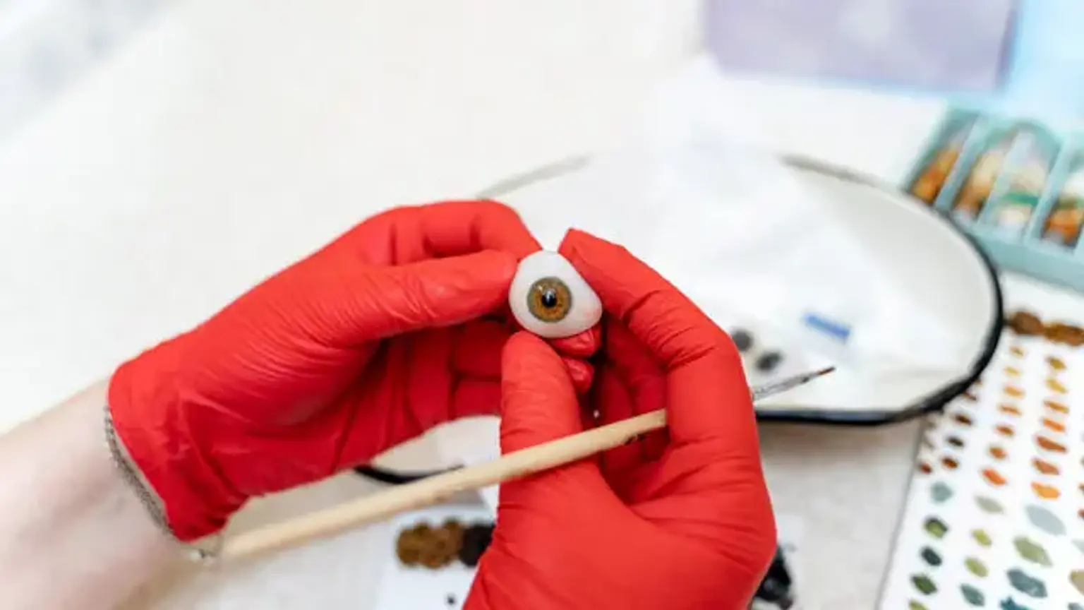
     

    Eye Enucleation definition

    Enucleation

    Enucleation refers to the removal of the complete globe from the orbit, as well as the separation of all connections from the orbit, including the transection of the optic nerve. With descriptions reaching back to 2600BC, it is one of the earliest operations in ophthalmology. Enucleation is one of the most difficult decisions to make and discuss with the patient. Trauma, a painful eye, an unattractive blind eye, intraocular cancer, and eye donation are the most common reasons for enucleation. Depending on the underlying diagnosis and health of the eye, procedures such as evisceration or exenteration may be recommended.

    The anophthalmic socket is difficult to manage, and choosing an implant and wrapping material should preferably be done as part of the pre-operative strategy. Synthetic, autologous, or human tissue supplied from an eye bank can all be used as wrapping materials.

    Peg implantation may be beneficial to certain individuals for better postoperative motility and cosmesis. However, because to the frequency of late problems such as infections, exposure, discharge, and peg loss, this operation has mostly been abandoned. Enucleation's ultimate goals are to eliminate sick tissue, increase patient comfort, replenish orbital volume, and offer the patient a satisfactory functional and attractive outcome. 


    Anatomy and Physiology

    Adequate volume replacement in the orbit, the creation of fornices bordered by conjunctival or mucous membranes, a well-fitting ocular prosthesis, and acceptable aesthetic and functional eyelids are all necessary for a satisfactory outcome following enucleation. Reduced circulation, metabolism, and insufficient volume replacement can cause orbital fat atrophy and cicatricial orbitopathy, resulting in post-enucleation socket syndrome symptoms (PESS). Damage to the lacrimal apparatus might result in a dry anophthalmic socket or discharge, as well as problems with prosthesis retention.

    Enucleation in children can be compounded further by the necessity to remove the eye before it has grown to full adult size. At two years of age, the eye reaches 85% of its axial length and continues to expand at a rate of 1% each year until it reaches full size. Patients who receive enucleation as a child may require implant exchange as a follow-up treatment as they mature. In juvenile patients, larger implants (16 to 20 mm) utilized at the time of first enucleation may reduce the need for additional implants.


    Indications

    Indications for enucleation

    Before addressing enucleation with the patient, the surgeon must consider their psychological requirements and wishes, as well as their visual potential and probable consequences. Intraocular malignancy, a blind painful eye, trauma, and the prevention of sympathetic ophthalmia are the most common reasons for enucleation. Microphthalmia in children and phthisis are two more possible causes. Autoenucleation is a rare occurrence that is usually associated with psychosis. The following is a list of them:

    Intraocular Malignancy

    • Choroidal melanoma
    • Retinoblastoma
    • Other neoplasms


    Trauma

    • Primary enucleation
    • Secondary enucleation for sympathetic ophthalmia
    • Autoenucleation (Oedipism)


    Blind Painful Eye

    • Neovascular glaucoma
    • Endophthalmitis and uveitis
    • Improve cosmesis


    Advantages

    Enucleation, unlike evisceration, enables for histologic inspection of the globe and optic nerve. This is especially significant in cases of biopsy-proven or suspected intraocular cancer, when it is critical to assess the tumor's margins and, if any, optic nerve invasion.

    One retrospective research found no statistically significant difference between enucleation and evisceration patients graded by both patients and masked observers when evaluating the aesthetics of enucleated and eviscerated sockets.

    Enucleation has traditionally been assumed to reduce the incidence of sympathetic ophthalmia by avoiding uveal antigen exposure during evisceration. Recent investigations, on the other hand, have shown no incidences of sympathetic ophthalmia after evisceration.


    Disadvantages

    Enucleations are frequently associated with a decrease in implant motility. Enucleation patients had statistically significantly lower implant motility than evisceration patients, according to one study. Patients who had their prostheses eviscerated or enucleated had no difference in prosthetic motility.


    Contraindications

    In situations with intraocular malignancy with signs of orbital spread, an enucleation is contraindicated; these patients usually require an exenteration. Patients with sympathetic ophthalmia may be a relative contraindication since the sympathizing eye may eventually have improved vision, and longitudinal studies have indicated no substantial benefit of enucleation versus evisceration in the prevention of sympathetic ophthalmia. Enucleation may be contraindicated in individuals who would suffer significant psychological anguish as a result of losing an eye.


    Preparation for surgery

    Preparation for eye surgery

    The surgeon should have a thorough conversation with the patient about the indications, risks, and advantages of enucleation, as well as other options. The patient must be informed of potential risks, which are covered in further detail in the section on complications. If the patient is using nonsteroidal anti-inflammatory drugs, aspirin, or anticoagulants, these can be stopped before surgery if medically possible. Any anticoagulant medication adjustments must be communicated to the patient's primary care provider.

    The patient's informed written permission is acquired normally before the operation date and reconfirmed on the day of the procedure to allow enough time to discuss any concerns. The surgeon, the theatre personnel, and the patient should all ensure that the surgery location is right. Before any medicine is delivered in the preoperative region, the surgeon should physically mark the side being operated on with the patient's permission and agreement.

    A peribulbar injection is used to administer a local anesthetic. Once the patient is under general anesthesia, it is recommended that a three-point check is performed to ensure that the correct eye is being removed: the surgeon reviews the consent form signed by the surgeon and the patient, the surgeon reviews the clinical notes that record the history, and the anesthesiologist, the circulating nurse, and the surgical nurse identify and agree with the correctly marked eye. After then, and only then, may the surgery begin.


    Surgical Technique

    Surgical Technique

    1. Once a sterile field is achieved, the eyelids are retracted with the lid speculum or a traction suture.
    2. A retrobulbar anesthetic injection is administered with a mixture of 2% lidocaine and 0.5% marcaine with epinephrine.
    3. A 360-degree conjunctival peritomy is performed, and Tenon fascia is carefully removed from the globe with blunt dissection between the rectus muscles.
    4. Each rectus muscle is isolated with a squint hook, and the Tenon capsule is detached from the muscles.
    5. The muscle is secured by a locking double-armed 6-0 suture posterior to the insertions. Displacement of the muscle is prevented by clamping the suture to the surgical drape. The muscle is then transected anterior to the suture.
    6. Tacking sutures are not used to displace the superior and inferior oblique muscles.
    7. The rectus muscle insertions are grasped to provide vertical traction on the globe. The optic nerve is pinched, and the nerve is transacted using enucleation scissors or snares placed into the posterior orbit. The stump of the inferior oblique is a good method for providing anterior traction.
    8. Enucleation scissors are used to sever the optic nerve.
    9. Wescott scissors are used to carefully dissect any remaining fibrous attachments.
    10.  Avoid manipulating orbital fat to avoid the risk of atrophy.
    11. Soak neurosurgical paddies/swabs in local anesthetic with epinephrine for a few minutes before pressing them into the orbit. Bipolar cautery can help with hemostasis, but it should be performed with caution.
    12. During enucleation, at least 7-10 mm of the optic nerve is removed in cases with intraocular malignancies. After enucleation, it's a good idea to photograph the globe and the length of the optic nerve.
    13. Send the globe to be examined pathologically.
    14. If you're going to put an orbital implant in, make sure it's the right size and wrap it properly before inserting it with the Carter introducer into the muscle cone, behind the Tenon capsule.
    15. The Tenon capsule is closed using buried and interrupted 5-0 absorbable sutures, and the rectus muscles are anchored to the implant.
    16. A running 6-0 or 7-0 absorbable suture is used to seal the conjunctiva, preventing invagination of the epithelium, which might lead to cyst development.
    17. Finally, a conformer is used to keep the fornices in place and avoid the formation of symblepharon. The fornices and lids are treated with antibiotic ointment.
    18. Postoperative pain is well controlled by a retro-orbital injection of liposomal bupivacaine.
    19. A 5-0 vicryl or 6-0 vicryl temporary tarsorrhaphy suture is usually put between the lateral, upper, and lower eyelids to decrease chemosis and keep the conformer from coming out. One to four weeks following surgery, the sutures are removed.
    20. To decrease postoperative edema, the orbit is covered with a light pressure patch for 24 hours.


    Post-Surgery

    eye Post-Surgery

    The patient might be given oral analgesics to use as needed. Infections may necessitate oral broad-spectrum antibiotic prophylaxis. The pressure patch can be left on for up to a week to help with postoperative edema, although it's usually taken off within 24 hours due to the inevitable seeping and soaking of the dressing. It will need to be removed so that a topical antibiotic and corticosteroid ointment may be applied to the conformer. Before handling the conformer, patients should wash their hands.

    • Orbital Implants

    Enucleation is frequently done in conjunction with implant placement, either as a main or subsequent operation. There have been many different types of implant materials employed. Nonporous materials include PMMA, silicone, and acrylic, whereas porous materials include hydroxyapatite and porous polyethylene. Porous materials provide for better vascularisation and tissue integration, which reduces migration and extrusion.

    The purpose of implant placement is to recover lost volume, enhance symmetry with the other eye, and allow prosthesis movement. Implants can be concealed or visible, and they can be integrated or nonintegrated. Regardless of the type of implant employed, the orbital contents will tend to compress towards the apex nasally and inferiorly over time. Superior sulcus deformity is most likely the result of this procedure.

    Prior to implantation, it is critical to size the orbital implant accurately. Sizers are utilized intraoperatively to choose an implant that is large enough for the Tenon capsule and conjunctiva layers to spread over its surface without stress. Any wrapping material will increase the implant diameter by 1 to 2 mm and lessen the likelihood of the implant becoming exposed.


    Complications

    Intraoperative

    The most feared intraoperative complication is the removal of the incorrect eye, which may be prevented with careful attention, effective communication, pre-surgery examinations, and labelling of the eye to be enucleated. A rectus muscle can be lost during surgery, however this can be avoided by carefully inserting the traction sutures. If the muscle is gone and retracts into the orbit, a thorough examination of the soft tissues can be carried out. The Tenon's fascia can be seen by grasping it with forceps in a hand-over-hand method.

    The retracted muscle may be identified by the inter-muscular septae that run between the extraocular muscles. The patient's anticoagulation status must be determined before to surgery and, if necessary, terminated during the perioperative period. Intraoperative ocular hemorrhage can be reduced with careful dissection, tissue manipulation, and cautery. Intraoperative bleeding can also be reduced by injecting anesthesia with epinephrine into the retrobulbar area.

    It may be essential to get a lengthy segment of the optic nerve in the presence of an intraocular tumor such as melanoma or retinoblastoma since the tumor may have progressed posteriorly down the optic nerve. It might be difficult to remove an acceptable length of the optic nerve when the globe is filled of malignancies.

    We employ the superomedial approach to the orbit to find the optic nerve and cut it under direct vision in these instances, which are mainly youngsters with big intraocular retinoblastomas. This eliminates the risk of unintentionally transecting the globe or getting an insufficient length of the optic nerve.


    Postoperative

    • Early Postoperative 

    With the use of compression bandages and the precautions outlined previously, postoperative orbital bleeding following enucleation is uncommon. If there is a large amount of blood loss, surgical investigation may be required, and different incisions can help reduce wound dehiscence and fat atrophy. Edema of the orbit is frequent following enucleation and normally goes away with time.

    A uncommon consequence is orbital infection, which can result in wound dehiscence, implant exposure, and extrusion. Increased chemosis and chronic discomfort in the socket are two possible symptoms. With integrated orbital implants, infection is more likely, and therapy with systemic antibiotics may be ineffective, forcing implant removal.

    Conjunctival prolapse and fornix shortening can occur if the conformer becomes dislodged. After cleaning, the patient should be urged to return the conformer in its original position. The above-mentioned temporary tarsorrhaphy sutures serve to limit the danger of conformer extrusion.

    • Late Postoperative

    The secondary effects of time, gravity, and the stretching of the soft tissues of the orbit by the prosthesis might result in a loose socket. It's a typical late complication after enucleation that causes the orbital implant to migrate lower and anteriorly. A larger, heavier prosthesis may give immediate comfort, but it will eventually promote further downward migration and deepening of the superior sulcus, as well as lower lid laxity.

    The post-enucleation socket syndrome or anophthalmic syndrome comprises the following:

    • Enophthalmos
    • Deep superior sulcus
    • Upper lid retraction/ptosis (one or both may occur)
    • Lower eyelid laxity
    • The fullness of the lower eyelid and shallowing of the inferior fornix (caused by the rotation of the orbital tissues that occur in an enucleated socket)
    • Posterior tilt of the prosthesis

    Enophthalmos can strike at any time. Many methods for restoring the orbital volume have been described. Rose et al. proposed a two-step volume replacement procedure, beginning with a dermis fat graft or implant and ending with a silastic block placed in the extraperiorbital area. Graft ulceration, fat atrophy, necrosis, wound dehiscence, granuloma development, hematoma, and graft surface keratinization are all possible complications after dermis fat transplants. Good surgical technique, together with cautious tissue handling and donor site selection, can help to lessen these risks.


    Follow up after surgery

    • Ride home. Make arrangements for a transportation from the hospital. You are not permitted to drive yourself home since driving after obtaining anesthesia might be harmful. Unless they have particular credentials, you must also be taken home by an adult other than a ride-sharing service.


    • Pain/medication. You may be given pain medicine, but most people can get by with over-the-counter pain remedies. In some circumstances, prescription antibiotics or steroids may be required.


    • Restrictions. Swimming, rigorous exercise, and other physically demanding activities are prohibited for a period of two to four weeks. For up to a month, you should avoid bending at the waist and moving heavy things. As soon as you feel well enough, you may drive and engage in other typical activities. Inquire with your surgeon about particular suggestions. Keep the bandage protecting the eye dry at all times. The bandage may itch or be uncomfortable at times, but it's critical to keep it on as long as your surgeon instructs. The bandage may usually be removed the next day.


    • Follow-up. A week following the operation, you'll have a follow-up exam. If you haven't already done so, your surgeon will remove the bandage and examine your eye to check how it's healing.


    Conclusion 

    Enucleation is a surgical operation in which the whole globe and its intraocular contents are removed but all other periorbital and orbital structures are preserved. Enucleation differs from evisceration, which involves removing the ocular contents from an unbroken sclera, and exenteration, which involves removing the complete orbital contents, including the globe and soft tissues.

    Enucleation necessitates the collaboration of a surgeon, an ophthalmic nurse, theatre personnel, and medical assistants. The team will select individuals who are candidates for enucleation and will follow safe surgical guidelines during the surgery. Prior to enucleation, they will be able to securely locate and verify the right surgical site. To guarantee optimum patient rehabilitation and results, the team will conduct patient education both before and after the enucleation surgery.