Introduction
Eyelid retraction is a condition where the eyelids are positioned abnormally, either too high or too low, exposing more of the eye than usual. While this may seem like a purely aesthetic concern, it can also cause significant functional issues like dryness, discomfort, and even visual impairment.
Eyelid retraction correction is a surgical procedure aimed at restoring normal eyelid positioning, improving both appearance and eye function. This article will walk you through the causes, symptoms, and treatment options available for eyelid retraction, providing a comprehensive overview of the correction process.
What is Eyelid Retraction?
Eyelid retraction refers to the abnormal elevation of the upper eyelid or downward displacement of the lower eyelid, resulting in excessive exposure of the eye. There are two main types:
Upper eyelid retraction: The upper eyelid moves too high, exposing the sclera (the white part of the eye).
Lower eyelid retraction: The lower eyelid pulls down too much, often leaving the inner eye exposed.
This condition can be caused by a range of factors, from medical conditions like thyroid disease (especially Graves' disease) to trauma, previous surgeries (e.g., blepharoplasty), or even age-related changes. Eyelid retraction can significantly impact both the appearance of the eyes and their function, causing symptoms like dryness, discomfort, and an inability to fully close the eyelids, which can lead to further eye issues.
Anatomy of the Eyelids and Orbital Region
Understanding the structure of the eyelids is key to understanding how eyelid retraction occurs. The eyelids are made up of several muscles and tissues, with the levator palpebrae superioris being the primary muscle responsible for raising the upper eyelid. The lower eyelid, on the other hand, is controlled by muscles that help maintain its position, preventing it from drooping or retracting too much.
In some cases, the muscles and connective tissues that hold the eyelids in place become weakened or stretched, leading to eyelid retraction. This can happen due to medical conditions like thyroid disease, where the muscles around the eye become inflamed, or after surgery, when the tissues may heal in an altered position. The orbital region—the area around the eyes—also plays a role in this condition, as the bones and soft tissues that support the eyelids are crucial for maintaining normal eyelid position.
Causes of Eyelid Retraction
Eyelid retraction can arise from a variety of causes, including:
Thyroid Disease (Graves' Disease): One of the most common causes, this condition leads to inflammation and swelling of the tissues behind the eyes, pushing the eyelids upwards.
Surgical Complications: Eyelid retraction is sometimes a complication after eyelid surgery (blepharoplasty) or other eye surgeries, where the tissues around the eyes may heal incorrectly.
Age-Related Changes: As we age, the muscles and tissues around the eyes naturally weaken, which can cause the eyelids to retract.
Trauma or Injury: Any physical damage to the eye area, whether from accidents or surgery, can result in scarring or muscle damage that leads to retraction.
Each cause requires a slightly different approach to treatment, and it’s essential to work with a skilled surgeon to identify the best course of action for correction.
Symptoms and Signs of Eyelid Retraction
Eyelid retraction can present itself in several ways, both physically and functionally. The most noticeable signs include:
Visible Sclera: An increased amount of the white part of the eye is exposed, especially when the patient is at rest or looking forward.
Eye Irritation: Due to excessive exposure, the eyes can become dry, irritated, or feel gritty. This happens because the eyelids no longer fully cover the eyes when blinking or sleeping.
Difficulty Closing Eyes: Patients with eyelid retraction may struggle to fully close their eyes, which can lead to discomfort, sensitivity to light, and even vision problems.
Tired or Bulging Appearance: The exposed sclera can make the eyes appear wide open or tired, contributing to an aged or distressed look.
The psychological impact can also be significant, with many patients reporting reduced self-esteem due to the altered appearance of their eyes. If left untreated, these symptoms can lead to chronic eye dryness, visual disturbances, and increased risk of eye infections.
When to Consider Eyelid Retraction Correction
If you experience any of the symptoms above, it may be time to consult with a medical professional. Eyelid retraction correction is typically considered when the condition interferes with daily life, either by causing discomfort or affecting vision.
Patients should also consider surgery if:
Functional Impairment: If eyelid retraction is preventing the eyelids from fully closing, it can lead to dry eyes, excessive tearing, and even corneal damage over time.
Cosmetic Concerns: Many patients seek correction for aesthetic reasons, particularly if the retracted eyelid gives them a perpetually tired or stressed appearance.
When Conservative Treatments Aren’t Effective: In some cases, non-surgical options like eye drops or artificial tears can help temporarily, but if these measures don’t provide relief, surgery may be the next step.
A qualified ophthalmologist or plastic surgeon specializing in eyelid procedures can help you determine the best time for surgery and whether you’re a good candidate.
Types of Eyelid Retraction Correction Procedures
Several surgical techniques are available to correct eyelid retraction, each designed to address specific causes and patient needs. The choice of procedure depends on factors such as the type and severity of retraction, the underlying cause, and the overall health of the patient.
Eyelid Muscle Repair: In cases where the eyelid muscles have weakened or stretched, the surgeon may repair or tighten these muscles to restore proper eyelid position.
Tendon Reattachment: For upper eyelid retraction, the tendon that controls the levator muscle may be repositioned to help lower the eyelid to its natural position.
Skin Grafting: In cases of severe retraction, particularly in the lower eyelid, skin grafting might be necessary to provide additional tissue and support.
Orbital Decompression: This is commonly used for eyelid retraction caused by thyroid disease. The procedure involves removing some of the bone and tissue from around the eye to relieve pressure and allow the eyelid to return to its normal position.
In some cases, a combination of these techniques may be used to achieve the best results.
Pre-Surgical Considerations
Before undergoing eyelid retraction correction, it’s important to have a thorough pre-surgical consultation. This typically involves:
Medical Evaluation: A complete eye examination will assess the extent of the eyelid retraction, its impact on vision, and overall eye health. Blood tests may also be conducted to rule out thyroid disease or other conditions that may be contributing to the problem.
Choosing the Right Surgeon: Ensure that your surgeon is board-certified and experienced in eyelid surgery. Look for a surgeon who specializes in both cosmetic and functional eyelid procedures for the best outcomes.
Discussion of Risks and Expectations: It’s essential to understand the potential risks, such as infection, scarring, or complications from anesthesia. Your surgeon will discuss the procedure’s potential benefits and set realistic expectations for the results.
Pre-Surgical Instructions: You’ll receive guidelines for preparing for surgery, such as avoiding certain medications (like blood thinners) and fasting before the procedure.
Taking these steps ensures a safer procedure and can significantly improve the chances of a smooth recovery.
Recovery Timeline and Post-Surgical Care
After surgery, proper care and patience are crucial for achieving the best possible results. Here's what the recovery process typically looks like:
First Few Days: Expect swelling, bruising, and mild discomfort, which is normal after eyelid surgery. Cold compresses can help reduce swelling, and prescribed pain medications will manage any discomfort.
First Week: Most patients can return to light activities within a few days, but you’ll need to avoid strenuous exercise or activities that could put strain on your eyes. Sutures are usually removed within a week.
Weeks 2-4: Swelling and bruising should significantly subside, though it can take several weeks for the full recovery to be visible. During this time, you should continue to follow post-surgery care instructions, such as using prescribed eye drops to keep your eyes moist and avoiding sun exposure.
Full Recovery: Final results, including improved eyelid position and reduced scarring, can take up to 3-6 months. Follow-up appointments with your surgeon are crucial to ensure proper healing and address any concerns.
Alternatives to Eyelid Retraction Surgery
While surgery is often the most effective treatment for eyelid retraction, there are non-surgical alternatives that can help manage mild cases or serve as a complement to surgery:
Artificial Tears: If dryness and irritation are the primary concerns, using artificial tears or lubricating eye drops may provide temporary relief.
Botox Injections: In some cases, Botox can be used to temporarily treat upper eyelid retraction. Botox works by relaxing the muscles around the eyelid, though the effects are temporary, lasting only a few months.
Eye Lubricating Ointments: Prescribed ointments can help keep the eyes moist and alleviate discomfort caused by eyelid retraction, particularly at night.
Eyelid Taping: Some people use eyelid taping to manually hold the eyelids in place, but this is a temporary solution and often impractical for long-term management.
Non-surgical options are typically used in less severe cases of eyelid retraction or as a preliminary step before surgery. It’s important to discuss these alternatives with your doctor to understand their effectiveness and limitations.
Risks and Complications of Eyelid Retraction Surgery
While eyelid retraction surgery is generally safe, like any surgical procedure, it comes with potential risks. These include:
Infection: Though rare, infection can occur after any surgery. It’s important to follow post-surgical care instructions and keep the area clean.
Scarring: Even though incisions are usually made in discreet locations, there is a possibility of visible scarring, especially if healing doesn’t go as planned.
Asymmetry: There’s a chance that the eyelids may not be perfectly symmetrical after surgery, though this is usually correctable with a secondary procedure.
Dry Eyes: Some patients experience dry eyes or difficulty fully closing their eyelids after surgery. This can be temporary, but in some cases, it may require additional treatment.
Overcorrection or Undercorrection: In some cases, the eyelids may be adjusted too much or not enough, necessitating further revision surgery.
Discussing these risks with your surgeon beforehand can help you make an informed decision and ensure that you’re prepared for the recovery process.
Effectiveness of Eyelid Retraction Correction
Eyelid retraction correction is generally highly effective, offering both functional and aesthetic improvements. The primary benefits include:
Cosmetic Improvement: One of the main reasons patients seek this surgery is to correct the appearance of their eyes. By repositioning the eyelids, patients can achieve a more rested, natural, and youthful look.
Functional Relief: Many patients find relief from discomfort, dry eyes, and difficulty closing the eyes. This can greatly enhance daily functioning and improve quality of life.
High Success Rate: Studies show that eyelid retraction correction procedures have a high success rate, with most patients experiencing significant improvements in both appearance and comfort.
Longevity of Results: Results from eyelid retraction surgery tend to be long-lasting. Once the eyelid is properly positioned, it typically stays in place, though natural aging processes may cause minor changes over time.
While results can vary depending on the severity of the condition and individual healing factors, most patients report satisfaction with both the cosmetic and functional outcomes of the surgery.
Cost of Eyelid Retraction Correction
The cost of eyelid retraction correction can vary widely depending on several factors, such as the complexity of the procedure, the surgeon’s experience, the geographical location of the clinic, and whether the surgery is performed for cosmetic or functional reasons. On average, the cost can range from $3,000 to $8,000 in the United States.
Insurance Coverage: If the surgery is deemed medically necessary (for example, if the eyelid retraction is impairing vision or eye function), insurance may cover part or all of the costs. However, if the surgery is purely cosmetic, the procedure will likely be out-of-pocket.
Additional Costs: Beyond the surgery itself, there may be additional costs, including pre-surgical consultations, post-surgery medications, follow-up visits, and any necessary revisions or touch-up surgeries.
Financing Options: Many clinics offer financing plans or payment options, allowing patients to pay for the surgery over time. It’s important to check with your provider to see what payment options are available.
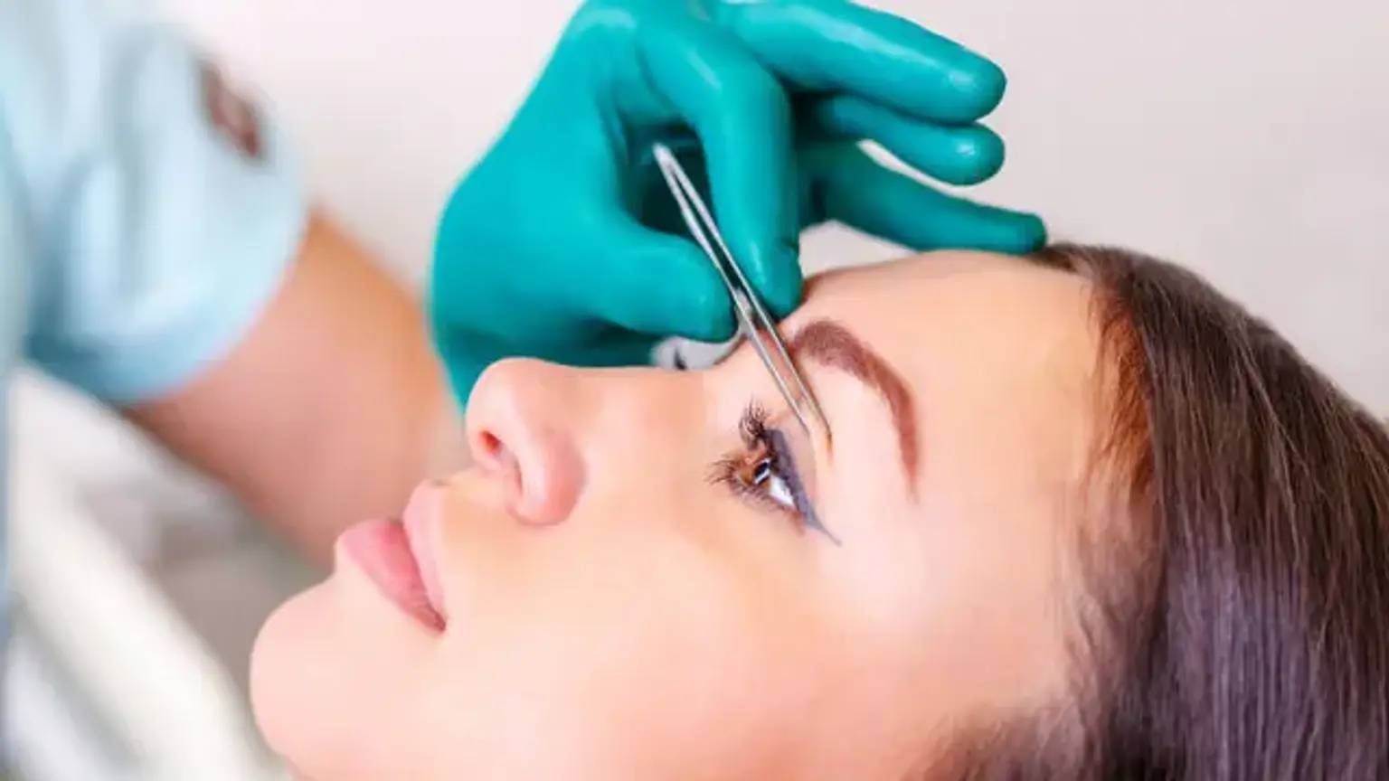
Eyelid Retraction Surgery Procedure: What to Expect
Eyelid retraction correction surgery typically involves restoring the eyelids to their normal position, which may require repairing muscles, repositioning tendons, or even removing excess tissue. Here’s what to expect during the procedure:
Anesthesia: The surgery is usually performed under local anesthesia with sedation, meaning you'll be awake but relaxed. In some cases, general anesthesia may be used, especially for more extensive procedures or if the patient prefers it.
Surgical Process: The surgeon will make small, discreet incisions, typically along the eyelid crease or at the edge of the eyelid. For upper eyelid retraction, the levator muscle may be tightened or repositioned. For lower eyelid retraction, the surgeon may perform a skin graft or reattach tissues to restore proper eyelid alignment.
Duration: The surgery typically lasts between 1 to 2 hours, depending on the complexity of the case. If a combination of techniques is required, it may take longer.
Precision: Surgeons use advanced techniques and tools to ensure minimal scarring and precise repositioning of the eyelid. Depending on the cause of retraction, the surgery will focus on either muscle repair, tendon reattachment, or skin grafting.
Choosing the Right Surgeon for Eyelid Retraction Correction
Selecting the right surgeon for your eyelid retraction correction is crucial for achieving the best possible results. Here are some factors to consider:
Board Certification: Ensure that the surgeon is board-certified in ophthalmology or plastic surgery, particularly in eyelid and orbital procedures.
Experience and Specialization: Look for a surgeon who specializes in eyelid surgery and has significant experience performing eyelid retraction corrections. A surgeon with expertise in both functional and cosmetic eyelid surgery will be able to tailor the procedure to your specific needs.
Before-and-After Photos: Ask to see before-and-after photos of previous patients who had similar procedures. This will give you an idea of the surgeon’s skills and the potential results you can expect.
Consultation: During your consultation, the surgeon should conduct a thorough examination, discuss your goals and expectations, and provide clear answers to any questions you may have. Trust and communication are key in ensuring that you’re comfortable with the chosen surgeon.
Patient Reviews: Reading reviews from other patients who have undergone similar procedures can help you gauge the surgeon’s reputation and patient satisfaction.
Global Popularity of Eyelid Retraction Correction
Eyelid retraction correction is performed worldwide, with a growing demand in many countries due to both the functional benefits and the aesthetic improvements it offers. Here’s an overview of its popularity across different regions:
United States and Canada: Eyelid surgery is a common procedure in North America, particularly among individuals with thyroid disease or those seeking to correct age-related changes. The high number of qualified surgeons and advanced surgical techniques contribute to the procedure’s widespread popularity.
Europe: In countries like Germany, France, and the UK, eyelid retraction correction is well-established, with a strong focus on both cosmetic and functional outcomes. Europe is also home to many world-renowned plastic and reconstructive surgeons specializing in eyelid procedures.
Asia: In countries such as South Korea and Japan, cosmetic eyelid surgery is extremely popular, driven by a culture that places high value on youthful, bright eyes. Although eyelid retraction correction is less common, demand is rising as awareness of the procedure grows.
Australia: Australia has a well-established medical community with many experienced surgeons offering eyelid correction surgeries. Patients often seek treatment for both functional relief and aesthetic reasons, and the procedure is becoming increasingly sought-after as people learn more about its benefits.
With advances in medical technology and greater access to information, eyelid retraction correction is becoming more globally recognized. As more people seek both cosmetic and functional improvements, the demand for skilled surgeons in this field continues to rise.
Common Frequently Asked Questions (FAQs)
Q: How long does the recovery take?
A: Full recovery takes 3 to 6 months, though most patients return to light activities within a week.
Q: Will the scars be noticeable?
A: Scarring is typically minimal, as incisions are placed in discreet areas. Over time, scars fade and are usually barely visible.
Q: Can eyelid retraction come back after surgery?
A: In most cases, the results are permanent. However, aging and other underlying conditions may affect the eyelids over time.
Q: Is the surgery painful?
A: There’s some discomfort during recovery, but pain is usually manageable with prescribed medications.
Patient Expectations and Outcomes
Setting realistic expectations is crucial for a successful eyelid retraction correction. While most patients experience significant improvement, results can vary depending on individual factors such as the severity of retraction and healing response.
Appearance: Most patients see a more rested, youthful look as the eyelids are restored to their natural position.
Functionality: The correction can improve the ability to fully close the eyelids, alleviating dryness and irritation.
Post-Surgical Appearance: Swelling and bruising may persist for several weeks, but results improve steadily over time.
Patients are typically satisfied with both cosmetic and functional improvements, especially when they follow post-surgical care closely.
Eyelid Retraction Correction for Special Cases
In certain cases, eyelid retraction may be linked to underlying medical conditions, such as thyroid disease or trauma. Here’s how these factors influence treatment:
Thyroid Disease (Graves' Disease): Eyelid retraction is common in people with thyroid disease. Orbital decompression and eyelid muscle repair can help correct retraction and improve eye appearance.
Trauma: Eyelid retraction caused by injury or previous surgeries may require more complex approaches, such as skin grafting or tendon repositioning, to restore the natural eyelid function and appearance.
Aging: As we age, eyelid muscles weaken, leading to retraction. Corrective surgery can rejuvenate the eyelid area, improving both aesthetics and function.
For all special cases, a tailored approach from an experienced surgeon is key to achieving the best results.
Conclusion
Eyelid retraction correction can significantly improve both the look and function of the eyes, offering relief from discomfort and enhancing appearance. Whether the cause is age, thyroid disease, or injury, this procedure provides lasting benefits.
If you’re considering eyelid retraction correction, consult with a qualified surgeon who specializes in eyelid surgery to discuss your specific needs, expectations, and possible outcomes. By doing so, you can make an informed decision and achieve the best possible results, restoring both your vision and confidence.