Fetal deformity
Last updated date: 13-Mar-2023
Originally Written in English
Fetal Deformity
Overview
Every year, an estimated 240 000 infants die within 28 days of birth owing to birth deformities. A further 170 000 children between the ages of one month and five years die as a result of birth deformities.
Congenital abnormalities, congenital disorders, and fetal malformations are all terms for birth defects. They are structural or functional abnormalities (for example, metabolic disorders) that arise during intrauterine life and can be recognized prenatally, at birth, or in certain cases only later in infancy, such as hearing problems. Congenital term refers to the presence at or before birth.
Although birth defects can be caused by one or more genetic, viral, nutritional, or environmental factors, determining the specific cause is sometimes challenging.
Some birth deformities are avoidable. Prevention techniques include vaccination, proper intake of folic acid or iodine through fortification of staple foods or supplements, and adequate care before and during pregnancy.
What is a Fetal Deformity?
A birth defect is an abnormality in your newborn baby's body. A baby in the United States is born with a birth defect every four and a half minutes. A defect that can damage nearly any portion of your baby's body is:
- Visible defect. Such as a missing arm or a birthmark.
- Internal (inside the body). Such as a kidney that hasn't grown properly or a ventricular septal defect (a hole between your baby's bottom chambers of the heart).
- Chemical disorder. A defect in a chemical reaction that results in developmental delay.
Your baby may be born with a single birth defect, such as a cleft lip (a gap in their upper lip), or several birth abnormalities, such as a cleft lip and cleft palate (a hole in the roof of their mouth), or even a cleft lip and cleft palate with brain, heart, and kidney disorders.
Your doctor will not be able to discover all birth problems when your kid is born. Some birth abnormalities, such as scoliosis, may not be seen until your kid is several months old. It might take years to detect a dysfunctional kidney.
How Common are Fetal Anomalies?
At birth, between 2% and 3% of newborns have one or more defects. This figure rises to 5% at the age of one (not all problems are found immediately after your child's birth). Birth defects impact one out of every 33 newborns born in the United States.
Types of Fetal Deformities
- Duodenal atresia. It is a small intestinal blockage. It can result in polyhydramios (excess fluid surrounding the fetus during pregnancy), which increases the risk of premature birth. It is occasionally linked to other genetic disorders.
- Dandy walker malformation. It is a condition in which the posterior fossa (a region in your baby's skull) and cerebellum grow abnormally (a section of the brain). This congenital abnormality can result in a range of issues.
- Limb abnormalities. They occur when the fetal amnion (the amniotic sack's inner lining) wraps around sections of the fetus (like a finger or foot).
- Microcephaly. It is a birth defect in which a baby's head is smaller than normal when compared to other newborns of the same gender and age. Babies with microcephaly frequently have smaller brains that did not develop correctly.
- Omphalocele. It is an abdominal (belly) wall birth deformity. The intestines, liver, or other organs of the newborn protrude through the belly button. The organs are surrounded by a thin, virtually translucent sac that is seldom opened or damaged.
- Atresia of the esophagus. It is a congenital defect in which a portion of a baby's esophagus (the tube connecting the mouth to the stomach) does not grow normally.
- Anencephaly. It is a significant birth condition in which a newborn is born with missing brain and skull parts. It's a kind of neural tube defect (NTD). As the neural tube develops and shuts, it contributes to the formation of the baby's brain and skull (upper portion of the neural tube), spinal cord, and back bones (lower part of the neural tube).
- Spina bifida. It is a spine problem that generally manifests itself during birth. It's a kind of neural tube defect (NTD). Spina bifida can occur anywhere along the spine if the neural tube fails to shut completely. When the neural tube fails to shut completely, the backbone that protects the spinal cord fails to develop and seal properly. This frequently leads in spinal cord and nerve injury.
- Down syndrome. Down syndrome babies have an extra copy of one of these chromosomes, chromosome 21. Trisomy is a medical word for having an extra copy of a chromosome. Trisomy 21 is another name for Down syndrome. This additional copy alters how the baby's body and brain develop, posing both mental and physical obstacles.
What Causes Birth Defects?
- Genetic
A small percentage of birth malformations are the result of genetic anomalies, such as chromosomal abnormalities (such as Down syndrome or trisomy 21) or single gene disorders (for example cystic fibrosis).
Consanguinity (when parents are close relatives) raises the likelihood of uncommon genetic birth deformities and nearly doubles the risk of neonatal and childhood mortality, intellectual impairment, and other deformities.
- Socioeconomic & demographic factors
Higher prevalence in resource-constrained families and nations. It is estimated that low- and middle-income nations account for around 94 percent of serious birth deformities. This increased risk is an indirect determinant related to pregnant women's likely lack of access to appropriate nutritional diets, greater exposure to agents or variables such as infection and alcohol, or poorer access to health care and screening.
The age of the mother is another risk factor for poor intrauterine fetal development. The likelihood of chromosomal abnormalities, including Down syndrome, increases with mother age.
- Environmental factors
Others develop as a result of environmental factors such as maternal infections (syphilis, rubella, Zika), radiation exposure, certain pollutants, maternal nutritional deficiencies (e.g., iodine, folate deficiency), illness (maternal diabetes), or certain medicines (alcohol, phenytoin).
- Unknown causes:
While complex genetic and environmental interactions have been proposed, the majority of birth abnormalities, such as congenital heart problems, cleft lip or palate, and club foot, have unknown causes.
How Do Genes Affect Birth Defects?
About 20% of birth defects are caused by genetic or hereditary factors. Genetic causes of birth defects fall into three general categories:
- Chromosomal abnormalities.
- Single-gene defects.
- Multifactorial.
Every human body cell has 46 chromosomes, each of which carries hundreds of genes. Each gene includes a blueprint that regulates the growth or function of a certain body part. People with too many or too few chromosomes will receive a confused message about anatomic development and function.
Down syndrome is an example of a disorder caused by an abnormal number of chromosomes. Individuals with Down syndrome have an extra copy of a certain chromosome due to an error during cell division (chromosome 21). This extra chromosome can result in a typical set of birth defects. Down syndrome can be distinguished by developmental delays, muscular weakness, a downward slant in the eyes, low-set and deformed ears, an abnormal crease in the palm of the hand, and congenital malformations of the heart and intestines.
A female with Turner syndrome loses part or all of one X chromosome. This can result in small height, intellectual problems, and the lack of ovaries in afflicted women.
Trisomy 13 (Patau Syndrome) and Trisomy 18 (Edwards Syndrome) are caused by having an extra copy of the 13th or 18th chromosome. These are uncommon, more catastrophic disorders that result in severe birth abnormalities that make survival after delivery impossible.
In addition to inheriting an additional or missing chromosome, single gene deletions or duplications can result in developmental abnormalities and birth problems. Cystic fibrosis is one example (a disorder that causes progressive damage of the lungs and pancreas).
Accidental damage can sometimes result in defective genes, a situation known as "spontaneous mutation." The majority of instances of achondroplasia (severe low height and deformed bones) are caused by fresh damage to the regulating gene. Furthermore, recombination mistakes can result in chromosomal translocations, which can result in complicated birth deformities.
How Do Environmental Factors Affect Fetal Defects?
Environmental factors may increase the risk of miscarriage or birth anomalies, or they may have no effect on your baby at all, depending on when the exposure happens throughout the pregnancy.
Following conception, your developing baby passes through two primary periods of development. The initial stage, known as the embryo stage, occurs within the first 10 weeks of conception. During this phase, the majority of the major body systems and organs develop. The second stage, also known as the fetal stage, is the remainder of the pregnancy. This fetal stage is a time of organ and fetus growth in general.
During the embryo stage, when organs are forming, your developing baby is most vulnerable to injury. Indeed, infections and medicines can do the most harm when they are exposed two to ten weeks after pregnancy.
Obesity and diabetes may raise your child's chance of birth abnormalities. Your healthcare professional may advise you to do all possible to control these issues before becoming pregnant.
Some medicines and recreational drugs have the potential to cause birth abnormalities, which are most severe when used during the first three months of pregnancy. Thalidomide, an anti-nausea medication used in the 1960s, caused birth deformities known as phocomelia (absence of most of the arm with the hands extending flipper-like from the shoulders).
Isotretinoin, often known as Accutane or Roaccutane, is another option. Isotretinoin is a retinoid, which is a synthetic version of vitamin A used to treat skin disorders. It is the cause of fetal retinoid syndrome. Fetal retinoid syndrome can include the following symptoms:
- Growth delay.
- Malformations of the skull and face.
- Abnormalities of the central nervous system.
- Abnormalities of the heart.
- Abnormalities of the parathyroid gland.
- Abnormalities of the renal, thymus gland.
The most often used medication that causes birth deformities is alcohol. Fetal alcohol syndrome refers to the common birth abnormalities induced by the mother's alcohol use:
- Learning disabilities.
- Developmental delay.
- Irritability.
- Hyperactivity.
- Poor coordination.
- Abnormalities of facial features.
Uterine constraint is another environmental factor that can lead to birth deformities. The fetus develops in the mother's uterus and is surrounded by amniotic fluid, which cushions it from extreme pressure (similar to being suspended in a bag of water). If the fiber sack that stores the fluid ruptures, fiber bands from the ripped sack might press on the fetus and produce amniotic band syndrome (which can result in partial contraction of or amputation of an arm or leg). An insufficient quantity of amniotic fluid can result in excessive pressure on the entire fetus, resulting in pulmonary hypoplasia (lack of development of the lungs).
About 30% of birth defects have been detected by medical science. That indicates that around 70% are still without a clear reason. As many as 50-70 percent of birth defects are sporadic, with no recognized etiology. Certain birth abnormalities can be increased by a combination of environmental and genetic factors.
How are Fetal Deformities Diagnosed?
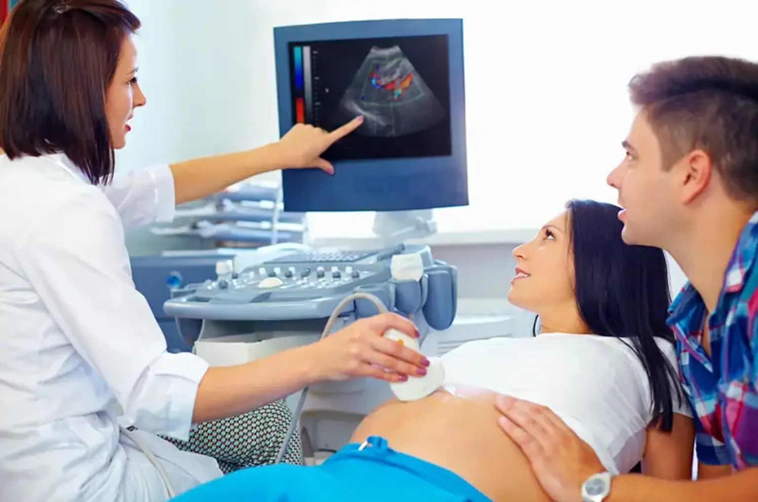
During pregnancy, you might choose to get ultrasounds or blood tests to screen for birth abnormalities and genetic conditions. If a screening test reveals an abnormality, a diagnostic test is frequently advised. Screenings throughout the first trimester look for abnormalities with your baby's heart as well as chromosomal diseases like Down syndrome. Among the screening tests are:
- Maternal blood test. Blood tests can detect protein levels or free fetal DNA in maternal blood. A fetal chromosomal abnormality is more likely if the findings are abnormal.
- Ultrasound. An ultrasound checks your baby's neck for extra fluid. This might indicate a higher likelihood of a heart problem or chromosomal issue.
Second trimester screenings check for problems with the structure of your baby’s anatomy. The tests include:
- Maternal serum screening. Blood testing in the second trimester can detect chromosomal abnormalities and/or spina bifida.
- Ultrasound. An abnormality ultrasound measures the baby's growth and looks for birth abnormalities.
If a screening test is abnormal, further testing may be required. Women with high-risk pregnancies are also provided such diagnostic tests. Among the tests are:
- Fetal echocardiography. This is a targeted ultrasound of your baby's heart that may be ordered in some high-risk pregnancies or if an anomaly ultrasound detects a heart abnormality. Not all cardiac abnormalities are detectable before birth.
- Fetal MRI. It may be requested in situations of possible birth abnormalities, especially those affecting the baby's brain or nervous system.
- Chorionic villus sampling. For this test, your healthcare professional will take a very little bit of the placenta. It will be checked for chromosomal or genetic abnormalities.
- Amniocentesis. A little amount of amniotic fluid will be collected by your healthcare provider. Cells are examined for chromosomal abnormalities and genetic diseases such as cystic fibrosis and Tay-Sachs disease. Amniocentesis can also detect infections like cytomegalovirus (CMV).
Many birth problems are not detected until your kid is delivered. They might be noticed right away, like a cleft lip, or they can be detected later. Keep an eye on your child's health and report any symptoms to your doctor. Additional testing may be required.
How is Fetal Deformity Prevented?
Preventive public health measures aim to reduce the occurrence of specific birth abnormalities by minimizing risk factors or strengthening protective ones. Important interventions and efforts include:
- Ensuring adolescent girls and mothers have a healthy diet that includes a wide variety of vegetables and fruit, and that they maintain a healthy weight.
- Ensuring an adequate dietary intake of vitamins and minerals, particularly folic acid in adolescent girls and mothers.
- Ensuring mothers avoid harmful substances, particularly alcohol and tobacco.
- Avoiding travel by pregnant women (and sometimes women of child-bearing age) to regions where there is a risk of infection.
- Minimizing or eliminating prenatal exposure to dangerous chemicals (such as heavy metals or insecticides).
- Diabetes is managed before and throughout pregnancy with counseling, weight management, nutrition, and insulin therapy as needed.
- Ensuring that any medicine or medical radiation (such as imaging rays) exposure of pregnant women is justified and based on a comprehensive health risk-benefit analysis.
- Vaccination, particularly against the rubella virus, is recommended for children and pregnant women.
- Improving and strengthening education of health professionals and others concerned in the prevention of congenital abnormalities
- Screening for infections, especially rubella, varicella and syphilis, and consideration of treatment.
Screening, Treatment & Care for Fetal Deformities
Screening
Preconception and peri-conception health care includes fundamental reproductive health practices, as well as medical genetic screening and counseling. Screening can take place at the following times:
This can help identify people who are at risk of developing specific disorders or passing a disorder on to their offspring. Screening includes acquiring family histories as well as carrier screening, which is especially important in areas where consanguineous marriage is widespread.
- Peri-conception screening
Maternal factors may raise risk, and screening findings should be used to provide risk-based care. Screening for young or advanced maternal age, as well as screening for alcohol, tobacco, or other hazards, may be included. During the first trimester, ultrasound may be used to test for Down syndrome and substantial structural abnormalities, and during the second trimester, it can be used to check for severe fetal deformities.
Maternal blood can be tested for placental markers to help in the prediction of the risk of chromosomal abnormalities or neural tube defects, as well as for free fetal DNA to screen for a variety of chromosomal abnormalities. In high-risk women, diagnostic techniques like as chorionic villus sampling and amniocentesis can be done to detect chromosomal abnormalities and infections.
- Neonatal screening
Newborn screening is a key step toward detection. This contributes to lower mortality and morbidity from birth defects by allowing for earlier referral and beginning of medical or surgical therapy.
Early detection of hearing loss allows for early repair and the possibility of improving language, speech, and communication abilities. Early detection of congenital cataracts in neonates allows for early referral and surgical repair, increasing the chance of sight.
Newborns may be examined for metabolic, hematologic, and endocrine abnormalities, many of which may not have obvious symptoms. The conditions that are checked for vary by country, based on prevalence and cost. Even in low- and middle-income countries, newborn screening is becoming more common.
Treatment & Care
Some congenital abnormalities are curable by medical or surgical intervention. Access to this treatment varies by nation and by health-care system level, while complex care is becoming more available in low- and middle-income countries.
Surgery combined with adequate follow-up care can frequently reduce mortality (as in the case of congenital heart problems) or morbidity (e.g., congenital talipes, cleft lip/palate) associated with structural birth defects. This aspect of treatment frequently underestimates its impact to lowering mortality and morbidity. Early detection at lower levels of the system improves outcomes through screening, referral, and management (at specialist centres in case of some issues like cardiac defects).
Certain metabolic, endocrine, and hematological problems can be treated medically, which can enhance quality of life. A notable example is congenital hypothyroidism, where early discovery and treatment allows for full physical and mental development to adulthood, but a missed diagnosis or absence of availability to a simple therapy increases the likelihood of major intellectual handicap.
Children with certain birth abnormalities may require long-term treatment, such as physical therapy, speech therapy, occupational therapy, and family and community support.
Conclusion
A Fetal deformity is a visible, internal, or chemically abnormal aspect of your newborn baby's body. The problem might be due to genetics, illness, radiation, or drug exposure, or it could be due to an unknown cause.
Fetal deformities can lead to long-term disability, putting more pressure on individuals, families, health-care systems, and society.
Heart problems, neural tube defects, and Down syndrome are the most frequent serious birth defects.