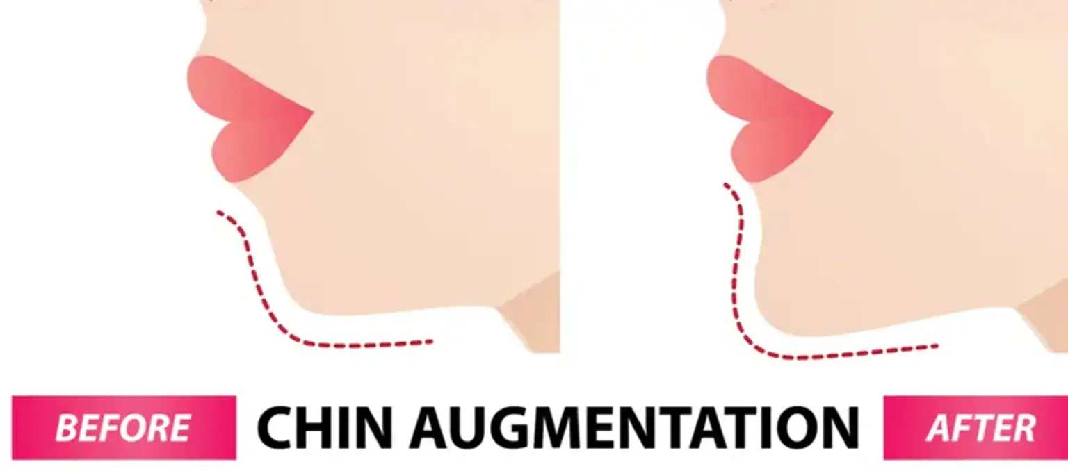
Front Chin Augmentation

Last updated date: 27-Apr-2023

Originally Written in English

Front Chin Augmentation

What is Chin Augmentation?

The chin is an unappreciated cosmetic component of the face; yet, it remains an important facial subunit that contributes significantly to overall facial beauty. A well-balanced and harmonious face requires a chin that is of proper size, shape, and contour.

The adjectives "weak" and "strong" chin are frequently used to characterize chins of a certain morphology that have psychological impacts. As a result, the art and science of surgically altering the chin, either alone or as part of an integrated change of the face osseocutaneous (bone and skin) morphology, is an essential component of orthognathic (surgical manipulation of facial skeleton parts) surgery.

Chin augmentation, also known as genioplasty, is a popular face plastic surgery technique for improving facial cosmetics. Genioplasty, or chin augmentation by osseous manipulation or implant augmentation, is an essential component of facial plastic surgery. When conducted with correct preoperative evaluation and technical execution, the outcomes can harmonize and restore equilibrium between the lower face's skeletal, soft tissue, and dental components. To properly assess the results of chin augmentation, the surgeon must undertake a complete face examination and have a solid grasp of the essential anatomy. 


Basic Anatomy

chin augmentation surgery

During chin augmentation surgery, the following structures are encountered:

  • Skin and subcutaneous tissue
  • Mentalis muscle
  • Mandibular periosteum
  • Gingivolabial mucosa (if the intraoral approach is used)
  • Mental nerve

Anatomic structures at risk:

  • The mental nerve
  • The mentalis muscle: will cause a ptotic (dropped) chin if not re-approximated properly.


Preoperative assessment

Front Chin Augmentation assessment

Gender, ethnicity, age, and medical comorbidities are all crucial aspects to consider when scheduling genioplasty procedure. Men have larger, square faces, generally with more projecting chins, whilst women have narrower faces.

In both the young and old populations, age can be an issue. Mandible surgery should be avoided in younger individuals since the lower facial skeleton will continue to expand. Because the dentition does not fully erupt until the age of 15, it is more vulnerable to harm during osteotomies. Furthermore, because of the probability of inadequate bone stock in older or edentulous individuals, alloplastic augmentation may be more appropriate.

Most significantly, genioplasty is an elective treatment that should only be performed on medically fit people. While smoking is not a contraindication, it does raise the risk of complications, such as delayed wound healing and graft failure if genioplasty needs a bone transplant. Following that, a thorough examination of the lower face and its connection to the rest of the face should be performed, taking into account the dental relationship, maxillomandibular bone morphology, and soft tissue envelope. The objective is to see if genioplasty alone or formal orthognathic surgery treating maxillary and/or mandibular dysmorphology will better meet the patient's cosmetic goals.


Dental Morphology:

Dental Morphology

The evaluation of occlusion and dental relation is critical in establishing whether or not a procedure is required, and if so, which operation will best address the patient's dysmorphology. The link between maxillary and mandibular dentition is established using angle classification.

Any chin abnormalities in individuals with normal class I occlusion can be addressed with isolated chin manipulation. Patients with class II or III occlusion, on the other hand, require further assessment to determine if they would benefit more from a combination of mandibular and maxillary osteotomies with or without genioplasty.

The presence of any dental compensation and history of past orthodontic therapy, if any, are crucial parts of the patient's teeth history since underlying skeletal dysmorphology may be identified during assessment.

Finally, poor or infected dentition should be addressed before considering genioplasty. 


Skeletal Evaluation:

The basic principle behind any alteration of the face skeleton remains cephalometric analysis (assessment of the dental and skeletal relations of a human skull). In most circumstances, however, a formal lateral cephalogram is not required. Most individuals can be assessed satisfactorily with a combined soft tissue and dental assessment. Still, cephalometric concepts remain vital in guiding treatment planning since they lay the foundation for numerous soft tissue connections. In complicated instances, formal osseous cephalometric examination is useful in understanding the connection between the skull base, maxilla, and mandible. 


Soft Tissue Analysis:

There are several methods available to assist with soft tissue analysis, and each surgeon has a favorite set of analyses that he/she utilizes to determine whether genioplasty is required and what kind of movement is required.

Every patient should be examined from both the frontal and sagittal views. A life-size image with bilateral sagittal, frontal, and oblique views might be useful.

Lip competency, as well as face height and symmetry, may be assessed from the front. Furthermore, the face should be examined with the lips in repose as well as smiling to see the dynamic soft tissue changes with motion.

  1. Lip competence To repair this deformity, patients with lip incompetence may choose the cosmetic advantages of osseous genioplasty over implant augmentation.
  2. Facial height The height of the bottom third of the face in relation to the height of the middle third of the face should be measured to determine if augmentation or reduction is needed.
  3. Facial symmetry Mandibular and chin asymmetries may need asymmetric motions and/or several osteotomies.

Following that, the profile image of the face is examined, and the following issues should be addressed:

  1. Labiomental fold the depression between the lower lip and the bottom region of the mandible is an essential feature to identify.
  2. Lip-chin relationship A simple line linking the most noticeable part of the upper and lower lip should hit the Pogonion (most protruding median point on the front side of the chin on a balanced face).
  3. Cervicomental angle The angle formed by the chin and the neck should be between 105 and 120 degrees.
  4. Nose-chin evaluation The nose and chin should complement each other.

Finally, check the skin of the lower face in both frontal and profile views, highlighting the quality, thickness, and flexibility, as well as any anomalies. Because these factors might influence final result, they should be discussed to manage a patient's expectations in the preparatory setting.


Treatment Approach

genioplasty surgery

Once the preoperative examination is complete, it is critical to arrange the information acquired in order to develop the best treatment plan.

As predicted, a portion of the decisions is influenced by each surgeon's subjective view and prior experience. Furthermore, because this is often an optional operation, each patient's preferences and goals should be taken into account.

There are two types of genioplasty surgery: osseous genioplasty and alloplastic implant augmentation. In general, most surgeons prefer to put implants rather than to conduct osteotomies.

With good patient and implant selection implant augmentation produces excellent outcomes and is simple to do.

Contrary to popular assumption, osseous genioplasty is not a difficult treatment to execute; in fact, it is a very flexible technique that allows mobility in all dimensions.

It can correct problems that an implant cannot, such as a chin that is excessively long, too short, or asymmetric. Furthermore, individuals who have had one or more alloplastic chin implant failures may benefit from an osseous genioplasty.

Because osseous genioplasty and alloplastic augmentation require different types of anesthesia, each patient's overall health, anesthesia tolerance, and anesthesia preference should be considered.

The vast majority of implant augmentation procedures may be carried out under local anesthetic, with or without sedation.

For best pain and airway control, osseous genioplasty should be done under at least intravenous sedation in a controlled setting by a qualified registered nurse anesthetist or anesthesiologist.


How Chin Augmentation is done?

alloplastic augmentation

There are several surgical procedures for osseous genioplasty and alloplastic augmentation. The surgical concepts and main processes are, in general, very similar. Minor subtleties and discrepancies are related to surgeon choice based on personal experience.


Osseous Genioplasty:

Osseous Genioplasty

Incision:

  • For osseous genioplasty, an intraoral incision is performed and preferable.
  • Following suitable sedation or anesthesia, the chin is injected with lidocaine (local anesthetic) with 1:100,000 epinephrine in and around the intended incision, as well as dissection and osteotomy sites.
  • The lower lip is then pulled outward to reveal the mental nerve through the mucosa. The incision should be kept between the visible nerve and the skin.
  • A watertight closure is achieved by making an incision and leaving a substantial cuff of mucosa and muscle. Electrocautery is used to make an incision in the mucosa and muscle.


Dissection:

  • A periosteal elevator is then used to expose the anterior surface of the chin while viewing and preserving the mental nerve passing through the foramen.
  • Extensive dissection should be avoided because soft tissue attachments aid to maintain the skeletal modification, decrease unanticipated soft tissue alterations, and reduce postoperative osseous resorption.
  • There is also no need to dissect above the mental nerve on either side, as this increases the risk of the nerve being too stretched or avulsed.


Osteotomy:

  • Following adequate retraction, a sterile pencil is used to outline the site of the osteotomy, which should be at least 5 mm below the apices of the canine teeth and 6 mm below the mental foramen to avoid damaging either the tooth root or the nerve.
  • The precise site and angle of the osteotomy will be determined by the intended movement.
  • First, a vertical groove in midline perpendicular to the proposed osteotomy is made using an oscillating saw to serve as a midline reference point.
  • A drill hole can be created in the midline of the distal chin at this point, and a screw can be partly inserted later to serve as a convenient retractor for the osteotomized chin piece.
  • Enough contact irrigation is advised throughout the osteotomy to avoid heating the bone and causing localized osteomyelitis.


Planned movement and fixation:

  • The distal chin is then moved into the appropriate position, with the amount and direction of movement determined by preoperative planning.
  • To position the distal segment, a screw can be partly put into a predrilled hole (as described above), and a wire twister can be used to keep the screw in place to serve as a retractor.
  • A straight titanium plate with three or four holes can be shaped and connected to both proximal and distal portions on either side of the osteotomy.
  • Prebent genioplasty plates, the size of which is determined by the degree of advancement, can also be employed. The positioning screw can be removed at this stage.


Refinements:

  • If extensive vertical or horizontal lengthening (> 5 mm) is desired, an interpositional graft (autograft, allograft, or off-the-shelf material such as hydroxyapatite) is molded and inserted into the defect.


Closure:

  • After thoroughly irrigating the wound to remove any debris, the muscle is reapproximated with a resorbable suture and the incision is closed with 4-0 chromic mattress sutures. 


Alloplastic Chin Augmentation:

Alloplastic Chin Augmentation

Implant choice:

  • The most widely utilized genioplasty implants nowadays are silastic and porous polyethylene.
  • The type of implant is mostly determined by the surgeon's preference and experience.
  • Some surgeons choose a two-piece porous polyethylene implant over a silastic implant because it promotes soft tissue ingrowth, reduces fibrous encapsulation, and minimizes shifting.
  • Porous polyethylene implants, on the other hand, maybe more difficult to put and remove due to soft tissue adhesion and ingrowth.
  • Depending on the severity of the insufficiency, a variety of implant forms and sizes are available.
  • Some chin implants go beyond the chin to modify the body of the mandible.


Incision:

  • Although both intraoral and submental incisions can be used, the submental incision is favored because it provides greater visibility as well as more precise shaping and implant positioning.


Dissection:

  • Wide dissection is conducted to increase vision and allow for more precise implant insertion in the subperiosteal plane.
  • Because silastic implants have a higher tendency to erode the underlying bone, they should be placed on the supraperiosteal plane.
  • However, because the risk of soft tissue damage and skin abnormalities is higher in the supraperiosteal plane, this method should be used with caution.


Implant placement and fixation/closure:

  • A two-piece porous polyethylene implant is shaped and molded to conform as closely as possible to the symphysis and, if desired, the mandible body.
  • It is critical to reduce the dead space between the implant and the underlying bone in order to avoid complications.
  • Titanium screws secure the implant to the mandible. The wound is irrigated and layered closed. 


What are the Complications of Chin Augmentation Surgery?

complications of chin augmentation surgery

Poor cosmetic outcomes, hematoma, infection, malposition, and nerve damage are all possible consequences during genioplasty.

Reoperation following genioplasty is very rare and generally includes implant replacement or removal and conversion of alloplastic genioplasty to an osteotomy.

According to recent retrospective research, patients who underwent osseous genioplasty had a somewhat greater satisfaction rate (90-95 percent) than those who underwent alloplastic augmentation (85-90 percent). Morbidity was the same for both procedures, and the complication profile was identical.

All procedures, regardless of technique, entail a risk of complications, and physicians doing cosmetic chin augmentation should get knowledgeable about the possibilities to determine what will work best for each patient.

The first step in minimizing complications and patient dissatisfaction is proper patient selection.

Once the patient has been determined to be a qualified medical candidate, it is critical to examine his or her objectives and expectations for the procedure.

Obviously, if the patient needs surgery to acquire a new job or a new boyfriend/girlfriend, the surgeon should reconsider conducting this treatment. Even if the cosmetic outcome is better than planned, the patient may regard the surgery as a failure if his or her ultimate aim is not met.

If the choice to proceed with chin augmentation is taken after medical and motivational aspects have been addressed, the patient should be properly informed about the surgical risks.

Complications from genioplasty can be broadly classified as follows:

  • Soft tissue complications.
  • Neurological complications.
  • Muscular complications.
  • Bone or tooth complications.
  • Technical errors.

Smoking, like with any cosmetic surgery, increases the chance of complications, thus patients should stop using nicotine products at least three weeks before surgery to prevent any further impact on recovery.

To reduce the risk of hematoma, anticoagulant drugs such as aspirin, warfarin, and vitamin E should be stopped at least 10 days before surgery (with approval from the patient's primary care physician). 


Soft tissue complications:

  • Hematomas are uncommon and typically treatable with needle aspiration.
  • Scar development is possible with an external technique; however, it is generally effectively masked if the incisions are positioned suitably in a submental crease.
  • Overgranulation of the buccal wound may occur when an intraoral technique is used; this can be addressed with local cautery.
  • Because wound dehiscence can occur with any method, the wound should be constantly watched for signs of infection. when the dehiscence is modest and in the absence of infection, both outer and intraoral wounds heal effectively.
  • Infection is another issue to consider. It has been observed that it occurs in around 5% to 7% of chin implant surgeries, although it can also occur after osseous genioplasty. When infections occur without a fluid collection or abscess, high-dose antibiotics administered early may save the implant. Many implants, however, must be removed with pocket irrigation and a loose re-approximation of the incision.
  • Capsular contracture (CC) surrounding an implant can give the chin an awkward, poorly shaped look. This deformity is highly difficult to treat and frequently necessitates a capsulectomy followed by the implantation of a bigger implant. 


Neurological complications:

  • The inferior alveolar nerve and the mental nerve, which normally exits below the bicuspid tooth but can alternatively be located below the cuspid or between the two premolars, are anatomic risks in this location.
  • Unfortunately, some sensory innervation to the incisor teeth, lower chin, or inferior border of the jaw will be lost as a result of an osteotomy. This happens when extra nerve fibers from the lingual and mylohyoid nerves enter the jaw to form a plexus.
  • The patient may experience transient postoperative hypoesthesia or dysesthesia of the chin, which happens in 3.4 percent to 12 percent of instances. These hazards should be discussed with the patient prior to surgery.
  • Lower lip numbness can also develop following implant insertion due to mental nerve strain, compression, or severing. This condition normally resolves on its own, but if no improvement is shown after two or three weeks, the implant should be withdrawn and the lower flange relocated inferiorly or clipped at its superior border to enable greater space for the nerve. If not handled within a two-month period, this issue may become permanent. 


Muscular complications:

  • When sealing the incision, great care should be taken to reattach the mentalis muscle, which lifts and compresses the chin against the anterior mandible and indirectly increases the lower lip.
  • Failure to do so might result in chin ptosis, lip ptosis, drooling, and increased lower teeth show. 


Bone complications:

  • Potential mandibular bone resorption is one of the most often reported bone-related complications following chin augmentation.
  • Although it is rarely noticeable cosmetically, one research found that resorption with a chin implant can occur at a rate as rapid as 0.1 mm each month.
  • This would be especially dangerous if the implant was placed high on the mandibular body, predisposing the patient to erosion into a tooth root, which can cause pain and other dental issues. 


Implant Malposition:

  • Implant malposition occurs when the implant is placed too low on the chin or migrates superior to the pogonion.
  • This condition may arise more frequently following an intraoral operation, and it can be corrected by replacing the implant via a submental route and anchoring it in place with sutures or screws.
  • The mentalis muscle must be reattached carefully to avoid a drooping "witch's chin" malformation. 


Under-augmentation Or Over-augmentation:

  • Both under- and over-augmentation are possible risks of cosmetic chin surgery, although over-augmentation is more unpleasant to patients.
  • Unintentional over-augmentation with an implant may occur if there are gaps between the front surface of the mandible and the overlying implant due to mandibular shape irregularities. 


Conclusion 

Chin augmentation

The chin is an overlooked aesthetic feature of the face; yet, it is a vital facial subunit that contributes greatly to overall facial attractiveness.

Chin augmentation, commonly known as genioplasty, is a common facial plastic surgery procedure used to improve facial aesthetics. Genioplasty, whether by implant augmentation or osteotomy, is a key component of face aesthetic transformation, whether performed alone or as part of formal orthognathic surgery. When performed with sufficient preoperative assessment and expertise, the results can harmonize and restore the balance between the bone, soft tissue, and dental components of the lower face.

Both implant and osseous genioplasty may be quite simple to do with proper recognition of underlying dysmorphology, preoperative examination, and surgical technique.