Gallbladder Cancer
Overview
Gallbladder cancer is the sixth most frequent gastrointestinal cancer, but it is the most prevalent biliary system cancer, accounting for 80-95 percent of biliary tract malignancies. This tumor is a severely deadly illness, with a 5-year overall survival rate of fewer than 5% and a mean survival time of fewer than 6 months.
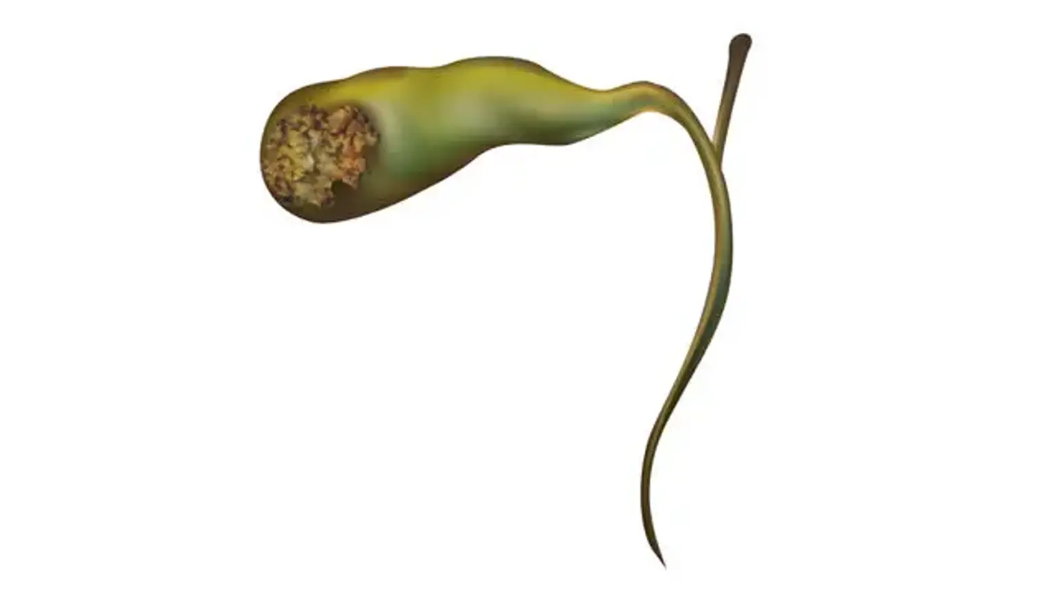
The gallbladder is a tiny hollow organ that stores and concentrates bile before releasing it into the small intestine. The pear-shaped gallbladder in humans is located underneath the liver, but the anatomy and position of the gallbladder vary greatly between animal species. It absorbs and stores bile generated by the liver via the common hepatic duct before releasing it into the duodenum via the common bile duct, where it aids in fat digestion.
An early diagnosis is critical since this cancer grows quietly if detected late. After a simple cholecystectomy for suspected gallbladder stone illness, 0.5-1.5 % of individuals are identified with gallbladder cancer. Laparoscopy should not be performed on patients who have a preoperative suspicion of gallbladder cancer.
Structure
The gallbladder is a hollow organ located in a shallow dip under the right lobe of the liver, which is grey-blue in color in life. When completely distended, the gallbladder in adults measures 7 to 10 centimeters (2.8 to 3.9 inches) in length and 4 cm in diameter. The gallbladder has a volume of around 50 milliliters.
The gallbladder has a pear shape, with the tip opening into the cystic duct. The gallbladder is made up of three sections: the fundus, the body, and the neck. The fundus is the spherical base that is tilted to face the abdominal wall. The body is in a depression on the lower liver's surface. The neck tapers and connects to the cystic duct, which is part of the biliary tree.
The gallbladder fossa is located under the intersection of hepatic segments IVB and V, against which the fundus and body of the gallbladder are located. The common bile duct is formed when the cystic duct joins the common hepatic duct.
Gallbladder lymphatic drainage follows the cystic node, which is placed between the cystic duct and the common hepatic duct. Lower hepatic lymph nodes receive lymphatics from the lower section of the organ. Finally, all of the lymph drains into celiac lymph nodes.
Gallbladder cancer definition
Gallbladder cancer (GC) is uncommon cancer that accounts for about half of all biliary tract cancers. Biliary cancers are extremely lethal, with a 5-year survival rate of 17.6 percent. Gallbladder cancer has a poor prognosis due to aggressive tumor biology, late presentation, complex anatomic location, and an advanced stage upon diagnosis. Palliative chemotherapy is used to treat locally advanced and metastatic illnesses. Early-stage treatment, on the other hand, has the potential to be curative, with surgical resection followed by adjuvant therapy.
Epidemiology
Because of the high frequency of gallstones and chronic gallbladder infections in such places, the incidence of gallbladder cancer is high among particular populations outside of the United States (e.g., South America, India, Pakistan, Japan, and Korea). In 2017, the American Cancer Society forecasts 11,740 new cases of gallbladder cancer and 3,830 fatalities in the United States, with a female predominance.
Gallbladder cancer incidence reduced in persons over the age of 50 but rose in the younger group. Gallbladder cancer is more prevalent among Whites, Southwestern Native Americans, and Mexican Americans than in African Americans.
Gallbladder cancer causes
The most serious risk of gallbladder cancer is chronic inflammation. A history of gallstones (cholelithiasis) is the biggest predictor of gallbladder cancer, and the risk increases with gallstone size, chronicity, and severity of symptoms. Porcelain gallbladder, or gallbladder calcification, is frequently associated with persistent cholelithiasis. This disease is commonly discovered by chance on imaging and frequently leads to cholecystectomy.
Gallbladder polyps, congenital biliary cysts, and aberrant pancreaticobiliary architecture are further risk factors that can contribute to chronic inflammation and gallbladder cancer. Salmonella typhi and helicobacter endemic areas reveal a relationship between chronic asymptomatic carriers and an increased risk of gallbladder cancer.
Furthermore, carcinogens that cause gallbladder cancer include (e.g., methyldopa, isoniazid), occupational exposure (e.g., methylcellulose, radon), and lifestyle factors (e.g., cigarette smoking, obesity, high carbohydrate intake). Gallbladder cancer can be caused by chronic primary sclerosing cholangitis and inflammatory bowel disease.
Pathophysiology
The current theory is that prolonged inflammation of the bile duct tissue causes a cascade of genetic alterations that leads to malignant transformation. The majority of gallbladder cancer histopathological alterations manifest as adenocarcinomas (90 % ). Following about 15 years of inflammation, this syndrome evolves from pre-neoplastic dysplasia to carcinoma in situ and, eventually, to invasive malignancy. Squamous cell carcinoma of the gallbladder is uncommon.
Gallbladder cancer symptoms
Gallbladder cancer is frequently discovered as an unintentional discovery on imaging or after a surgical operation. Early-stage gallbladder cancer is frequently detected following cholecystectomy and examination of the surgical pathology material.
Patients with gallbladder cancer are frequently asymptomatic or express nonspecific symptoms such as abdominal discomfort, nausea or vomiting, indigestion, weakness, anorexia, loss of appetite, weight loss, and jaundice, which can easily be misdiagnosed as cholecystitis. Cancer-related biliary obstruction causes jaundice, clay-colored feces, cola-colored urine, and cutaneous itch.
In addition, Mirizzi syndrome (common hepatic duct blockage caused by an impacted stone in the gallbladder neck owing to extrinsic compression) has been linked to gallbladder cancer. Because of its unresectability at presentation, it has a dismal prognosis. Other signs of advanced cancer, such as weight loss and general malaise, may also be present.
Physical examination may reveal jaundice, right upper quadrant discomfort, or the Courvoisier sign (a non-tender palpable gallbladder with jaundice), which is more likely to occur as a result of persistent progressive malignant blockage rather than intermittent gallstone obstruction. On physical examination, hepatomegaly, abdominal palpable mass, ascites, and intestinal obstruction suggest an advanced metastatic stage.
Gallbladder cancer diagnosis
Patients with obstructive jaundice, in particular, will require a full blood count, a basic chemical panel, and a liver function test. The results might suggest an unspecific cholestatic pattern produced by bile blockage that necessitates decompression. The earliest imaging investigations are typically ultrasonography (US) and computed tomography (CT).
It is necessary to determine if gallbladder cancer can be removed surgically in order to plan treatment. The tests and procedures used to detect, diagnose, and stage gallbladder cancer are frequently performed concurrently. The following tests and procedures are possible:
- Physical exam and health history: An examination of the body to look for general indicators of health, including the detection of disease-related symptoms such as tumors or anything else that appears strange. A history of the patient's health habits, as well as previous diseases and treatments, will be collected.
- Liver function tests: A test that examines a blood sample to determine the number of specific chemicals produced into the blood by the liver. A higher-than-normal level of a chemical can indicate liver illness caused by gallbladder cancer.
- Blood chemistry studies: A method in which a blood sample is examined to determine the levels of specific compounds produced into the blood by the body's organs and tissues. A chemical in an unusual (higher or lower than normal) concentration might be a symptom of sickness.
- CT scan (CAT scan): A method that takes a sequence of detailed photographs from various perspectives of locations within the body, such as the chest, abdomen, and pelvis. A computer coupled with an x-ray machine creates the images. To make the organs or tissues show up more clearly, a dye may be injected into a vein or ingested. This is also known as computed tomography, computerized tomography, or computerized axial tomography.
- Ultrasound exam: A technique in which high-energy sound waves (ultrasound) bounce off inside tissues or organs, creating echoes. The echoes combine to generate an image of bodily tissues known as a sonogram. An abdominal ultrasound is used to diagnose gallbladder cancer.
- PTC (percutaneous transhepatic cholangiography): A technique for x-raying the liver and bile ducts. A tiny needle is introduced into the liver via the skin behind the ribs. An x-ray is obtained after dye is injected into the liver or bile ducts. If a blockage is discovered, a thin, flexible tube known as a stent may be placed in the liver to drain bile into the small intestine or a collecting bag outside the body.
- ERCP (endoscopic retrograde cholangiopancreatography): A technique for x-raying the ducts (tubes) that transport bile from the liver to the gallbladder and from the gallbladder to the small intestine. Gallbladder cancer can cause these ducts to shrink and obstruct or delay the flow of bile, resulting in jaundice. An endoscope (a thin, illuminated tube) is inserted into the first section of the small intestine through the mouth, esophagus, and stomach.
The endoscope is then used to implant a catheter (a smaller tube) into the bile ducts. An x-ray is obtained after a dye is introduced into the ducts through the catheter. If a tumor has clogged the ducts, a tiny tube may be placed into the duct to unclog it. To maintain the duct open, this tube (or stent) may be kept in situ. Tissue samples may also be collected.
- MRI (magnetic resonance imaging) with gadolinium: A technique that uses a magnet, radio waves, and a computer to create a sequence of detailed images of locations within the body. A gadolinium-containing material is injected into a vein. Gadolinium accumulates around cancer cells, making them seem brighter in the image. This method is also known as nuclear magnetic resonance imaging.
- Endoscopic ultrasound (EUS): An endoscope is put into the body, commonly through the mouth or the rectum. An endoscope is a narrow, tube-like tool with a light and a viewing lens. A probe at the end of the endoscope is used to create echoes by bouncing high-energy sound waves (ultrasound) off inside tissues or organs. The echoes combine to generate an image of bodily tissues known as a sonogram. Endosonography is another name for this surgery.
- Laparoscopy: A surgical technique that examines the organs inside the abdomen for symptoms of illness. Small incisions (cuts) are made in the abdominal wall, and a laparoscope (a thin, illuminated tube) is introduced through one of them. Other instruments may be put into the same or different incisions to undertake procedures such as organ removal or tissue sample collection for biopsy. The laparoscopy determines if the cancer is limited to the gallbladder or has spread to adjacent tissues, and whether it can be removed surgically.
- Biopsy: The excision of cells or tissues so that a pathologist may examine them under a microscope for symptoms of malignancy. Following surgery to remove the tumor, a biopsy may be performed. If the tumor cannot be removed surgically, a biopsy with a thin needle may be performed to extract cells from the tumor.
Treatment for gallbladder cancer
Because of the advanced illness at the time of diagnosis, neoadjuvant treatment is not always a possibility, and it is not considered standard of care in resectable patients. Early clinical trial referral should be considered.
For individuals with Stage II or less and no contraindications, surgery is the sole curative option (see evaluation). Gallbladder cancer surgical resection will comprise cholecystectomy with a marginal hepatectomy and regional lymphadenectomy or common bile duct resection (extended organs may require removal). The advice is to return for additional examination and re-resection of gallbladder cancer identified incidentally on cholecystectomy pathological specimens with stage T2 or above.
Postoperative chemotherapy should be administered within 8 to 12 weeks of surgery, and baseline laboratory and imaging tests should be performed to re-stage the illness prior to starting medication. Patients having a resected pathology specimen report of T2 or above, node-positive, and margin positive should be administered adjuvant therapy, ideally for six months with concurrent adjuvant chemoradiation or four months with concomitant adjuvant chemoradiation.
Lymph node assessment is a vital component of radical resections for gallbladder cancer and has been proven in a retrospective trial to increase survival. Although there is no agreement on the minimum number of lymph nodes necessary for appropriate staging, one research found that excision and histologic assessment of at least 6 lymph nodes enhances risk categorization.
This emphasizes the significance of a comprehensive lymphadenectomy of the porta hepatis and a careful assessment of pathology materials in patients with gallbladder cancer.
In the treatment of unresectable illness, the surgical involvement is mainly restricted to tumor biopsy for diagnosis and possibly biliary decompression treatments.
Differential Diagnosis
- Acalculous cholecystitis
- Acalculous cholecystopathy
- Ampullary carcinoma
- Bile duct strictures
- Bile duct tumors
- Biliary colic
- Biliary disease
- Biliary obstruction
- Cholangitis
- Cholecystitis
Gallbladder cancer staging
Gallbladder cancer staging tests and treatments are frequently performed at the same time as the diagnosis.
- There are three ways that cancer spreads in the body.
- Cancer may spread from where it began to other parts of the body.
- The following stages are used for gallbladder cancer:
- Stage 0 (Carcinoma in Situ)
- Stage I
- Stage II
- Stage III
- Stage IV
- For gallbladder cancer, stages are also grouped according to how cancer may be treated. There are two treatment groups:
-
- Localized (Stage I)
- Unresectable, recurrent, or metastatic (Stage II, Stage III, and Stage IV)
There are three ways that cancer spreads in the body.
Cancer can spread through tissue, the lymph system, and the blood:
- Tissue. Cancer spreads from where it started into neighboring places.
- The lymphatic system: The malignancy spreads from the site of origin by infiltrating the lymphatic system. Cancer spreads to other regions of the body via the lymph vessels.
- Blood. Cancer spreads from the site of origin by entering the bloodstream. Cancer spreads to other regions of the body via the blood arteries.
Cancer may spread from where it began to other parts of the body.
Metastasis occurs when cancer spreads to another section of the body. Cancer cells break out from the original tumor and migrate via the lymph system or bloodstream.
- The lymphatic system cancer enters the lymphatic system, moves via the lymph vessels, and eventually develops a tumor (metastatic tumor) in another portion of the body.
- Blood. Cancer enters the bloodstream, travels through the blood vessels, and eventually develops a tumor (metastatic tumor) in another portion of the body.
The initial tumor and the metastatic tumor are both cancers. If gallbladder cancer spreads to the liver, for example, the cancer cells in the liver are actually gallbladder cancer cells. It is gallbladder cancer that has spread to the liver, not liver cancer.
The following stages are used for gallbladder cancer:
- Stage 0 (Carcinoma in Situ)
The mucosa (innermost layer) of the gallbladder wall is observed to be abnormal in stage 0. These aberrant cells have the potential to develop into cancer and spread to surrounding normal tissue. Cancer in situ is another term for stage 0 cancer.
Stage I
Cancer has grown in the mucosa (innermost layer) of the gallbladder wall at stage I and may have progressed to the muscle layer.
Stage II
Stage II is divided into stages IIA and IIB, depending on where cancer has spread in the gallbladder.
- Cancer has progressed through the muscle layer to the connective tissue layer of the gallbladder wall on the side of the gallbladder, not near the liver in stage IIA.
- Cancer has progressed from the muscle layer to the connective tissue layer of the gallbladder wall on the same side as the liver in stage IIB. Cancer hasn't gone to the liver yet.
Stage III
Stage III is divided into stages IIIA and IIIB, depending on where the cancer has spread.
- In stage IIIA, cancer has spread through the connective tissue layer of the gallbladder wall and one or more of the following is true:
-
- The cancer has progressed to the serosa (layer of tissue that covers the gallbladder).
- The cancer has progressed to the liver.
- Cancer has progressed to a neighboring organ or tissue (for example, the stomach, small intestine, colon, pancreas, or bile ducts outside the liver).
- Cancer has grown in the mucosa (innermost layer) of the gallbladder wall and may have spread to the muscle, connective tissue, or serosa (layer of tissue that covers the gallbladder), as well as to the liver or one neighboring organ or structure in stage IIIB (such as the stomach, small intestine, colon, pancreas, or the bile ducts outside the liver). One to three neighboring lymph nodes have been infected with cancer.
Stage IV
Stage IV is divided into stages IVA and IVB.
- Cancer has progressed to the portal vein or hepatic artery, or to two or more organs or tissues other than the liver, at stage IVA. One to three neighboring lymph nodes may have been infected with cancer.
- Cancer at stage IVB may have spread to neighboring organs or tissues. The disease has spread:
-
- to four or more nearby lymph nodes; or
- to other parts of the body, such as the peritoneum and liver.
Gallbladder cancer prognosis
As previously noted, the prognosis for gallbladder cancer is often bleak. The stage at when the tumor was discovered, the exact location of the tumor, operability/response to treatment, and if and where it had spread are all factors that influence the prognosis. The recurrence rate of gallbladder cancer is significant, and the 5-year survival rate is low.
Certain factors affect the prognosis (chance of recovery) and treatment options.
The prognosis and treatment options depend on the following:
- The stage of cancer (whether cancer has spread from the gallbladder to other places in the body).
- Whether or whether cancer can be totally eliminated by surgery.
- The classification of gallbladder cancer (how the cancer cell looks under a microscope).
- Whether the cancer was just discovered or has recurred (come back).
Complications
Tumor recurrence, which may cause visceral discomfort due to intra-abdominal involvement, is one of the disease's complications. Obstructive jaundice might result from a regional recurrence. Clinicians should consider recurring illnesses in patients who have unexplained weight loss, obstructive jaundice, or increased intra-abdominal discomfort.
Conclusion
Gallbladder cancer is infrequent and usually develops later in life. When cancer arises, it usually affects the glands that line the surface of the gallbladder (adenocarcinoma). Gallstones are suspected to play a role in the development of cancer. Large gallbladder polyps and a heavily calcified "porcelain" gallbladder are other risk factors.
Gallbladder cancer can result in biliary discomfort, skin discoloration (jaundice), and weight loss. A big gallbladder can be felt in the belly. Liver function tests, notably GGT and ALP, may be high, with ultrasonography and CT scans being the preferred medical imaging studies. The gallbladder is removed to treat gallbladder cancer.
Cancer of the gallbladder can also be discovered incidentally following surgical removal of the gallbladder, accounting for 1–3 percent of all malignancies discovered in this manner. Gallbladder polyps are typically benign growths or lesions that resemble growths that occur in the gallbladder wall, and they are only related with cancer when they get big. Cholesterol polyps, which are frequently linked with cholesterolosis ("strawberry gallbladder," a change in the gallbladder wall caused by high cholesterol), frequently cause no symptoms and are thus frequently identified in this manner.
While most gallbladder disorders are minor, gallbladder cancer (GC) is an uncommon malignancy that accounts for about half of all biliary tract cancers. Biliary cancers are extremely lethal. The healthcare team, including all doctors and mid-level practitioners, must be informed of the probability of this unfavorable occurrence occurring over the course of these patients' care.