Gastric cancer

Last updated date: 14-Apr-2023

Originally Written in English

Gastric cancer

Overview

Gastric cancer is the fifth most common cancer and the third main cause of cancer deaths globally, despite a global drop since the mid-century. In the United States, the incidence of stomach cancer has dropped during the last several decades, whereas the incidence of gastroesophageal cancer has climbed.

Gastric adenocarcinoma is classified into two types: intestinal (well-differentiated) and diffuse (undifferentiated), each with its own morphologic appearance, etiology, and genetic profile. Surgical resection with appropriate lymphadenectomy is the only possibly curative therapy option for people with stomach cancer.

Perioperative therapy to enhance a patient's survival are supported by current research. Regrettably, patients with incurable, locally advanced, or metastatic illness may only be provided life-prolonging palliative care.
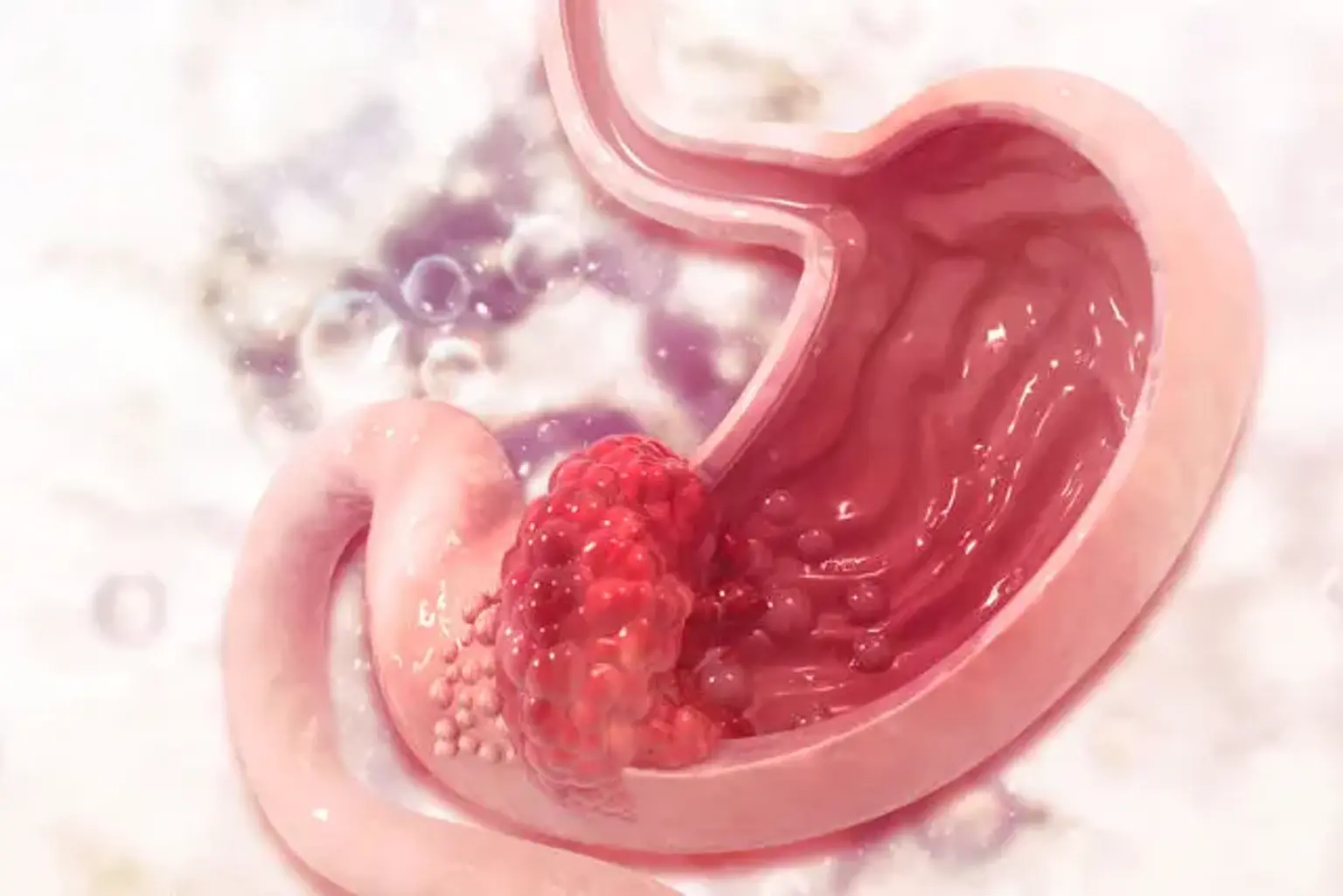
 

Gastric cancer definition

Gastric cancer is a kind of cancer that arises from the stomach lining. The majority of stomach malignancies are gastric carcinomas, which can be further subdivided into several subtypes, including gastric adenocarcinomas. In addition, lymphomas and mesenchymal tumors can form in the stomach.


Anatomy

Anatomy of Gastric cancer

Management of stomach cancer requires a thorough understanding of gastric anatomy: 

In the upper abdomen, the stomach is a J-shaped organ. It is a component of the digestive system that processes nutrients (vitamins, minerals, carbohydrates, fats, proteins, and water) in food and aids in the elimination of waste. Food travels from the neck to the stomach via the esophagus, a hollow, muscular tube. Partially digested food enters the small intestine and subsequently the large intestine after exiting the stomach.

The stomach wall is composed of five layers of tissue. The layers of the stomach wall are, from innermost to outermost, mucosa, submucosa, muscle, subserosa (connective tissue), and serosa. As it progresses, gastric cancer originates in the mucosa and travels to the outer layers.

The larger sac's peritoneum covers the front side of the stomach. A piece of the smaller sac hangs behind the stomach. There is little or no serosal coating at the gastroesophageal junction.

The anterior gastric surface on the right is next to the left lobe of the liver and the anterior abdominal wall. The spleen, the left adrenal gland, the superior region of the left kidney, the ventral component of the pancreas, and the transverse colon are all located on the left side of the stomach.

The location of stomach cancer is characterized based on its connection to the stomach's long axis. Approximately 40% of malignancies grow in the lower portion of the organ, 40% in the middle area, and 15% in the upper part; 10% involve more than one part of the organ. The majority of the decline in gastric cancer incidence and death in the United States has been due to cancer in the lower portion of the stomach; nevertheless, the incidence of adenocarcinoma in the cardia has gradually increased.


Epidemiology

Globally, the incidence of gastric cancer is steadily decreasing. However, the speed has varied among areas such as China and Japan. The drop in gastric cancer may be related to the detection and treatment of viral causes, as well as lifestyle changes of dietary and environmental risk factors, although it remains widespread in places of the world where fresh food storage and water quality are poor.

The great majority of stomach cancers occur in underdeveloped nations, with males being twice as likely as women and black men being more likely than white men. White western civilization with a better socioeconomic standing has the lowest occurrence.

Migration studies have supported evidence for the influence of lifestyle changes on the development of stomach cancer, since the second and third generations born in the United States have lower rates. Prior understandings of stomach cancer substantially support the idea that dietary, social, and medical variables, rather than hereditary susceptibility, have been established in Japanese migrants.

The histological patterns of stomach cancer have also changed epidemiologically; conversely, the intestinal gastric type is gradually declining but remains more prevalent (70 % ). It is more common in men over the age of 50, and it is connected to environmental variables. The diffuse or infiltrative kind, on the other hand, is less common (30%) but diagnosed at a younger age in both sexes and has a poorer prognosis.

The growing frequency of distal esophageal carcinoma in the United States coincides with a significant anatomic shift from distal to proximal stomach cancer. In Western nations, the most prevalent locations are the proximal lesser curvature, heart, and esophagogastric junction (EGJ), although non-proximal continues to prevail in Japan. Gastric cancer has a far better prognosis in Japan than in the United States, owing partly to endoscopic screening programs that aid in the detection of early lesions and possibly curable stages.


Etiology 

Nutritional factors such as high salt (salt-preserved food), N-nitroso compounds consumption (dietary source), smoking, a low vitamin A and C diet, consuming large amounts of smoked or cured foods, a lack of refrigerated foods, and contaminated drinking water have all been linked to an increased risk of gastric cancer. Adenocarcinomas of the distal esophagus, proximal stomach, and junction are related with a higher risk of BMI, increased calorie consumption, gastroesophageal reflux, and smoking.

Rubber production, tin mining, metal processing, and coal mining all enhance the danger. Helicobacter pylori infection has an associated risk of 46% to 63%, whereas Epstein-Barr virus infection is estimated to be 5% to 10% globally. Radiation exposure and previous gastric surgery have also been identified as risk factors.

Aspirin and other nonsteroidal anti-inflammatory drugs have been linked to a decreased incidence of gastroesophageal junction cancer and other gastrointestinal malignancies (HR 0.79 for each year of NSAID use). Alcohol use has not been shown to be a risk factor, and some data suggest that, despite scant evidence, daily wine drinking may be beneficial. Gastric cancer has not been linked to chronic "iatrogenic" histamine-2-receptor antagonist or proton pump inhibitor use.

Kind A blood has around 20% higher stomach cancer cases than blood types O, B, or AB and is particularly related with the diffuse type. Pernicious anemia, an autoimmune chronic atrophic gastritis, increases the incidence of intestinal-type stomach cancer by up to sixfold. Gastric polyps, benign gastric ulcers, and hypertrophic gastropathy are all risk factors for stomach cancer.

The majority of stomach cancers are random, however 5% to 10% of patients have a family history of gastric cancer. Hereditary diffuse gastric cancer , gastric adenocarcinoma and proximal polyposis of the stomach , and familial intestine gastric cancer (FIGC) are the three principal syndromes responsible for up to 3% to 5% of hereditary familial gastric cancer. Other hereditary cancer syndromes include as follows:

  • Hereditary non-polyposis colon cancer (HNPCC 13% lifetime risk, predominantly intestinal type)
  • Familial adenomatous syndrome (FAP, 10% risk)
  • Peutz Jeghers syndrome (PJS, 29% risk)
  • Juvenile polyposis syndrome (JPS, 21%)
  • Li-Fraumeni syndrome
  • Hereditary breast and ovarian cancer syndrome
  • Phosphatase and tensin homolog (PTEN) or hamartoma tumor (Cowden's) syndrome.

All of them, however, are uncommon causes of stomach cancer. Screening recommendations for genetic disorders linked with stomach cancer are proposed based on their risk. In terms of cancer research, the World Health Organization has designated H. pylori as a definite gastric carcinogen and determined a link between processed meat intake and stomach cancer.


Pathophysiology

Pathophysiology of Gastric cancer

According to Lauren's histopathologic categorization, there are two primary histologic types of gastric adenocarcinoma. The most common is the "intestinal type," so named because of its morphologic resemblance to adenocarcinomas of the gastrointestinal system. The less frequent diffuse-type gastric cancer is distinguished by a lack of intercellular adhesions, which disrupts glandular structure creation.

The lack of intercellular adhesions in individuals with a hereditary form of diffuse-type gastric cancer is caused by a germline mutation (HDGC) in the cell adhesion protein E-cadherin (CDH1). Asymptomatic CDH1 carriers may require preventive gastrectomy before the age of 30, and women are also at risk of developing early breast cancer. There are no obvious precancerous lesions in the diffuse form.

One concept for the "intestinal-type" of gastric cancer involves a transition from chronic gastritis caused by H. pylori, pernicious anemia, or high-salt diets to a loss of parietal cells, resulting in chronic atrophic gastritis. Compensatory hypergastrinemia in atrophic gastritis causes persistent inflammation, which leads to intestinal metaplasia, dysplasia, and, finally, cancer.

Several studies have found a sixfold rise in H. pylori infection in individuals with gastric cancer, particularly adenocarcinoma of the distal stomach, which includes both intestinal and diffuse kinds. As previously stated, H. pylori causes inflammation, which leads to stomach atrophy and subsequent metaplasia, ending in cancer. Furthermore, the majority of people with H. pylori infection develop ulcers rather than cancer. 


Gastric cancer symptoms

Gastric cancer symptoms

In the United States, the majority of individuals come with advanced-stage symptoms. Non-specific weight loss, persistent stomach discomfort, dysphagia, hematemesis, anorexia, nausea, early satiety, and dyspepsia are the most typical presenting signs of gastric cancer. Patients with locally advanced or metastatic illness typically appear with considerable abdominal discomfort, possible ascites, weight loss, exhaustion, visceral metastases on imaging, and a gastric-outlet blockage.

A palpable abdominal mass suggesting advanced illness is the most prevalent physical examination result. The patient may also show symptoms of metastatic lymphatic dissemination, such as Virchow's node (left supraclavicular adenopathy), Sister Mary Joseph node (peri-umbilical nodule), and Irish node (left axillary node). Krukenberg's tumor (ovary mass), Blumer's shelf (cul-de-sac mass), ascites (peritoneal carcinomatosis), and hepatomegaly are all symptoms of direct peritoneal metastasis (often diffuse disease burden).

Dermatological (diffuse seborrheic keratosis or acanthosis nigricans), hematological (microangiopathic hemolytic anemia and hypercoagulable state), renal (membranous nephropathy), and autoimmune (polyarteritis nodosa) manifestations are uncommon clinical findings, and none are specific to gastric cancer.


Diagnosis

Diagnosis of Gastric cancer

Patients who come with any symptoms suggestive of stomach cancer should get an upper endoscopy instead of a barium examination (except for limited plastic presenting as leather-flask appearance). Although upper endoscopy is more intrusive and expensive, it provides tissue diagnosis of esophageal, gastric, or duodenal abnormalities by direct biopsy.

Any suspected stomach ulcer should be biopsied many times for greater diagnostic accuracy (sensitivity of one (70%) versus sensitivity of seven (98%)). Only in locations with a high cancer incidence has upper endoscopy successfully diagnosed early stages of gastric cancer, with greater curable rates following resection (Japan).


The American Joint Committee on Cancer has proposed a new staging system based on tumor, node, and metastasis (TNM) with 5-year overall survival (5-y OS) depending on pathological stage and management.

Chest and abdominal imaging are used in staging pre-operative examinations to rule out metastases and determine surgical resectability. Although abdominopelvic computed tomography is used to rule out gross metastatic illness, it does not properly assess T, N, and tiny peritoneal metastases, with an overall accuracy of 42% to 82 percent.

Endoscopic ultrasonography offers a higher diagnostic accuracy of tumor depth (57 percent to 88 percent) and lymph node status (30 percent to 90 percent), and hence aids in proper staging; nevertheless, it is operator dependant. Biopsies should be conducted to confirm suspicious solitary or oligometastatic locations; similarly, if malignant ascites is suspected, paracentesis should be performed. A computed tomography (CT) of the chest is preferable over a simple radiograph.

If earlier staging evaluations for metastatic disease are negative, positron emission tomography paired with computed tomography imaging may assist identify the resectability of stomach tumors in some circumstances (T2N0).


Serum indicators (carcinoembryonic antigen, glycoprotein CA 125 antigen, carbohydrate antigen 19-9, and cancer antigen 72-4) have limited value and can be raised for a variety of reasons. In the absence of apparent spread, staging laparoscopy with peritoneal cytology examination is indicated before surgery, particularly for clinical stages higher than T1b, and it is suggested for patients undergoing preoperative medication. Positive peritoneal cytology in the absence of recognizable peritoneal spread is an independent predictor of high recurrence following curative resection, and surgery is thus not advised.

The Food and Drug Administration (FDA) in the United States has approved immunotherapy for patients with microsatellite instability in solid tumors, including gastric cancer, and can assess the possibility of immunotherapy in patients with metastatic illness who progressed on conventional therapy. Gastric cancers that test positive for Epstein-Barr virus (EBV) have a better prognosis; nonetheless, EBV staining is not yet indicated in normal clinical care.


Gastric cancer treatment

Surgery

The surgical approach in gastric cancer depends on the location, size, and locally invasive characteristics of the tumor.

Types of surgical intervention in gastric cancer include the following:

  • If negative margins need total gastrectomy, this procedure is performed.
  • Esophagogastrectomy for cardia and gastroesophageal junction tumors
  • Subtotal gastrectomy for distal stomach cancers
  • Lymph node dissection: There is debate about the extent of dissection; the National Comprehensive Cancer Network (NCCN) prefers D2 dissections over D1 dissections; a pancreas- and spleen-preserving D2 lymphadenectomy provides more staging information and may provide a survival benefit while avoiding excess morbidity when possible.


Chemotherapy

Antineoplastic agents and combinations of agents used in managing gastric cancer include the following:

  • Platinum-based combination chemotherapy: First-line regimens include epirubicin/cisplatin/5-FU or docetaxel/cisplatin/5-FU; 
  • Trastuzumab in combination with cisplatin and capecitabine or 5-FU: For patients who have not received previous treatment for metastatic disease
  • Ramucirumab is being studied for the treatment of advanced stomach cancer or gastroesophageal junction adenocarcinoma in individuals who have unresectable or metastatic disease after treatment with a fluoropyrimidine- or platinum-containing regimen.
  • Pembrolizumab for gastric or GE junction cancer in patients with PD-L1 expression and disease progression on or after 2 or more prior lines of treatment, including fluoropyrimidine- and platinum-containing chemotherapy, and, if indicated, HER2/neu-targeted therapy.
  • Nivolumab in conjunction with fluoropyrimidine- and platinum-containing chemotherapy for advanced or metastatic gastric cor GE junction carcinoma as first-line treatment
  • Trastuzumab is used to treat patients with locally progressed or metastatic HER2-positive gastric or GE junction adenocarcinoma who have previously undergone a trastuzumab-based treatment.


Neoadjuvant, adjuvant, and palliative therapies

Potentially useful therapies in gastric cancer include the following:

  • Neoadjuvant chemotherapy
  • Intraoperative radiotherapy (IORT)
  • Adjuvant chemotherapy (eg, 5-FU)
  • Adjuvant radiotherapy
  • Adjuvant chemoradiotherapy
  • Palliative radiotherapy
  • Palliative-intent procedures (eg, wide local excision, partial gastrectomy, total gastrectomy, simple laparotomy, gastrointestinal anastomosis, bypass)


Differential Diagnosis

  • Acute gastritis
  • Atrophic gastritis
  • Bacterial gastroenteritis
  • Chronic gastritis
  • Esophageal cancer
  • Esophageal stricture
  • Esophagitis
  • Non-Hodgkin lymphoma
  • Peptic ulcer disease
  • Viral gastroenteritis


Follow-up tests may be needed

Some of the tests used to identify the cancer or determine the stage of the disease may need to be repeated. Some tests will be repeated to see how effective the therapy is. The findings of these tests may be used to make decisions on whether to continue, adjust, or discontinue therapy.

Some of the tests will continue to be performed after the therapy has concluded. The findings of these tests might indicate whether or not your condition has altered or whether the cancer has returned (come back). These exams are often known as follow-up testing or check-ups.

Other tests may also be done:

  • Carcinoembryonic antigen (CEA) assay and CA 19-9 assay: A method that examines a sample tissue to determine the levels of various compounds produced by organs, tissues, or tumor cells in the body. When some compounds are discovered in high concentrations in the body, they are associated to particular forms of cancer. These are known as tumor markers. Increased levels of carcinoembryonic antigen (CEA) and CA 19-9 may indicate that stomach cancer has returned following therapy.


Prognosis

Stomach cancer prognosis is correlated with tumor extent and includes both nodal involvement and direct tumor spread outside the gastric wall. The grade of the tumor may potentially give some prognostic information.

Localized distal gastric cancer can be cured in more than half of patients, although early-stage illness accounts for just 10% to 20% of all cases detected in the United States. The remaining gastric cancer patients have metastasis in either local or distant areas.

The overall 5-year survival rate for these individuals ranges from practically nil for disseminated disease to approximately 50% for distal, resectable localized illness. Even individuals with obvious localized illness had a 5-year survival rate of approximately 10% to 15% in patients with proximal gastric cancer. While therapy for individuals with diffused gastric cancer may result in symptomatic relief and some life extension, long-term remissions are unlikely.


Complications

Loss of appetite and weight loss can occur as a result of gastric cancer. Ascites can result from stomach cancer, which causes fluid buildup in the abdomen, causing the patient to feel pressure on their belly and shortness of breath. A consequence of advanced gastric cancer is metastasis. The lungs, liver, and bones are common locations for gastric cancer metastases. Some individuals may experience side effects from radiation treatment or chemotherapy.


Conclusion 

Gastric cancer is a condition in which malignant (cancer) cells grow in the stomach lining. Gastric cancer incidence corresponds with socioeconomic position and is obviously affected by environmental/geographical variables.

Gastric cancer patients require the medical knowledge of an interdisciplinary team, as well as the assistance of a supporting team (nutritionist, social worker, nurses, geneticists, and palliative care providers). Before deciding on the appropriate procedure for stomach cancer and extension lymph node dissection, members and patients should consider the disagreement around the effectiveness of perioperative chemotherapy or radiation alone or in combination with conflicting standards of care.

Trastuzumab should be added to fluoropyrimidine/platinum combination chemotherapy regimens (triplets reserved for selected patients) over single-agent therapy in all advanced unresectable and metastatic disease, and if HER2-overexpressing, trastuzumab should be added to a fluoropyrimidine/platinum combination chemotherapy regimens (triplets reserved for selected patients). Ramucirumab, a VEGFR antibody, had the highest effectiveness in the second-line setting, either alone or in combination with paclitaxel.