Gastroscopy
Overview
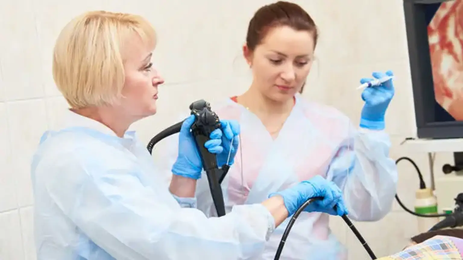
A gastroscopy is a technique that uses a thin, flexible tube called an endoscope to examine the oesophagus (gullet), stomach, and first section of the small intestine (duodenum). It is also known as Esophagogastroduodenoscopy (EGD) and is one of the most popular procedures performed by gastroenterologists.
Typically, a gastroscopy is performed to explore symptoms such as indigestion, nausea, or trouble swallowing. It can detect inflammation, ulcers, polyps, and other growths.
Gastroscopy is often used to treat diseases such as stomach ulcers, to enlarge a small oesophagus (a procedure called as dilatation), or to remove a foreign material. Doctors can obtain tissue samples (biopsies) and remove polyps from anything strange they detect.
Definition
Gastroscopy is a procedure that involves examining the upper gastrointestinal system, including the esophagus, stomach, and upper section of the colon, to confirm or rule out the existence of medical diseases such as stomach ulcers and gastritis.
The treatment is carried done using a flexible telescope called an endoscope, which is softly inserted into your mouth and progressed into your gastrointestinal system. When patients complain of symptoms such as heartburn, stomach discomfort, or bloating, the test is usually performed. Gastroscopy is also the most effective method of screening for stomach cancer.
Anatomy and Physiology
- Oesophagus:
The esophagus is found posterior to the trachea and extends from the cricoid cartilage to the cardiac opening of the stomach. It has a diameter of 4 to 6 mm and a length of 9 to 10 cm in a term newborn to around 25 cm in an adult. The transition from the esophagus to the stomach epithelium is marked by a shift in mucosa color from pale to reddish-pink (Z line).
- Stomach:
In an adult, the stomach is normally positioned beneath the diaphragm, around 40 cm distal to the incisors. Gastric cardia refers to the part of the stomach where the esophagus enters. The fundus is the area of the stomach above the esophagus and stomach junction. It can be seen via a retroflexed endoscopic view.
The stomach body is the bulk of the stomach. The incisura separates the gastric body from the antrum and runs along the minor curvature of the stomach. Endoscopically, the transition from the body to the antrum is characterized by a change from rugae to flat mucosa. The pylorus is the muscular aperture between the stomach's lower end and the duodenal bulb.
- Duodenum:
The duodenum is a tube that runs from the pylorus to the duodenojejunal angle. The duodenal bulb is a swollen area immediately distal to the pylorus. The duodenum then forms a C-shaped loop and twists endoscopically posteriorly and to the right for 2.5 cm, inferiorly for 7.5 to 10 cm (descending part), anteriorly and to the left for roughly 2.5 cm, and eventually links to the jejunum at the level of the Treitz ligament.
Uses of Gastroscopy
A gastroscopy can be used to check symptoms or confirm a diagnosis, or it can be used to treat a condition.
1. Checking symptoms:
If you have symptoms that point to a problem with your stomach, oesophagus (gullet), or the first portion of your small intestine, a gastroscopy may be indicated (duodenum).
Problems that are sometimes investigated using a gastroscopy include:
- abdominal (tummy) pain or stomach ache
- heartburn or indigestion
- persistently feeling and being sick
- swallowing difficulty or pain during swallowing (dysphagia) a decrease in the quantity of red blood cells (iron deficiency anaemia), which can be caused by continuous internal bleeding.
- significant bleeding, which may have resulted in a great pain in your belly, vomiting blood, or very black or "tar-like" feces.
2. Diagnosing Conditions:
A gastroscopy is also used to help confirm (or rule out) suspected conditions, such as:
- Stomach ulcers (also known as peptic ulcers) are open sores that form on the lining of the stomach and small intestine as a result of gastro-oesophageal reflux disease (GORD) - heartburn, which occurs when stomach acid leaks back up into the esophagus.
- Coeliac disease – it is a frequent digestive ailment in which a person reacts negatively to gluten in meals.
- Barrett's oesophagus - abnormal cells on the lining of the oesophagus.
- portal hypertension – a condition in which the blood pressure inside the liver is unusually high, causing bulging veins (varices) to form on the stomach and oesophagus lining.
- stomach cancer and oesophageal cancer
The endoscope (a thin, flexible tube that is inserted down your throat) can be used to take tiny samples of tissue for testing in addition to inspecting the oesophagus, stomach, and duodenum. This is referred to as a biopsy.
3. Treating conditions:
A gastroscopy can also be used to treat issues with the esophagus, stomach, and duodenum.
A gastroscopy, for example, can be used to:
- Halt bleeding within the stomach or oesophagus, such as bleeding from a stomach ulcer or swollen veins (varices) broaden a restricted oesophagus causing discomfort or swallowing difficulties - this can be caused by GORD, oesophageal cancer, or oesophageal radiation.
- Remove malignant tumors, non-cancerous growths (polyps), or foreign objects deliver nourishment — when a person is unable to eat normally, a gastroscopy can assist doctors in guiding a feeding tube into the stomach
Contraindications of Gastroscopy Use
Contraindications for EGD include the following:
- Possible perforation
- Medically unstable patients
- Unwilling patients.
- Anticoagulation, pharyngeal diverticulum, or head and neck surgery (relative contraindications).
- Peritonitis.
- Toxic megacolon in an unstable patient.
- Abdominal and iliac aorta aneurysm.
Diagnostic EGD is considered a low-risk technique for bleeding in anticoagulant-treated individuals and can thus be conducted without adjusting anticoagulants before to the operation. However, if polypectomy is being considered or is a possibility, the patient's coagulation profile should be normalized. Patients with significant coagulation disorders may also be at risk for retropharyngeal hematoma.
Certain therapeutic treatments (for example, dilations, percutaneous endoscopic gastrostomy [PEG], polypectomy, endoscopic sphincterotomy, EUS-guided fine-needle aspiration [FNA], laser ablation, and coagulation) are considered high-risk for bleeding, and anticoagulation may need to be adjusted.
Equipment Used
- Gastroscopes:
Standard gastroscopes have a 10 mm diameter and a 2.8 mm instrument canal. Endoscopes with a diameter of less than 6 mm should be used for regular endoscopy in children weighing less than 10 kg. In severe acute upper GI bleeding, a gastroscope with a large operative channel measuring 3.8 to 4.2 mm is beneficial. To screen for pre-malignant gastric or duodenal lesions, high-definition gastroscopes with optical zoom should be accessible.
- Accessories:
Tissue sampling necessitates the use of biopsy forceps (standard and jumbo). Rat tooth forceps, alligator forceps, a retrieval net, a polypectomy snare, overtubes of esophageal and gastric lengths, and a foreign body protective hood should be present for recovery of a foreign body during esophagogastroduodenoscopy (EGD). If therapeutic operations are planned, more equipment may be necessary.
How Do You Prepare for a Gastrointestinal Endoscopy?
Routine endoscopy in children and adults is often done as an outpatient procedure under parenteral or general anesthesia. Endoscopy is occasionally required at the hospital bedside or in an operating room.
- Diet:
Fasting is required prior to the elective upper endoscopy procedure. According to the American Society of Anesthesiologists (ASA), patients should fast for at least 2 hours after consuming clear drinks and 6 hours after consuming light meals. In an emergency or when gastric emptying is hindered, the risk of pulmonary aspiration of stomach contents must be evaluated to decide (1) the dose of sedation, (2) if endotracheal intubation should be considered to protect the airway, or (3) whether the surgery should be postponed.
- Medications:
Most drugs may be continued and are normally given with a little sip of water before the endoscopy, however diabetic meds must be altered owing to the fasting time prior to the operation. For choices on the management of anti-thrombotic drugs or the use of antibiotic prophylaxis in at-risk patients prior to endoscopy, the American Society for Gastrointestinal Endoscopy (ASGE) guidelines should be followed.
- Sedation and Monitoring:
Most patients are sedated not just to reduce pain but also to create amnesia during the treatment. Pre-procedural examination is required for all patients undergoing upper endoscopy to determine their risk for sedation and to handle any complications connected to pre-existing health disorders.
The type of sedation used varies, from conscious sedation administered by the proceduralist to supervised anesthetic care administered by an anesthesiologist, and preferences for one type of sedation over another are mostly determined by training and available local resources. Many endoscopists use propofol intravenous sedation for regular upper endoscopy.
General anesthesia may be necessary for therapeutic endoscopic treatments such as foreign body removal or in patients whose cooperation is not expected, including very young children. ASGE guidelines advocate regular vital sign monitoring in addition to clinical observation for changes in cardiopulmonary state throughout all sedation-assisted endoscopic operations.
- Informed consent
Patients, parents, or legal guardians should provide informed consents before the Esophagogastroduodenoscopy (EGD) and for the administration of sedation.
What Happens During a Gastroscopy?
Gastroscopy is typically performed as an outpatient 'day case.' It is a routine test that is performed on a regular basis. The operator may spray local anesthesia into the back of your mouth or offer you an anaesthetic lozenge to consume. To assist you relax, you may be given a sedative. This is often administered through injection into a vein on the back of your hand. The sedative can make you drowsy, but it does not totally put you to sleep. It is not used as a general anaesthetic.
On a couch, you lie on your side. You are instructed to place a plastic mouth guard between your teeth. This guards your teeth and prevents you from biting the endoscope. After that, the operator will instruct you to swallow the first piece of the endoscope. Modern endoscopes are relatively thin, however this may be troublesome for certain people.
The operator then gently slides it down your gullet (oesophagus), through your stomach, and into the first portion of your gut (small intestine) known as your duodenum. The video camera on the endoscope's tip feeds images to a screen. The operator watches the screen for abnormalities of the oesophagus, stomach and duodenum. Air is passed down a channel in the endoscope into the stomach to make the stomach lining easier to see. This may cause you to feel full and want to belch.
Depending on the reason for the test and what they find, the operator may collect one or more tiny samples (biopsies) of the interior lining of the stomach. This is a painless procedure. Biopsy samples are transported to a laboratory for testing and examination under a microscope. The endoscope is then carefully removed.
A gastroscopy typically lasts 10 minutes. You should, however, allocate at least two hours for the entire visit. This is to provide time for the sedative to take effect (if you have one), for the gastroscopy itself, and for recovery. A gastroscopy can be fairly unpleasant, although it normally does not harm.
What Can I Expect After a Gastroscopy?
After a half-hour or so of rest, most people are ready to go home.
If you've taken a sedative, it may take a little longer for you to be ready to go home. Normally, the sedative will make you feel pretty comfortable and calm. You should not, however, drive, operate equipment, or consume alcohol for 24 hours after taking the sedative. You'll need someone to accompany you home and stay with you for 24 hours until the effects wear off completely. After 24 hours, most people are able to resume routine activities.
The operator prepares a report and transmits it to the doctor who ordered the gastroscopy. The results of any sample (biopsy) may take a few days, which may cause the report to be delayed. Before you depart, the operator may also tell you what he or she witnessed. However, if you have taken a sedative, you may not recall what you were told later. As a result, you may want to bring along a cousin or close friend who can recall what was said.
Is It safe?
An endoscopy is a very safe procedure. Rare complications include:
- Bleeding: If the process includes taking a sample of tissue for testing (biopsy) or treating a digestive system disease, your risk of bleeding issues following an endoscopy is enhanced. Such bleeding may necessitate a blood transfusion in rare situations.
- Infection: The majority of endoscopies include an inspection and biopsy, and the risk of infection is negligible. When additional procedures are performed as part of your endoscopy, the risk of infection increases. The majority of illnesses are mild and treatable with medications. If you are at a higher risk of infection, your doctor may prescribe prophylactic antibiotics before your treatment.
- Tearing of the gastrointestinal tract: A rip in the esophagus or another component of the upper digestive tract may necessitate hospitalization and, in some cases, surgery to heal. This consequence is extremely rare, occurring in approximately one out of every 2,500 to 11,000 diagnostic upper endoscopies. The risk rises if further operations, such as esophageal dilatation, are undertaken.
You can reduce your risk of complications by carefully following your doctor's instructions for preparing for an endoscopy, such as fasting and stopping certain medications.
When To Call Your Doctor?
Signs and symptoms to watch for after your endoscopy include:
- Fever.
- Chest pain.
- Shortness of breath.
- Bloody, black or very dark colored stool.
- Difficulty swallowing.
- Severe or persistent abdominal pain.
- Vomiting, especially if your vomit is bloody or looks like coffee grounds.
Call your doctor immediately or go to an emergency room if you experience any of these signs or symptoms.
Conclusion
Gastroscopy, also known as upper endoscopy, is the examination of the upper digestive system with a thin flexible tube (endoscope).
To observe these places, the tube is placed into the mouth and proceeds through the food pipe (oesophagus), stomach, and first section of the small intestine (duodenum). The endoscope has a light and video camera that sends images to a monitor where a doctor may view them.
Typically, a gastroscopy is performed to explore symptoms such as indigestion, nausea, or trouble swallowing. It can detect inflammation, ulcers, polyps, and other growths.
Gastroscopy is often used to treat diseases such as bleeding ulcers, to enlarge a small oesophagus (a procedure called as dilatation), or to remove a foreign material. Doctors can obtain tissue samples (biopsies) and remove polyps from anything strange they detect.
Complications of a gastroscopy are exceedingly rare, such as perforation (puncture) of your stomach or intestine wall or significant bleeding (requiring a blood transfusion, for example).
Other treatments or surgeries performed through the gastroscope may carry a higher risk, depending on the disease being treated and the operation suggested. Inquire with your doctor or the physician doing the gastroscopy about the risks associated with any subsequent procedures or surgeries.
Sedation may be used during gastroscopy. Sedation risks are infrequent, however they might include trouble breathing and irregular heart rhythms.
Patients with serious heart or chest illness may be more prone to severe sedative effects. These problems are typically prevented by giving oxygen throughout the surgery and monitoring the blood oxygen level.