Glaucoma

Last updated date: 07-Apr-2023

Originally Written in English

Glaucoma

Overview

Glaucoma is the second greatest cause of irreversible blindness in the United States, and it primarily affects older persons. Adult glaucoma is classified into four types: primary open-angle and angle-closure glaucoma, secondary open-angle and angle-closure glaucoma.

The most frequent kind in the United States is primary open-angle glaucoma (POAG). Glaucoma is described as an acquired loss of retinal ganglion cells and axons inside the optic nerve, also known as optic neuropathy, which causes a distinctive optic nerve head appearance and a gradual loss of vision. This pattern of peripheral vision loss can also be used to differentiate it from other types of vision loss.

Unless evidence of early glaucoma are detected during a regular eye exam, the patient with POAG is frequently asymptomatic until the optic nerve damage is severe. Acute angle-closure glaucoma, on the other hand, can appear unexpectedly and cause a more rapid deterioration in vision, as well as corneal edema, eye discomfort, headache, nausea, and emesis.
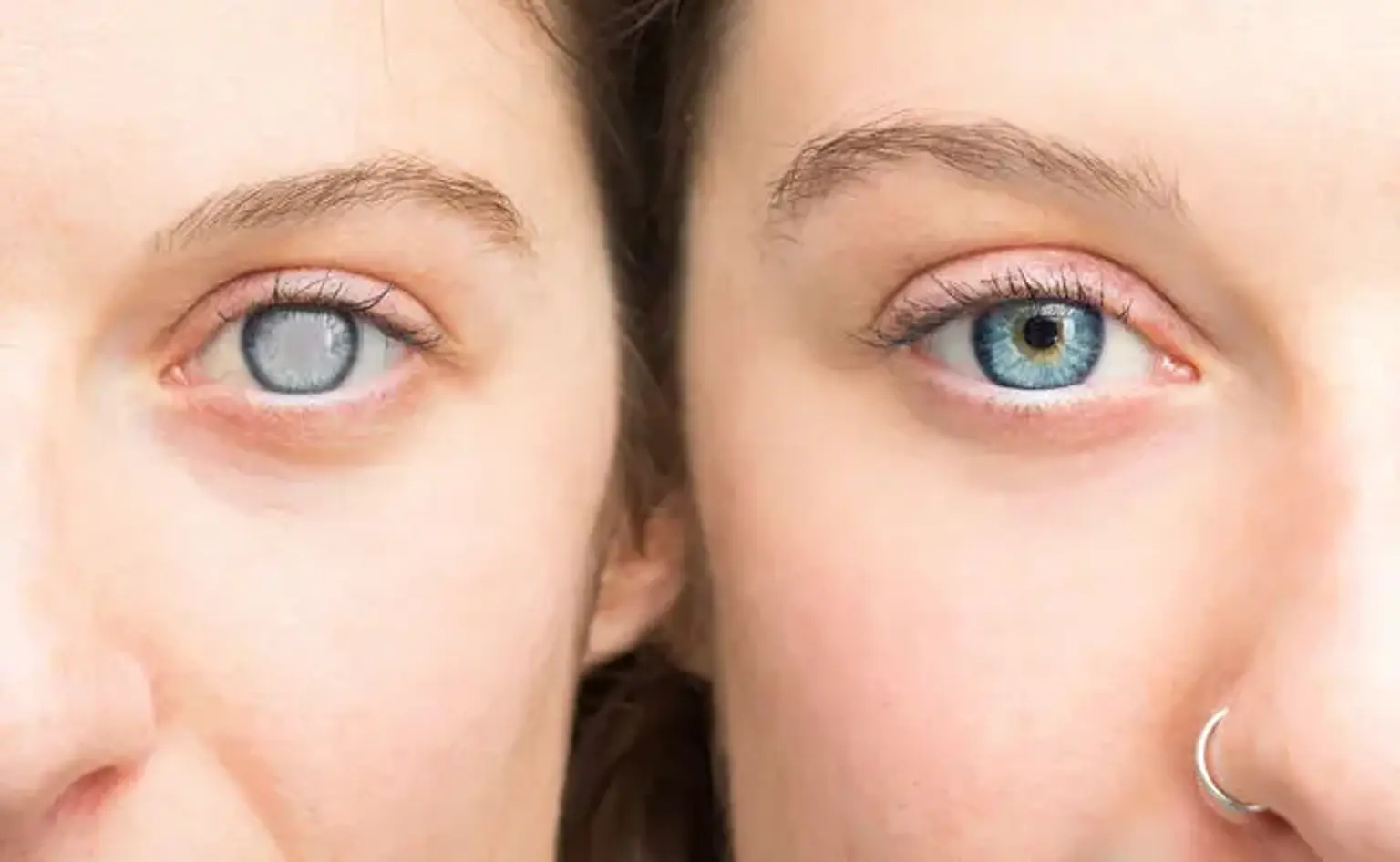
Secondary glaucoma is frequently associated with a previous eye injury or disease condition, resulting in increased intraocular pressure (IOP) and optic neuropathy.

While there are congenital, infantile, and developing glaucomas, as well as a juvenile variation of POAG, the four glaucoma types described above commonly affect persons over the age of 40. The reason is often associated with increased intraocular pressure, however no definite cause-and-effect link has been established. Glaucoma development might possibly have a hereditary component. Monozygotic twin pairings had a greater proportion of concordance, but not pairs, suggesting that environmental variables impact illness development.

Glaucoma cannot currently be avoided or cured, but its development can be managed to assist prevent additional vision loss by medication, glaucoma laser therapy, or incisional glaucoma procedures.


Epidemiology of Glaucoma

Epidemiology of Glaucoma

Glaucoma-related optic neuropathy affects an estimated 60 million individuals worldwide. The open-angle form is more common among the African population. When compared to other demographic groups, persons of African descent are up to 15 times more likely to go blind from open-angle glaucoma. The Inuit community has the highest prevalence of angle-closure, and it has also been proven to effect a higher rate in women than males, as well as individuals of Asian descent, with these populations often having a shallower anterior chamber.

Open-angle glaucoma accounts for 90% of glaucoma cases in the United States. Closed-angle glaucoma accounts for fewer than 10% of glaucoma cases in the United States, but as many as half of glaucoma cases in other nations (particularly East Asian countries).

Age is a key risk factor in the ongoing loss of retinal ganglion cells in all kinds. Other risk factors for developing glaucoma include a glaucoma family history (mother, father, brother, sister, or children), medical conditions such as diabetes, high blood pressure, heart disease, eye trauma, anatomical differences such as thinner corneas, a history of retinal detachment, eye tumors or inflammation, and long-term corticosteroid use.


Causes of Glaucoma

Causes of Glaucoma

Ocular hypertension (raised pressure within the eye) is the most major risk factor in most glaucomas, however in other groups, only half of persons with primary open-angle glaucoma have elevated ocular pressure. Ocular hypertension, defined as intraocular pressure beyond the conventional threshold of 21 mmHg (2.8 kPa) or even 24 mmHg (3.2 kPa), is not always a serious disease, although it does raise the chance of developing glaucoma.

One research discovered an 18% conversion rate within five years, implying that fewer than one in every five persons with increased intraocular pressure may have glaucomatous visual field loss during that time. It is debatable whether everyone with increased intraocular pressure should be treated for glaucoma; presently, most ophthalmologists support treating individuals with additional risk factors.

Dietary:

There is no conclusive evidence that vitamin deficits induce glaucoma in people. As a result, taking vitamins orally is not a suggested therapy. Caffeine raises intraocular pressure in those with glaucoma but has no effect on healthy people.

Ethnicity:

Because of their smaller anterior chamber depths, many persons of East Asian heritage are predisposed to developing angle closure glaucoma, with the majority of cases of glaucoma in this community involving some kind of angle closure. In Canada and Greenland, Inuit populations have been shown to have higher incidences of glaucoma than White ones.

Genetics:

Glaucoma is related with a number of rare congenital/genetic eye abnormalities. Failure of the normal third-trimester gestational atrophy of the hyaloid canal and tunica vasculosa lentis is occasionally linked with additional abnormalities.

Other causes:

Other causes of glaucoma, known as "secondary glaucoma," include long-term use of steroids (steroid-induced glaucoma), conditions that severely restrict blood flow to the eye, such as severe diabetic retinopathy and central retinal vein occlusion (neovascular glaucoma), ocular trauma (angle-recession glaucoma), and inflammation of the middle layer of the pigmented vascular eye structure (uveitis).


Classifications of Glaucoma

Classifications of Glaucoma

Primary glaucoma:

  • Primary angle closure glaucoma:

It is produced by contact between the iris and the trabecular meshwork, which obstructs the aqueous humor's outflow from the eye. This contact between the iris and the trabecular meshwork (TM) may gradually compromise the meshwork's function until it can no longer keep up with aqueous production and the pressure increases. Prolonged contact between the iris and the TM results in the production of synechiae in more than half of all instances (effectively "scars").

These permanently impede aqueous outflow. Pressure may rapidly build up in the eye in rare circumstances, producing discomfort and redness (symptomatic, or so-called "acute" angle closure). In this case, eyesight may become fuzzy, and halos around bright lights may be visible. A headache and vomiting are possible symptoms.

  • Primary open-angle glaucoma:

It occurs when the optic nerve is damaged, resulting in a gradual loss of vision field. This is connected with increased intraocular pressure. Although not all persons with primary open-angle glaucoma have raised ocular pressure above normal, further lowering of eye pressure has been demonstrated to arrest development even in these situations.

The increased pressure is generated by a blockage in the trabecular meshwork. Because the small channels are obstructed, pressure builds up in the eye, resulting in subtle, progressive vision loss. Peripheral vision is impaired initially, but if not addressed, the full eyesight will be gone.


Developmental glaucoma:

  • Primary congenital glaucoma
  • Infantile glaucoma
  • Glaucoma associated with hereditary or familial diseases


Secondary glaucoma:

  • Inflammatory glaucoma:
  1. Uveitis of all types.
  2. Fuchs heterochromic iridocyclitis.


  • Phacogenic glaucoma:
  1. Angle-closure glaucoma with mature cataract.
  2. Phacoanaphylactic glaucoma secondary to rupture of lens capsule.
  3. Phacolytic glaucoma due to phacotoxic meshwork blockage.
  4. Subluxation of lens.


  • Glaucoma secondary to intraocular hemorrhage:
  1. Hyphema
  2. Hemolytic glaucoma, also known as erythroclastic glaucoma


  • Traumatic glaucoma:
  1. Angle recession glaucoma: Traumatic recession on anterior chamber angle.
  2. Postsurgical glaucoma.
  3. Aphakic pupillary block.
  4. Ciliary block glaucoma.


Drug-induced glaucoma:

  • Corticosteroid induced glaucoma
  • Alpha-chymotrypsin glaucoma. Postoperative ocular hypertension from use of alpha chymotrypsin.


Glaucoma of miscellaneous origin:

  • Associated with intraocular tumors
  • Associated with retinal detachments
  • Secondary to severe chemical burns of the eye
  • Associated with essential iris atrophy


Neovascular glaucoma:

A rare form of glaucoma that is difficult or practically impossible to cure and is frequently caused by proliferative diabetic retinopathy (PDR) or central retinal vein blockage (CRVO). Other disorders that cause ischemia of the retina or ciliary body may also cause it. Individuals with insufficient blood flow to the eye are especially vulnerable to this condition.

Neovascular glaucoma develops when new, abnormal vessels form in the angle of the eye, obstructing drainage. People suffering from this illness begin to progressively lose their vision. The condition might arise suddenly at times, especially after cataract surgery.


Toxic glaucoma:

It is open-angle glaucoma with an inexplicable large increase in intraocular pressure caused by an unknown etiology. Intraocular pressure can occasionally reach 80 mmHg (11 kPa). It is distinguished by ciliary body inflammation and extensive trabecular oedema, which can sometimes extend to Schlemm's canal. The existence of a deep and clean anterior chamber, as well as the absence of aqueous misdirection, distinguishes this condition from malignant glaucoma. Furthermore, the corneal look is less blurry. Following neuroretinal breakdown, visual acuity might deteriorate.


Diagnostic work-up of Glaucoma

Diagnostic work-up of Glaucoma

A fundoscopic examination, visual field tests, tonometry, optical coherence tomography, and gonioscopy are used to assess patients. Other useful tests include visual acuity to determine whether or not vision is impaired, pachymetry to assess corneal thickness, and retinal scans to monitor for progressive changes in the retinal nerve fiber layer.

Glaucoma is diagnosed based on the above-mentioned testing modalities and the distinctive clinical signs of progressive optic neuropathy or visual field abnormalities. There is no one gold standard test for glaucoma diagnosis. To establish an accurate diagnosis and staging of glaucoma, providers must detect the typical optic nerve appearance, risk factors, and combine the findings of ancillary tests.

Currently, the American Academy of Ophthalmology advises routine thorough eye examinations for individuals with glaucoma risk factors on an individual basis, taking into account age, risk factors, race, and family history.


Visual defects in Glaucoma

Visual defects in Glaucoma

Damage to the retinal nerve fiber layer (RNFL) causes visual field abnormalities in glaucoma. Primary open angle glaucoma is the most common cause of field defects. Many distinct patterns may be noticed in the visual field due to the RNFL's unique architecture. The majority of early glaucomatous alterations are evident in the center visual field, primarily in Bjerrum's region, 10-20° from fixation.

Following are the common glaucomatous field defects:

  • Generalized depression: In the early stages of glaucoma and many other disorders, patients experience generalized depression. Isopter contraction causes mild restriction of the central and peripheral vision fields, which is classified as generalized depression. When all of the isopters show identical depression to the same point, this is referred to as a contraction of the visual field. Relative paracentral scotomas are places where the patient cannot see smaller and darker targets. Targets that are larger and brighter can be viewed. Normal tension glaucoma is characterized by small paracentral depressions, primarily superonasal (NTG). Cataract can also cause a widespread depression of the whole field.


  • Baring of blind spot: "Baring of blind spot" means exclusion of blind spot from the central field due to inward curve of the outer boundary of 30° central field. It is only an early non-specific visual field change, without much diagnostic value in glaucoma.


  • Small wing-shaped Paracentral scotoma: Small wing-shaped Paracentral scotoma within Bjerrum's area is the earliest clinically significant field defect seen in glaucoma. It may also be associated with nasal steps. Scotoma may be seen above or below the blind spot.


  • Siedel's sickle-shaped scotoma: Paracentral scotoma joins with the blind spot to form the Seidel sign.


  • Ring or Double arcuate scotoma: Two arcuate scotomas join together to form a Ring or Double arcuate scotoma. This defect is seen in advanced stages of glaucoma.


  • Roenne's central nasal step: It is created when two arcuate scotomas run in different arcs to form a right angled defect. This is also seen in advanced stages of glaucoma.


  • Peripheral field defects: Peripheral field defects may occur in early or late stages of glaucoma. Roenne's peripheral nasal steps occur due to contraction of peripheral isopter.


  • Tubular vision: Because macular fibers are the most resistant to glaucomatous damage, central vision stays undamaged until the disease progresses to the terminal stages. Tubular vision, also known as tunnel vision, is characterized by the loss of peripheral vision while retaining central vision, resulting in a restricted circular tunnel-like field of vision. It can be noticed in the last stages of glaucoma. Another illness that produces tubular vision is retinitis pigmentosa.


  • Temporal island of vision: It is also seen in end stages of glaucoma. The temporal islands lie outside of the central 24 to 30° visual field, so it may not be visible with standard central field measurements done in glaucoma.


Is Glaucoma curable?

Is Glaucoma curable?

Glaucoma treatment is tailored to the specific kind and severity of the disease. However, at this time, there is no treatment that will restore any of the visual loss that has already happened; it can only assist to avoid more damage and vision loss. Visual field testing and mapping of vision loss are useful tools for tracking illness development.

Medications to reduce ocular pressure are usually used to treat open-angle glaucoma at first. Prostaglandin analogs, beta-blockers, carbonic anhydrase inhibitors, an alpha-2 agonist, miotic drugs, and, more recently, rho-kinase inhibitors and nitric-oxide donor pharmaceuticals are among the medication types.

In certain circumstances, laser trabeculoplasty is employed as the primary therapeutic option. If medicinal therapy is ineffective, treatments such as laser trabeculoplasty, trabeculectomy, inserting a drainage valve/tube shunt, or laser treatment to the ciliary body to minimize aqueous production can be employed to improve IOP control.

MIGS (minimally invasive glaucoma surgery) is another emerging option for those with mild-moderate glaucoma. MIGS offers a better overall safety profile than traditional trabeculectomy and tube shunts, a faster recovery period, and has been shown to be successful for IOP reduction to the mid-high teens range. MIGS installation has also been shown in studies to minimize the amount of blood pressure drugs necessary to maintain goal IOP levels.

Normal-tension glaucoma can be treated with intraocular pressure-lowering drugs, as well as any other underlying medical disorders. Prostaglandin analogs, alpha-2 agonists, carbonic anhydrase inhibitors, and miotics are examples of medications.

Beta-blockers are contentious due to concerns of reduced optic nerve head perfusion, particularly in light of the possible worsening of the early AM nadir in blood pressure. If medicinal treatment fails, laser trabeculoplasty or filtration surgery may be used if vision loss continues to advance. Patients with normal-tension glaucoma were demonstrated to reduce or stabilize their field loss following a 30% drop in IOP in the Collaborative normal-tension glaucoma research.

Angle-closure is a medical emergency that must be handled as such. This is due to the fact that pressures can be high enough to produce glaucoma, ischemic nerve injury, or retinal vascular occlusion. Patients can take medications to relieve their eye pressure as early as possible, but they typically require a laser operation known as laser peripheral iridotomy. This laser makes a tiny hole in the iris to alleviate pupillary block, causing the pressure gradient between the posterior and anterior chambers to normalize and resolving iris bombe and opening the anterior chamber drainage angle.

Laser iridoplasty and, less typically, laser pupilloplasty can be used to flatten the peripheral iris. Because the ciliary body can suffer ischemic damage during an attack and have decreased production for a few weeks, it is important to have a follow-up gonioscopy to ensure the angle has reopened and to comment on the percentage of the angle with peripheral anterior synechia from the acute or prior subacute attacks.

After the acute crisis has passed, individuals are at high risk of having an attack in the contralateral eye and should be evaluated for gonioscopy and, if narrow, preventive iridotomy in the opposite eye.

Secondary glaucoma should be treated for the underlying cause of glaucoma, with drugs to reduce intraocular pressure perhaps added depending on the underlying reason.


Prognosis of Glaucoma

Prognosis of Glaucoma

Glaucoma is a serious condition that, if left untreated, can result in irreversible visual loss. The greater the pressure, the greater the chance of optic nerve damage. However, with therapy, most individuals have a fair prognosis. Low intraocular pressures can arrest the course of this condition by preventing additional visual field loss.


Conclusion

Glaucoma

Glaucoma is a group of eye diseases that cause damage to the optic nerve or retina, resulting in visual loss. The most frequent form is open-angle (wide angle, chronic simple) glaucoma, in which the drainage angle for fluid within the eye stays open, whereas closed-angle (narrow angle, acute congestive) glaucoma and normal-tension glaucoma are less prevalent.

There is no discomfort with open-angle glaucoma since it grows slowly over time. Peripheral vision may begin to deteriorate first, followed by central vision, eventually leading to blindness if not addressed. Closed-angle glaucoma can manifest gradually or abruptly. Severe eye discomfort, blurred vision, a mid-dilated pupil, redness of the eye, and nausea may accompany the abrupt onset. Once glaucoma has caused vision loss, it is irreversible. Glaucoma-affected eyes are referred to as glaucomatous.

Glaucoma risk factors include growing age, high intraocular pressure, a family history of glaucoma, and usage of steroid treatment. A number of 21 mmHg or 2.8 kPa over atmospheric pressure (760 mmHg) is commonly used for ocular pressures, with higher pressures posing a larger danger. However, some people can have high eye pressure for years without experiencing any injury.

Optic nerve injury can occur with normal pressure, which is known as normal-tension glaucoma. The delayed escape of aqueous humor through the trabecular meshwork is thought to be the mechanism of open-angle glaucoma, whereas the iris prevents the trabecular meshwork in closed-angle glaucoma. A dilated eye examination is used to make a diagnosis. The optic nerve frequently exhibits an inordinate degree of cupping.

It is possible to halt or stop the progression of disease using medication, laser therapy, or surgery if addressed early. The purpose of these therapies is to reduce ocular pressure. There are several types of glaucoma medications available. Laser therapies for both open-angle and closed-angle glaucoma may be beneficial. In those who do not react adequately to alternative treatments, a variety of glaucoma operations may be employed. Closed-angle glaucoma treatment is a medical emergency.