Hemostatis

Last updated date: 02-Mar-2023

Originally Written in English

Hemostasis

Overview

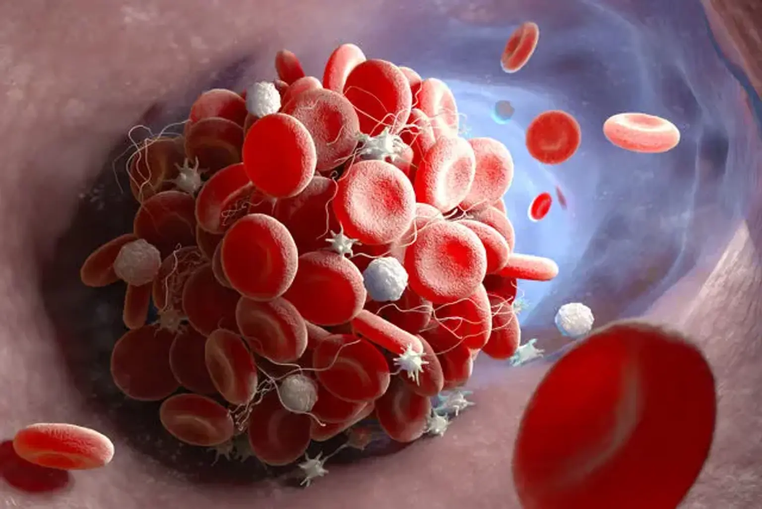
Hemostasis is the process that results in the cessation of bleeding from a blood vessel. It is a multi-step procedure with several interconnected steps. This cascade results in the development of a "plug" that seals the injured region of the blood vessel, thereby regulating the bleeding. It all starts with harm to the blood vessel lining.


Hemostasis definition

Hemostasis, also known as hemostasis, is a mechanism that prevents and stops bleeding by keeping blood within a broken blood artery (the opposite of hemostasis is hemorrhage). It is the first step in the healing of a wound. Coagulation is the process through which blood changes from a liquid to a gel. Intact blood arteries are critical in reducing the tendency of blood to clot.

Endothelial cells in intact arteries use a heparin-like molecule and thrombomodulin to prevent blood clotting and nitric oxide and prostacyclin to prevent platelet aggregation. When endothelial cells are injured, they stop secreting coagulation and aggregation inhibitors and instead release von Willebrand factor, which initiates hemostasis maintenance following injury.


Hemostasis physiology

The coagulation process involves a number of biological components. The mechanisms connected with the endothelium, platelets, and hepatocytes are particularly notable.

Endothelium. Clotting factors III and VIII are derived from endothelial cells, whereas clotting factor IV is derived from plasma. Factors III, IV, and VIII all undergo K-dependent gamma-carboxylation of their glutamic acid residues, allowing them to bind to calcium and other ions during the coagulation cascade.

Platelets. These are non-nucleated disc-like cells that develop from bone marrow megakaryocytes. They are approximately 2 to 3 microns in size. Plasma membrane, open canalicular system, spectrin and actin cytoskeleton, microtubules, mitochondria, lysosomes, granules, and peroxisomes are some of their distinguishing structural components. These cells secrete substances that aid in clotting and platelet aggregation.

Hepatocytes. The liver is responsible for the majority of the proteins that act as clotting factors and anticoagulants.


Function

Stability of the heart. Under normal conditions, a delicate balance exists between the procoagulant and anticoagulant pathways. This mechanism ensures hemorrhage control when needed and the end of pro-coagulant pathway activity beyond the injury site/or in the absence of bleeding. When this balance is disrupted in any way, it might lead to thrombotic/bleeding issues. Wound healing is also aided by the hemostatic system.

The cardiovascular system. Peripheral arteriolar dilation is caused by PGA1 and PGA2. Vasodilation is caused by prostacyclin, while constriction is caused by thromboxane A2. Prostacyclin causes vasodilation by inhibiting platelet aggregation, whereas thromboxane A2 and endoperoxides enhance platelet aggregation and cause vasoconstriction. The degree of platelet plug formation is determined by the balance of prostacyclin and thromboxane A2. As a result, prostaglandins have a significant impact on transient hemostasis.


Mechanism

Hemostasis is the presence of blood outside of the body or blood vessels. It is the body's inherent mechanism to stop bleeding and blood loss. Three phases occur in rapid succession during hemostasis. The first response is a vascular spasm, which occurs when blood arteries tighten to allow less blood to be lost. Platelets join together to form a temporary closure to cover the rupture in the vessel wall in the second step, platelet plug formation. Coagulation, or blood clotting, is the third and final phase.

Coagulation strengthens the platelet blockage by forming fibrin threads that act as "molecular glue." Platelets play an important role in the hemostatic process. They enable the formation of the "platelet plug," which occurs very immediately when a blood artery is broken.

Platelets begin to bind to the sub-endothelium surface of a blood vessel's epithelial wall within seconds of its epithelial wall being broken. It takes around sixty seconds for the first fibrin strands to intertwine with the wound. After a few minutes, fibrin has completely formed the platelet block. Three systems keep the body in a state of hemostasis:

Vascular spasm (Vasoconstriction) 

Vasoconstriction is the blood vessel's first response to injury and is produced by vascular smooth muscle cells. The smooth muscle cells are regulated by the vascular endothelium, which sends intravascular signals to modulate their contracting properties. When a blood artery is injured, an immediate reflex is elicited by local sympathetic pain receptors, which aids in the promotion of vasoconstriction.

The injured vessels constrict (vasoconstrict), reducing the volume of blood flow through the area and limiting blood loss. When collagen is exposed at the site of injury, it enhances platelet adhesion to the injury site. Platelets expel cytoplasmic granules containing serotonin, ADP, and thromboxane A2, all of which increase the effect of vasoconstriction. 

As the amount of injury increases, the spasm reaction becomes more effective. In smaller blood vessels, vascular spasm is far more effective. 


Platelet plug formation

Platelets bind to injured endothelial cells to create a platelet plug (primary hemostasis) and then degranulate. This mechanism is controlled by thromboregulation. Von Willebrand factor (vWF), a glycoprotein present in plasma, stimulates plug formation. Platelets are important players in the hemostatic process. When platelets come into contact with wounded endothelial cells, they alter form, release granules, and eventually become sticky.'

Platelets express various receptors, some of which are involved in platelet attachment to collagen. Platelets that are activated express glycoprotein receptors that interact with other platelets, causing aggregation and adhesion. Platelets secrete cytoplasmic granules that include adenosine diphosphate (ADP), serotonin, and thromboxane A2.

ADP attracts additional platelets to the damaged area, serotonin is a vasoconstrictor, and thromboxane A2 aids in platelet aggregation, vasoconstriction, and degranulation. As more chemicals are released, more platelets clump together and release their chemicals, resulting in a platelet clog and a positive feedback loop. Platelets alone are responsible for stopping the bleeding of our skin's unseen wear and tear on a regular basis. This is known as primary hemostasis.


Clot formation

Once the platelets have formed the platelet plug, the clotting factors (a dozen proteins that circulate through the blood plasma in an inactive condition) are activated, resulting in the synthesis of Fibrin from inactive fibrinogen plasma protein. As a result, a Fibrin mesh is formed all around the platelet plug to keep it in place; this process is known as "Secondary Hemostasis."

During this process, some red and white blood cells become trapped in the mesh, causing the primary hemostasis plug to harden; the resulting plug is known as a thrombus or a clot. As a result, the term "blood clot" refers to a secondary hemostasis plug that has blood cells trapped within it.

Though this is frequently a beneficial step in wound healing, it has the potential to cause serious health problems if the thrombus becomes detached from the vessel wall and travels through the circulatory system; if it reaches the brain, heart, or lungs, it could result in stroke, heart attack, or pulmonary embolism, respectively. However, healing a wound would be impossible without this mechanism.


Related Testing

Indications. The assessment of platelet function as well as its dysfunction has become vital in the current era in multiple clinical scenarios; several examples are:

  • For patients with clotting or bleeding disorders
  • For patients after cardiac stenting or stroke to monitor the activity of the antiplatelet agents
  • For perioperative evaluation.


Platelet Specific. Various tests have undergone development for platelet testing; they include:

  • Bleeding time (BT)
  • Light transmission platelet aggregation
  • Impedance platelet aggregation
  • Global thrombosis test
  • PFA-100/200
  • VerifyNow system
  • Thromboelastography (TEG)
  • Flow cytometric analysis of platelet function


Coagulation Cascade Specific. There has been the development of various tests that evaluate specific events in the coagulation cascade.

  • They aid in determining where the shortage exists in the intrinsic, extrinsic, or ultimate common pathways, as well as identifying qualitative or quantitative deficiencies in individual clotting components.
  • The prothrombin time, which was discovered in 1935, evaluates the function of the extrinsic and common coagulation cascades.
  • Activated partial thromboplastin time evaluates both the intrinsic and common coagulation pathways.
  • Thrombin time assesses fibrin development in the final common pathway of coagulation.
  • The reptilase time and other fibrinogen assays are used to evaluate the fibrin production phase.
  • Mixing studies, factor activity assays, and factor inhibitor assays are further tests for determining the presence of inhibitors or antibodies, as well as factor deficiency.


Pathophysiology

Virchow’s triad of hypercoagulability, vascular stasis, and vascular trauma, described in 1856, remains a true predictor of thrombosis.

  • Hypercoagulability
  • Stasis
  • Trauma


Etiologies. The physiology of coagulation undergoes alteration due to various factors, including:

  • Genetics
  • Medications
  • Procoagulant
  • Anticoagulation defects of the coagulation cascade
  • Quantitative defects of the integral components of the coagulation
  • Qualitative defects of the integral components of coagulation.


Clinical Presentations. With the altering of hemostatic physiology, various clinical outcomes including:

  • Pulmonary embolism
  • Deep vein thrombosis
  • Stroke
  • Myocardial infarction


Coagulopathies. Few of the disorders of coagulation include:

  • Anti-thrombin 3 deficiency,
  • Protein C deficiency,
  • Hyperhomocysteinemia,
  • Anti-phospholipid antibody syndrome


Risk Factors. Some acquired factors influencing the coagulation include:

  • Pregnancy
  • Trauma
  • Malignancy-related hypercoagulable state
  • Hormone replacement therapy
  • Inflammation
  • Infection
  • Heparin-induced thrombocytopenia


Issues of Concern

Hyper-coagulation

The hemostatic cascade is intended to limit hemorrhage while also acting as a protective mechanism. This process can be activated mistakenly while the blood is still within the lumen of the blood artery and there is no bleeding. This scenario results in thrombosis, a pathologic phenomenon that can have disastrous consequences by blocking blood flow and causing ischemia and even infarction of the tissues fed by the occluded blood arteries.

A physiologic process becomes a pathologic process, resulting in morbidity and/or mortality. Antiphospholipid antibody syndrome, Factor 5 Leiden mutation, Protein C deficiency, Protein S deficiency, Prothrombin gene mutation, and other conditions are examples.


Hypo-coagulation

When any component of this hemostatic cascade fails to work properly, it can result in poor hemostasis and the inability to control hemorrhage; this can result in substantial blood loss, hemorrhage, and problems caused by the inhibited blood supply to essential organs. Von Willebrand disease, hemophilia, disseminated intravascular coagulation, clotting factor deficiency, platelet disorders, collagen vascular disorders are a few examples.


Iatrogenic Coagulopathy

Medicine is currently in the era of widespread use of antiplatelet agents such as aspirin, clopidogrel, ticagrelor, and anticoagulants such as warfarin, heparin, low molecular weight heparin, rivaroxaban, apixaban, dabigatran, fondaparinux, and others for a variety of commonly encountered clinical conditions such as cardiac stenting/percutaneous coronary intervention, atrial fibrillation, deep

The manner in which these drugs alter the functionality of the various components of the clotting cascade can aid patients in managing their clinical problems. However, in cases of incorrect dosage, noncompliance, or pharmaceutical combinations, it can cause bleeding/thrombosis, resulting in considerable morbidity and fatality.


Clinical Significance

There are various hypercoagulable and hypercoagulable conditions resulting from defects in the coagulation pathways. The full extent is beyond the scope of this topic. Here are several examples:

  • Cardiovascular. There has been an increase in the incidence of bleeding while on antiplatelet and anticoagulant agents for recent myocardial infarction, stroke, cardiac stents, peripheral vascular stenting, atrial fibrillation, pulmonary embolism, deep venous thrombosis, and a variety of other conditions, leading to the development and use of reversal agents.
  • Renal. End-stage renal disease, for example, can cause uremic platelet dysfunction, which can be treated with dialysis and renal replacement therapy.
  • Immunological. Replenishing deficient clotting factors, removing antibodies against clotting factors, and using medications to enhance or improve clotting cascade functionality—these newer developments have led to significant advances in the field of medicine and provided treatment options for a variety of difficult-to-manage clinical scenarios. Blood products such as packed red blood cells, platelets, and clotting factors are also used to help with management. To replenish the missing clotting factors, prothrombin complex concentrate and various formulations are available.
  • Pharmacological. Prudent use of antiplatelet agents such as aspirin, clopidogrel, as well as anticoagulant agents such as heparin, warfarin, or rivaroxaban can result in an increased risk of blood loss form different sites of the body.


Health problems associated with hemostasis

Hemostasis is the natural clotting of blood in reaction to an injury. However, your body might clot excessively, a condition called as hypercoagulability. This can result in a large number of blood clots forming spontaneously and obstructing normal blood flow. Thrombosis occurs when blood clots develop within your blood arteries. When thrombosis occurs on a regular basis, the disorder is known as thrombophilia.


Thrombophilia (hypercoagulability or too much clotting)

Hypercoagulability occurs when your blood clots excessively or quickly. This is problematic because clots can form or become trapped in various parts of your body, causing serious, life-threatening complications. Here are some examples of these issues:

  • Stroke.
  • Deep vein thrombosis (DVT), can then cause a pulmonary embolism.
  • Heart attack.

Many forms of cancer can induce hypercoagulability, and certain uncommon disorders can also be inherited. That is, they are either inherited from your parents or occur as a result of a chance mutation in your DNA. Thrombophilia is a condition that affects people with several illnesses. Here are a few instances of such conditions:

  • Protein C deficiency.
  • Prothrombin gene mutation.
  • Factor 5 Leiden mutation.

Inherited thrombophilia diseases are less prevalent than "acquired" illnesses, which generally emerge later in life. Acquired conditions include the following:

  • Antiphospholipid syndrome.
  • Inflammatory bowel disease.
  • Severe reactions to infections, such as sepsis.

Most thrombophilia drugs make it more difficult for your blood to clot in some manner. Antiplatelet, anticoagulant, and fibrinolytic (fibrin-breaking or clot-busting) medications are examples of these.


Hypercoagulability (not enough clotting)

When your blood does not clot properly, every injury becomes even more hazardous. Even mild injuries might result in significant blood loss if appropriate coagulation is not present. It also means you're more likely to sustain damage to your organs and blood vessels, which can lead to internal bleeding.

Certain cancers, such as leukemia, can produce excessive bleeding. This is due to the fact that they frequently include a deficiency of platelets in your body or anti-clotting action. Conditions that prevent your blood from clotting are frequently inherited. Here are some instances of genetic conditions:

  • Hemophilia.
  • Von Willebrand disease.
  • Inherited thrombocytopenia (low platelet count).

Medications that restrict or block your body's anti-clotting processes, increase your body's capacity to create platelets, or add more of particular clotting components to your blood are often used to treat disorders that prevent your blood from clotting. You can also receive platelet transfusions if your body requires them.


Artificial hemostasis

Hemostasis is the process of stopping blood leakage from a vessel or organ of the body. The phrase is derived from the Ancient Greek roots "heme," which means "blood," and "stasis," which means "halting." Hemostasis may be traced back to ancient Greece, when it was first utilized in the Battle of Troy. It all began with the understanding that severe bleeding would ultimately result in death.

The Greeks and Romans utilized vegetable and mineral styptics on major wounds until Greece took over Egypt in 332BC. Many further improvements in general medicine were made at this period as a result of the study of Egyptian mummification practices, which led to a better understanding of the hemostatic process. Many of the veins and arteries that flow throughout the human body, as well as the directions in which they move, were discovered during this time period.

Doctors at the time understood that if they were clogged, blood would be unable to exit the body. Nonetheless, it took until the introduction of the printing press in the fourteenth century for medical notes and ideas to spread westward, allowing the concept and practice of hemostasis to grow.


Conclusion 

Hemostasis, is a mechanism that prevents and stops bleeding by keeping blood within a broken blood artery (the opposite of hemostasis is hemorrhage). It is the initial step in the healing of a wound. Coagulation is the process through which blood changes from a liquid to a gel. 

Intact blood arteries are critical in reducing the tendency of blood to clot. Endothelial cells in intact arteries use a heparin-like molecule and thrombomodulin to prevent blood clotting and nitric oxide and prostacyclin to inhibit platelet aggregation.

When endothelial cells are injured, they cease secreting coagulation and aggregation inhibitors and instead release von Willebrand factor, which initiates hemostasis maintenance following damage.