Introduction
A hip fracture is a serious injury that commonly occurs due to falls, accidents, or even osteoporosis. It typically involves the breaking of the upper part of the femur (thigh bone) near the hip joint. Hip fractures are particularly prevalent in older adults, as their bones become more brittle with age.
Surgery is often necessary to repair the fracture and restore function. This procedure is essential for improving mobility, reducing pain, and preventing further complications, such as blood clots or infections. There are several surgical options, but the primary goal is to help the patient regain independence and quality of life as quickly as possible.
Types of Hip Fractures and Their Surgical Treatment
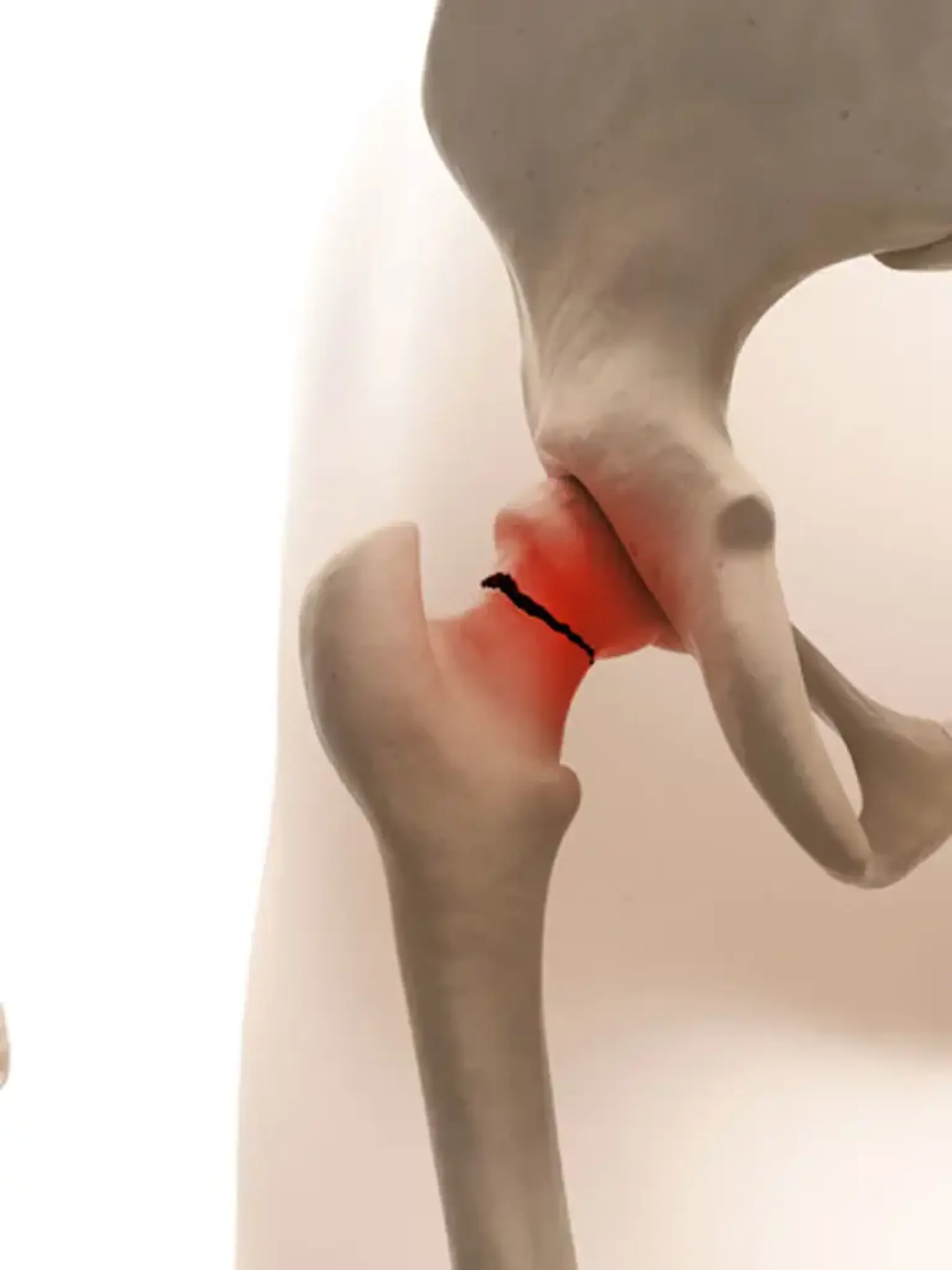
Hip fractures are classified into several types based on where the bone breaks. The most common are:
Femoral neck fractures: These occur just below the ball of the hip joint.
Intertrochanteric fractures: These happen between the hip joint and the femur’s shaft.
Subtrochanteric fractures: These occur below the trochanter (the bony prominence on the upper femur).
The treatment approach varies depending on the type of fracture, age, health, and activity level of the patient. Common surgical options include:
Total Hip Replacement (THR): Often recommended for older adults or those with severe arthritis, a total hip replacement involves removing the damaged parts of the hip and replacing them with artificial components.
Hip Pinning: This less invasive procedure is often used for younger, healthier individuals with stable fractures. Metal pins are inserted to hold the bone in place while it heals.
Hemiarthroplasty: In this surgery, only the ball part of the hip joint is replaced, often when the fracture involves the femoral head.
Understanding the Surgical Procedure
Before surgery, patients will undergo a preoperative assessment to ensure they are in optimal health for the procedure. This may include blood tests, imaging studies like X-rays, and consultations with the surgeon and anesthesiologist.
During the surgery, patients are typically under general anesthesia, ensuring they are unconscious and pain-free. The surgeon will make an incision over the hip area, then repair or replace the broken parts. If the fracture is stable, hip pinning may be used, while more extensive fractures often require a total hip replacement or hemiarthroplasty. The surgery typically takes 1-2 hours, depending on the complexity of the fracture.
Preparing for Hip Fracture Surgery
Proper preparation is key to ensuring a successful surgery and recovery. Prior to the operation, the patient will undergo several tests to assess their health. These tests might include checking for underlying conditions like diabetes or heart disease, as these can affect the surgery and healing process.
In the days leading up to the surgery, patients may be advised to:
Avoid eating or drinking for a set period of time to reduce the risk of complications during anesthesia.
Adjust medications if necessary, especially blood thinners or anticoagulants.
Prepare the home environment by making it easier to move around post-surgery, such as setting up a safe, clutter-free space with necessary supplies.
Psychologically, it’s essential to be mentally prepared for the recovery process, as it can take weeks to months before normal activities can be resumed. Emotional support from family and friends is crucial for ensuring a smooth recovery journey.
Managing Pain After Hip Fracture Surgery
Pain management after surgery is critical to recovery. While some discomfort is normal, it can be controlled through:
Medications: Doctors typically prescribe painkillers like acetaminophen or opioids for the first few days. Non-steroidal anti-inflammatory drugs (NSAIDs) may also be recommended.
Ice Packs and Elevation: Applying ice to the hip can help reduce swelling and relieve pain.
Physical Therapy: Gentle movements and exercises can help manage stiffness and pain over time, improving joint function.
Patients should communicate openly with their doctor about pain levels to ensure proper management and avoid complications.
Recovery Timeline and What to Expect
The recovery process after hip fracture surgery varies depending on the type of surgery and the patient's health. Generally, the recovery occurs in stages:
Immediate Post-Op (1-2 Days): After surgery, patients are monitored in the hospital for a few days. Pain management, early mobility, and preventing complications like blood clots are the priorities.
Weeks 1-6: During the first month, patients often use a walker or crutches and gradually increase activity. The focus is on regaining mobility and strength.
Months 2-6: Physical therapy is intensified to help patients regain full hip function. Patients can expect to slowly return to normal activities, though full recovery can take up to six months.
Physical Therapy and Rehabilitation After Surgery
Rehabilitation plays a crucial role in the recovery of hip fracture surgery patients. Early physical therapy focuses on:
Strengthening muscles around the hip joint and improving balance.
Increasing range of motion to prevent stiffness.
Regaining weight-bearing capacity—patients will gradually progress from non-weight-bearing to partial and full weight-bearing.
Most patients begin physical therapy as early as the day after surgery. A tailored plan is created based on the patient's age, fracture type, and overall health. Consistent participation in physical therapy is key for a successful recovery.
Preventing Complications Post-Surgery
After hip fracture surgery, there are a few potential complications to be aware of:
Infections: Surgical site infections are rare but can occur. Keeping the incision clean and following care instructions is essential.
Blood Clots: Patients may be at risk of developing blood clots in their legs. To prevent this, doctors may prescribe blood thinners and encourage early mobilization.
Dislocations: In cases where the hip replacement is performed, dislocations can occur, especially during the early recovery phase. Proper movement and care during recovery can minimize this risk.
Follow-up appointments with the surgeon are vital to detect any complications early and ensure proper healing.
Factors Influencing Recovery Time
Several factors influence how quickly a patient recovers from hip fracture surgery:
Age: Older adults often face a longer recovery period due to lower bone density, muscle weakness, and other age-related conditions.
Overall Health: Conditions like diabetes, heart disease, and obesity can slow healing. Patients with fewer comorbidities typically recover faster.
Surgery Type: Patients who undergo total hip replacement might have a longer recovery period compared to those who have less invasive surgeries like hip pinning.
Activity Level: Active individuals may regain mobility faster than those with a sedentary lifestyle.
Understanding these factors helps set realistic expectations for recovery. Patients should work closely with their healthcare providers to manage any underlying health conditions and optimize recovery.
Lifestyle Modifications and Post-Surgery Care
Adapting your lifestyle after hip fracture surgery is crucial to ensure a smooth recovery:
Home Adjustments: Create a safe environment by removing trip hazards and adding handrails to bathrooms or stairs. Consider using mobility aids such as walkers or canes.
Avoiding Falls: Patients should be cautious while moving around, especially in the early stages of recovery. Non-slip footwear and good lighting are essential.
Diet and Nutrition: A balanced diet rich in calcium and vitamin D is important for bone healing. Proper nutrition also supports overall strength and immune function.
Hydration and Rest: Staying hydrated and getting plenty of rest accelerates healing and helps the body recover from surgery.
Making these adjustments helps minimize the risk of complications and supports the overall recovery process.
Preparing for Hip Fracture Surgery
Proper preparation is key to ensuring a successful surgery and recovery. Prior to the operation, the patient will undergo several tests to assess their health. These tests might include checking for underlying conditions like diabetes or heart disease, as these can affect the surgery and healing process.
In the days leading up to the surgery, patients may be advised to:
Avoid eating or drinking for a set period of time to reduce the risk of complications during anesthesia.
Adjust medications if necessary, especially blood thinners or anticoagulants.
Prepare the home environment by making it easier to move around post-surgery, such as setting up a safe, clutter-free space with necessary supplies.
Psychologically, it’s essential to be mentally prepared for the recovery process, as it can take weeks to months before normal activities can be resumed. Emotional support from family and friends is crucial for ensuring a smooth recovery journey.
Mental and Emotional Well-Being During Recovery
The recovery process from hip fracture surgery is not just physical; it can also take a mental and emotional toll. It's common to feel frustrated or anxious during recovery, especially when progress is slower than expected.
Coping with Temporary Limitations: Patients may need support in managing their temporary loss of mobility and independence. It's important to focus on small milestones and celebrate progress.
Support Systems: Family members, caregivers, and support groups play a crucial role in the recovery process. Encouragement and assistance with daily tasks can help reduce stress and improve mental well-being.
Seeking Professional Help: If depression or anxiety arises, speaking to a counselor or therapist can be beneficial. Mental health is just as important as physical health during recovery.
Maintaining a positive mindset and seeking emotional support can significantly improve the quality of life during the recovery period.
Rehabilitation Success Stories
Hearing about successful recoveries can be incredibly motivating for patients undergoing hip fracture surgery. Many individuals, even those over the age of 80, experience significant improvement in mobility and quality of life after surgery.
Case Study 1: Mary, a 74-year-old woman, underwent total hip replacement after a fall caused by osteoporosis. Despite her age and health challenges, after six months of rehabilitation, she regained her independence and returned to walking, gardening, and enjoying her daily activities.
Case Study 2: John, 60, had a femoral neck fracture and underwent hip pinning. With the help of physical therapy, John was able to return to his active lifestyle within four months, including playing tennis and hiking.
These stories highlight the importance of rehabilitation, motivation, and following the doctor’s guidelines for a successful recovery.
Common Concerns and Frequently Asked Questions (FAQs)
Patients often have concerns as they approach and undergo hip fracture surgery. Addressing these questions can provide reassurance:
How long will I need to stay in the hospital? Typically, patients stay in the hospital for 2-3 days after surgery. The length of stay depends on how well you recover initially.
When can I walk again? With physical therapy, most patients can start walking with assistance within a few days. Full weight-bearing and walking independently typically take several weeks to months.
Will I need a cane or walker long-term? Many patients use walking aids for the first 6-12 weeks, but the need for these aids decreases as they regain strength and mobility.
Is hip fracture surgery safe? While all surgeries carry risks, hip fracture surgery is generally very safe, especially when performed by an experienced orthopedic surgeon. The benefits—reduced pain and improved mobility—usually outweigh the risks.
Addressing these questions can help set expectations and ease any anxiety patients may have about the procedure and recovery process.
Long-Term Health Considerations After Hip Fracture Surgery
While the initial goal of surgery is to relieve pain and improve mobility, patients should also consider long-term health management after recovery.
Bone Health: It’s essential to continue bone-strengthening practices, including weight-bearing exercises, calcium, and vitamin D supplementation, especially for those with osteoporosis.
Joint Care: For patients who have undergone hip replacement surgery, maintaining a healthy weight and avoiding high-impact activities can extend the lifespan of the artificial joint.
Preventing Future Falls: The risk of future falls remains elevated after a hip fracture. Regular strength training, balance exercises, and a fall prevention strategy at home are important steps to reduce the risk of additional fractures.
By focusing on long-term health, patients can enjoy a better quality of life while minimizing the risk of future complications.
Financial Considerations and Insurance Coverage
The cost of hip fracture surgery can vary significantly depending on the procedure, location, and healthcare system. For many, it is important to understand the financial aspects of the surgery and recovery process:
Insurance Coverage: Most insurance plans, including Medicare, cover hip fracture surgery and rehabilitation. However, it’s essential to confirm specifics with the insurer before the surgery, especially if opting for a particular type of surgery or rehabilitation center.
Out-of-Pocket Costs: In some cases, patients may need to cover certain expenses such as rehabilitation, medications, or home care. Understanding these costs upfront can help with financial planning.
Government Assistance: For those without adequate insurance, some governments and nonprofit organizations offer financial assistance or subsidies to help with medical bills and rehabilitation expenses.
Being prepared financially can reduce stress during the recovery process and help ensure the patient receives the necessary care.
Technological Advancements in Hip Fracture Surgery
Recent advancements in surgical techniques and technology have significantly improved the outcomes of hip fracture surgeries. Key developments include:
Minimally Invasive Surgery: Techniques like minimally invasive hip replacement use smaller incisions, resulting in less blood loss, quicker recovery, and smaller scars.
Robotic-Assisted Surgery: Some hospitals are now using robotic systems to improve precision during hip replacement surgeries, which can lead to better alignment and faster recovery times.
3D Imaging: Surgeons use 3D imaging to create more accurate pre-surgical plans, especially in complex fractures, enhancing the precision of implant placement.
These innovations are reducing recovery times and improving the quality of care for hip fracture patients.
The Global Appeal of Hip Fracture Surgery
Hip fractures are a global health concern, and the surgery to treat them is common across various healthcare systems worldwide. The popularity of hip fracture surgery is increasing due to the aging population, particularly in developed countries.
In Developed Countries: In countries like the United States, Canada, and the UK, hip fractures are frequently treated with surgery, with hospitals offering cutting-edge technologies like minimally invasive procedures.
In Developing Countries: While surgery is increasingly available, there may be disparities in access to the latest techniques, surgical expertise, and post-operative care. However, the demand for hip fracture surgery is growing as healthcare systems improve and populations age.
Global Collaboration: Many international organizations, including the World Health Organization (WHO), are focusing on improving bone health and reducing the global burden of fractures. This includes educating the public, enhancing early detection, and providing access to advanced surgical care.
The global approach to treating hip fractures continues to evolve, improving outcomes and access to life-changing surgeries for people worldwide.
The Role of Nutrition in Recovery
Nutrition plays a vital role in recovery after hip fracture surgery. A diet rich in essential nutrients supports bone healing and overall recovery:
Calcium and Vitamin D: These nutrients are crucial for bone repair. Foods like dairy products, leafy greens, and fortified cereals are excellent sources.
Protein: Adequate protein intake helps with muscle repair and tissue healing. Lean meats, eggs, and legumes are good options.
Hydration: Staying hydrated is important for reducing fatigue, improving energy levels, and aiding in the healing process.
Patients should work with a nutritionist to develop a meal plan that supports their specific recovery needs.
The Importance of Follow-Up Care
Post-surgery follow-up care is essential to ensure proper healing and monitor for any complications:
Routine Check-ups: Regular visits to the surgeon help track recovery progress, detect issues like infections or joint dislocations, and adjust rehabilitation plans.
X-rays and Imaging: Follow-up X-rays may be needed to confirm that the bones are healing properly and that the surgical implants are in place.
Physical Therapy Adjustments: As recovery progresses, therapy plans may need to be adjusted to focus on strengthening muscles or improving flexibility.
These appointments ensure that recovery is on track and complications are managed early.
Conclusion
Hip fracture surgery is a transformative procedure that plays a crucial role in restoring mobility and quality of life for individuals who experience this common injury, particularly in older adults. With advancements in surgical techniques, pain management, and rehabilitation, the recovery process has become faster and more effective, allowing many patients to return to their daily activities with greater independence.
While recovery can take time and requires careful attention to factors like nutrition, physical therapy, and follow-up care, the benefits far outweigh the challenges. By addressing both the physical and emotional aspects of recovery, patients can regain strength, confidence, and vitality in the months following surgery.
As global healthcare systems continue to evolve and technology advances, the future of hip fracture surgery looks even more promising. With improved surgical options, faster recovery protocols, and increased focus on prevention, patients worldwide can look forward to better outcomes and healthier aging.
Ultimately, a positive mindset, support from loved ones, and dedication to rehabilitation can lead to successful, life-changing recoveries from hip fracture surgery.