Histiocytosis
Overview
Histiocytosis, also known as Langerhans Cell Histiocytosis (LCH) and formally known as Histiocytosis X, is a rare disorder involving particular cells that ordinarily play critical roles in the immune system. While the etiology of LCH is uncertain, it frequently behaves like cancer and is therefore treated by cancer specialists.
Histiocytosis is a broad term for a collection of diseases defined by an abnormal increase in the number of immune cells known as histiocytes. Monocytes, macrophages, and dendritic cells are examples of these.
A histiocyte is a type of immune cell that can be found in many regions of the body, including the bone marrow, bloodstream, skin, liver, lungs, lymph glands, and spleen. Histiocytes go into tissues where they are not ordinarily found and cause harm to those tissues in histiocytosis. Tumors can arise as a result of these proliferating immune cells, which can impact numerous regions of the body.
Histiocytosis's actual cause is unknown. Recent research suggests that it is caused by the growth and enlargement of an aberrant Langerhans cell, which then leads to the accumulation of other immune system cells, resulting in collections or tumors in various parts of the body. Some forms are inherited.
When LCH affects only one part of the body, it is known as a single system disease; when it affects more than one part of the body, it is classified as a multisystem disease. In children, histiocytosis mainly affects the bones and can occur in a single or numerous locations. The skull is commonly affected. Children over the age of five are more likely to have a single system disease with only bone involvement. Young children, particularly infants, are at a higher risk of developing the multisystem disease.
How Popular is Histiocytosis?
Langerhans cell histiocytosis is a rare condition. It affects 1 to 2 infants per million each year. In children aged 15 years, the incidence is 4 to 5 cases per million per year. The Langerhans disease affects roughly 1 to 2 people per million per year in adults. It can happen at any age, but it is more common in people under the age of 15.
Causes of Histiocytosis
The cause of LCH is unknown and remains a point of conflict; however, most experts agree that LCH is either a reactive or neoplastic process. Multiple cytokines are involved in LGH, there is high survival in isolated lesions, and spontaneous remissions can occur. These traits lend themselves to a reactive process.
LGH, on the other hand, can cause organ invasion. The common condition is related with an elevated mortality rate, it usually responds to chemotherapy, and at least one study has found a relationship with the BRAF gene mutation. These traits are favorable to a neoplastic progression.
Classifications of Histiocytosis
The disease spectrum is caused by clonal accumulation and proliferation of cells that resemble epidermal dendritic cells known as Langerhans cells, also known as dendritic cell histiocytosis. These cells, in conjunction with lymphocytes, eosinophils, and normal histiocytes, form the typical LCH lesions present in practically any organ. Canine histiocytic disorders are characterized by a similar range of diseases.
Clinically, LCH is classified into three types: unifocal, multifocal unisystem, and multifocal multisystem.
- Unifocal LCH:
Unifocal LCH, also known as eosinophilic granuloma (an older term that is now known to be a misnomer), is a condition characterized by a growing proliferation of Langerhans cells in a single organ, causing damage known as lesions. It usually has no extraskeletal involvement, but a lesion in the skin, lungs, or stomach is not uncommon. It might manifest as a single lesion in an organ or as a huge number of lesions in the same organ.
A multifocal unisystem variant occurs when multiple lesions are spread across one organ. When discovered in the lungs, it should be separated from Pulmonary Langerhans cell hystiocytosis, a subtype of disease that is most frequent in adult smokers. When it is located in the skin, it is referred to as a cutaneous single system Langerhans cell (LCH).
In some rare circumstances, this variant can cure without the use of therapy. This involvement of the primary bone helps to distinguish eosinophilic granuloma from other types of Langerhans Cell Histiocytosis (Letterer-Siwe or Hand-Schüller-Christian variant).
- Multifocal unisystem LCH:
Multifocal unisystem LCH is most commonly seen in children and is characterized by fever, bone lesions, and diffuse eruptions, most commonly on the scalp and in the ear canals. In 50% of instances, the pituitary stalk is involved, which frequently leads to diabetes insipidus. The Hand-Schüller-Christian triad is characterized by diabetes insipidus, exophthalmos, and lytic bone lesions. Peak onset occurs between the ages of 2 and 10 years.
- Multifocal multisystem LCH:
Letterer-Siwe disease, also known as multifocal multisystem LCH, is a rapidly progressive condition in which Langerhans Cell cells grow in several tissues. It is most common in children under the age of two, and the prognosis is dismal: even with rigorous chemotherapy, the five-year survival rate is only 50%.
- Pulmonary Langerhans cell Histiocytosis (PLCH):
Pulmonary Langerhans cell histiocytosis (PLCH) is a kind of LCH that is nearly exclusively found in cigarette smokers. It is currently thought to be a type of smoking-related interstitial lung disease.
PLCH develops when an abundance of monoclonal CD1a-positive Langerhans (immature histiocytes) proliferate the bronchioles and alveolar interstitium, and this flood of histiocytes recruits granulocytes like eosinophils and neutrophils and agranulocytes like lymphocytes further destroying bronchioles and the interstitial alveolar space that can cause damage to the lungs.
It is hypothesized that bronchiolar destruction in PLCH is first attributed to the special status of Langerhans cells that elicit cytotoxic T-cell responses, and this is supported by research that has demonstrated an excess of CD4+ T-cells in early PLCH lesions that have early activation markers. Some people recover entirely after quitting smoking, but others experience long-term consequences such as lung fibrosis and pulmonary hypertension.
Symptoms & Signs of Histiocytosis
LCH provokes a non-specific inflammatory response, which includes fever, lethargy, and weight loss. Organ involvement can also cause more specific symptoms.
- Bone: Painful bone swelling is the most common sign of both unifocal and multifocal disease. The skull is the most commonly affected bone, followed by upper extremity long bones and flat bones. It is unusual to find infiltration in the hands and feet. Pathological fractures can result from osteolytic lesions.
- Skin: A rash ranging from scaly erythematous lesions to red papules noticeable in intertriginous areas is common. Up to 80% of LCH patients develop severe scalp eruptions.
- Bone marrow: Pancytopenia with superadded infection is typically associated with a bad prognosis. Anemia can be caused by a variety of sources and does not always indicate bone marrow infiltration.
- Lymph node: The liver is enlarged in 20% of cases, the spleen is enlarged in 30%, and the lymph nodes are enlarged in 50% of cases with Histiocytosis.
- Endocrine glands: The hypothalamic pituitary axis is frequently implicated. Diabetes insipidus is the most prevalent kind. In most cases, anterior pituitary hormone insufficiency is permanent.
- Lungs: Some people are asymptomatic and were found by chance due to lung nodules on radiography; others have a persistent cough and shortness of breath.
Less commonly, the gastrointestinal tract, the central nervous system, and the oral cavity are involved.
Diagnosis of Histiocytosis
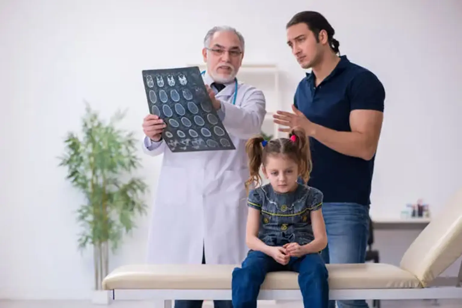
Tissue biopsy confirms the diagnosis histologically. A biopsy slide stained with hematoxylin and eosin will exhibit Langerhans Cell characteristics such as a defined cell border and pink granular cytoplasm. More specific findings include the presence of Birbeck granules on electron microscopy and immuno-cytochemical markers such as CD1 positive. Routine blood tests, such as a complete blood count, liver function test, U&Es, and a bone profile, are first performed to evaluate the degree of the disease and rule out other causes.
Chest X-rays may show micronodular and reticular abnormalities in the lungs, as well as cyst development in severe instances. Small, cavitated nodules with thin-walled cysts may be seen on MRI and high-resolution CT. A brain MRI scan can reveal three types of lesions: tumorous/granulomatous lesions, nontumorous/granulomatous lesions, and atrophy. Tumourous lesions are often observed in the hypothalamic-pituitary axis, with space-occupying lesions that may or may not include calcifications. There is a symmetrical hyperintense T2 signal with a hypointense or hyperintense T1 signal extending from grey matter into white matter in non-tumorous lesions. MRI reveals a hyperintense T1 signal in the globus pallidus in the basal ganglia.
Treatment of Histiocytosis
Medical care:
The best way to treat Langerhans cell histiocytosis (LCH) has yet to be determined. In an ideal world, the distinctions between normal and pathologic Langerhans cells (PLCs) would be exploited to guide disease therapy. However, a lack of knowledge has impeded targeted therapy. Some argue that LCH therapy should be cautious and confined to people who have constitutional symptoms such discomfort, fever, failure to thrive, and important organ dysfunction, as well as those who are at risk of CNS involvement.
Treatment decisions for histiocytosis are influenced by the kind, location, and severity of the illness, the organs involved, biologic findings, the genetics of the disease, the degree of risk involved, and a variety of other considerations. The disease's significant variability, as well as the fact that 10-20% of LCH patients have spontaneous regression, complicates comparisons of existing nonspecific therapy. Several medications, including cancer chemotherapeutic treatments, have been shown to be effective in the treatment of LCH.
Patients with high-risk illness must be treated for at least one year with a combination of vincristine, prednisone, and mercaptopurine. In resistant or recurring instances, agents such as vinblastine, prednisone, cytarabine, cladribine, and clofarabine might be given as needed. Treatment with clofarabine or cytarabine (the latter at greater dosages) should be explored in individuals with pituitary involvement and CNS illness.
an increase in the treatment period from 6 to 12 months, the use of repeated induction therapy in patients who did not respond well to initial induction with vinblastine and steroids, and the treatment of refractory disease in a risk organ with cladribine and cytarabine—the 5-year survival rate improved from 92 percent to The 5-year survival rate in a specific group of patients with refractory disease in a risk organ rose from 60% to 92%.
Experience has shown that the pharmacological combinations typically utilized in children, as described above, are significantly more harmful and ineffective in the adult population. In this demographic, cytarabine is both effective and well tolerated.
In LCH, radiation treatment is successful. Doses of 750-1500 cGy are commonly used, resulting in good local control of single lesions or metastasis, which can occur in important locations or cause irreversible harm. Radiotherapy with fractionated dosages has also been employed.
Summary of suggested therapeutic approach:
While there are no treatment guidelines for LCH, the following are proposed therapies, not recommendations, based on available literature, with these treatments to be taken alone or in combination as needed.
Treatment for a single bone lesion may include one or more of the following:
- Limited curettage or resection
- Local corticosteroid injection
- Radiation therapy
- Systemic treatment
- Treatment for multiple bone lesions+/- nonrisk site includes the therapies listed above along with administration of vincristine plus prednisone (1 year).
Treatment for limited-skin-lesion LCH includes the following:
- Local therapies - Surgical resection, topical steroids, nitrogen mustard, imiquimod, phototherapy
- Systemic treatments - Steroids, methotrexate, 6-mercaptopurine, thalidomide, cladribine, cytarabine, vincristine, vinblastine
- Treatment for LCH with single lymph node involvement includes excisional biopsy.
Treatment for primary pulmonary LCH includes the following:
- Systemic therapy
- Cladribine
- Lung transplant
Treatment for high-risk multisystem LCH includes the following:
- Vincristine + prednisone + 6-mercaptopurine (1 year)
- Cytarabine
Treatment for CNS-risk lesions, including, but not limited to, bone lesions of the mastoid, sphenoid, orbit, clivus, or temporal bone, includes the following:
- Surgery (if possible)
- Radiation therapy
- Systemic therapy - Clofarabine, cytarabine, cladribine, vincristine + prednisone
- Salvage therapy, depending on the prior treatments used, can be carried out via a number of options, including higher doses of some agents. Treatments include the following:Cytarabine, Cladribine, Clofarabine, Radiation therapy.
- Stem cell transplantation
Consultations
While there are no treatment guidelines for LCH, the following are proposed therapies, not recommendations, based on available literature, with these treatments to be taken alone or in combination as needed.
Long-Term Monitoring:
Long-term monitoring of patients with Langerhans cell histiocytosis (LCH) and its potential consequences and morbidities is required. Long-term problems can occur in even low-risk patients, including discomfort, development delay, neurological illnesses, pituitary malfunction, including diabetes insipidus, hearing loss, and sclerosing cholangitis. Furthermore, the long-term negative effects of therapy necessitate close monitoring.
LCH can cause significant damage. Polyendocrinopathies caused by pituitary injury, as well as neurodegenerative disorders of unknown origin, are serious concerns. The latter can arise years after the sickness has been resolved. There has been evidence of progressive cerebellar atrophy.
Ataxia, dysmetria, dysarthria, tremor, speech issues, vision impairments, kinetic malfunctions, and behavioral dysfunction all necessitate a thorough neurologic assessment and follow-up. This involves using neurologic measures like the ataxia rating scale on a regular basis, as well as ophthalmologic checks, neuroendocrine testing, metabolic profiles, MRI, and relevant referrals.
Complications of Histiocytosis
Late sequelae are prevalent; consequently, oncology follow-up is critical. Diabetes insipidus, growth failure, delayed puberty, tooth loss, mandibular bone loss, hearing loss, secondary malignancies, neurologic/cerebellar impacts, liver disease, and pulmonary fibrosis are all complications.
Prognosis of Histiocytosis
More over half of children under the age of two who have disseminated Langerhans cell histiocytosis will die from the condition, whereas those with localized disease may live longer. A full recovery can be predicted for people who have been treated and have no new lesions after 12 to 24 months. When the lungs are involved, the outlook is bleak. Patients with isolated skin involvement and a single lymph node involvement, on the other hand, have an excellent prognosis.
Unfortunately, nearly 50% of patients with Langerhans cell histiocytosis are prone to a variety of complications which include the following:
- Musculoskeletal disability.
- Skin scarring.
- Diabetes insipidus.
- Hearing impairment.
- Neuropsychiatric problems like depression, anxiety, and intellectual impairment.
- Pulmonary impairment.
- Secondary malignancies like lymphoblastic leukemia and sodi tumors.
- Growth retardation.
- Liver cirrhosis.
Conclusion
Langerhans cell histiocytosis (LCH) is an idiopathic disorder characterized by uncontrolled Langerhans (antigen-presenting) cell proliferation. The condition resembles both an abnormal reactive response and a cancerous process. It may first manifest as a rash. It can spread throughout the body, affecting the bone marrow, lungs, liver, spleen, lymph nodes, gastrointestinal tract, and pituitary gland. The prognosis varies according on the patient's presentation and organ involvement.
LCH can develop at any age, however it is most frequent between the ages of one and fifteen. The presentation, treatment, and prognosis vary greatly. Chemotherapy is sometimes used to treat LCH, as it is for other types of cancer. Many persons with the disease seek treatment from cancer experts such as oncologists and haematologists. As a result, referral and long-term follow-up with an oncologist are critical in the management of LCH.
Late problems are prevalent, so it's critical to keep in touch with your oncologist. Diabetes insipidus, delayed puberty, tooth loss, mandibular bone loss, hearing loss, secondary malignancies, neurologic/cerebellar complications, liver problems, and pulmonary fibrosis are some of the complications.