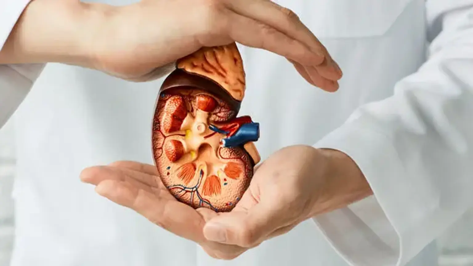
Hydronephrosis
Last updated date: 12-Mar-2023
Originally Written in English
Hydronephrosis
Hydroureter and hydronephrosis are common conditions seen in primary care, emergency medicine, and nephrology and urology clinics. Hydronephrosis is a condition in which the renal collecting system of one or both kidneys dilate and distend due to a blockage of urinary outflow distal to the renal pelvis (i.e., ureter, urinary bladder, and urethra). Hydroureter refers to ureter dilation caused by a blockage in the urinary outflow.
The urinary system is a complex, multi-component organ system whose major job is to maintain body homeostasis by regulating bodily fluid volume, electrolyte balance, and the excretion of metabolic end products through urine. It comprises the kidneys, ureters, urinary bladder, and urethra anatomically. Each kidney has an outer cortex and an inner medulla, which combine to produce renal pyramids that extend into the renal pelvis, where the ureter continues. Hydronephrosis and hydroureter can occur alone or together. They affect people of all ages. Acute or chronic, physiologic (particularly prevalent in pregnant women) or pathologic, unilateral or bilateral, the appearance might be acute or chronic.
Hydronephrosis Definition
Hydrostatic dilatation of the renal pelvis and calyces as a result of a blockage to urinary flow downstream is known as hydronephrosis. Alternatively, hydroureter refers to ureter dilation, while hydronephroureter refers to the complete upper urinary tract dilation (both the renal pelvicalyceal system and the ureter).
Epidemiology
The reasons and manifestations differ depending on the age range. The primary cause of birth defects in neonates and children is structural anomalies. Every year, 1 percent of the estimated 6 percent of total new births born with significant birth disorders have congenital malformations of the kidney and urinary tract. Hydronephrosis is becoming more common in newborns and babies, with the most common cause being obstruction of the ureteropelvic junction. In the United States, this occurs in up to 1 in every 100 babies born and is managed conservatively. By the age of two, the majority of cases had resolved spontaneously. Vesicoureteral reflux, which accounts for 15% to 20% of newborn and infantile hydronephrosis, is a less prevalent disease. Other common anatomical anomalies include obstruction of the ureterovesical junction, posterior urethral valves, and stricture.
In young adults, nephrolithiasis is the most frequent cause of hydroureteronephrosis. Kidney stones afflict over 700,000 adults in the United States each year, or nearly one in every twelve people. The prevalence of kidney stones in the United States is 9%, according to the National Health and Nutrition Examination Survey. It is 10 percent among men and 7 percent among women. Kidney stones are highly linked to the white race, overweight, and diabetes. The most common causes in the older population are prostate hyperplasia and neoplasia, pelvic and retroperitoneal malignancies, and kidney stones.
Hydronephrosis is a condition that affects up to 85% of pregnant women. The cause is assumed to be mechanical compression of the ureters due to the enlarged uterus and the effects of progesterone. It is most commonly seen in the second trimester and can last up to 8 to 12 weeks after delivery. If a patient develops discomfort and renal failure, ureteral stenting is the preferred treatment.
Due to pregnancy and gynecologic neoplasm, hydronephrosis is more prevalent in women between the ages of 20 and 60. It becomes increasingly frequent in males over the age of 60 as a result of prostate diseases and sequelae.
Hydronephrosis Pathophysiology
The blockage of urine before the renal pelvis causes hydronephrosis. The obstruction causes dilatation of the nephron tubules and flattening of the tubule lining within the kidneys, resulting in renal calyces enlargement.
Acute or chronic hydronephrosis can occur. In acute hydronephrosis, kidney function returns to normal. Chronic hydronephrosis, on the other hand, results in chronic kidney function reduction even when the obstruction is cleared.
The inability to transfer urine from the kidney to the bladder causes increased pressure within the kidney structures if a blockage occurs anywhere along the upper urinary system. Obstructing stones and ureteropelvic junction (UPJ) obstruction produced by intrinsic constriction of the ureters or an overlaying artery are two common causes of upper tract obstruction.
Increased pressure can also be caused by an obstruction in the lower urinary tract, which causes urine to reflux into the kidney. Bladder malfunction (such as the neurogenic bladder) and urethral blockage (such as posterior urethral valves in male newborns) are common reasons, as well as compression (such as prostatic hypertrophy in older male adults).
Causes of Hydronephrosis
Hydronephrosis can be caused by a variety of aberrant pathophysiological events. Hydronephrosis is caused by structural abnormalities of the junctions of the kidney, ureter, and bladder that can develop during fetal development. Although several of these congenital disorders have been recognized as inherited, the benefits of correlating genetic testing to early diagnosis have yet to be demonstrated. Injury, surgery, or radiation treatment could all result in structural problems.
Anatomical anomalies are the most frequent causes of hydronephrosis in children. Vesicoureteral reflux, urethral stricture, and stenosis are examples. Kidney stones are the most frequent cause of hydronephrosis in young adults. The most frequent cause of hydronephrosis in older persons are benign prostatic hyperplasia (BPH) and intrapelvic malignancies such as prostate cancer.
Other developmental problems that do not occur entirely during the fetal stage, such as an improperly situated vein, artery, or tumor, can also cause compression of one or both ureters. Due to the uterus's expansion, bilateral compression of the ureters can develop during pregnancy. Changes in hormone levels during this period may influence bladder muscle contractions, aggravating the problem further.
Kidney stones, blood clots, and retroperitoneal fibrosis are examples of obstructions that can occur due to a variety of factors.
The obstruction can develop anywhere from the urethral meatus to the renal calyces, and it can be partial or complete. Hydronephrosis can also be induced by the retrograde stream of urine from the bladder back into the kidneys (vesicoureteral reflux), which can be resulted from some of the factors listed above, as well as compression of the bladder outlet into the urethra due to prostate enlargement or fecal impaction in the rectum (which sits directly behind the prostate), as well as abnormal bladder detrusor muscular activity due to neurological impairment (neurogenic bladder) or other muscular diseases.
Symptoms of Hydronephrosis
Whether the blockage is acute or chronic, partial or total, unilateral or bilateral, the signs and symptoms of hydronephrosis vary. Acute hydronephrosis (as induced by a kidney stone) can cause extreme pain in the flank region (between the hips and ribs), which is known as renal colic. This type of discomfort has been referred to as "Dietl's crisis" in the past. Hydronephrosis that occurs slowly over time, on the other hand, usually causes either mild pain or no pain. Nausea and vomiting are other possible symptoms. Because of the distension of the bladder, a blockage at the urethra or bladder outlet can produce pain and pressure. When the flow of urine is blocked, it is more likely to develop urinary tract infections, which can lead to the formation of stones, fever, and blood or pus in the urine. If the obstruction is complete, it can lead to postrenal acute kidney injury (obstructive nephropathy).
Blood tests can reveal electrolyte imbalances including hyponatremia or hyperchloremic metabolic acidosis, as well as compromised kidney function (elevated urea or creatinine). Urinalysis may reveal an increased pH as a result of secondary nephron loss in the afflicted kidney, which reduces acid excretion. A palpable abdominal or flank bulge caused by an enlarged kidney may be detected during a physical examination in a slim patient.
Hydronephrosis Diagnosis
Hydronephrosis Ultrasound
Prenatal diagnosis is possible, and in fact, standard screening ultrasounds taken during pregnancy are used to discover the majority of instances in pediatric patients. However, over half of all prenatally detected hydronephrosis is temporary and disappears by the time the baby is born, while the remaining 16% has persistent hydronephrosis but is not linked with urinary tract blockage. For these children, reversal of the hydronephrosis occurs spontaneously, usually by age 3. A pathological disease can be found postnatally in the remaining 36% of patients with prenatal hydronephrosis.
Laboratory Tests
The patient's age, as well as whether the hydronephrosis was discovered accidentally or prenatally, and whether it is coupled with other symptoms, all influence the diagnostic workup. Blood tests (such as creatinine measurements) are frequently recommended; however, they must be interpreted with caution. Even with severe unilateral hydronephrosis, overall kidney function can be preserved because the unaffected kidney compensates for the blocked kidney.
Urinalysis is frequently done to check for the presence of blood (which is common in kidney stones) or infection indications (such as a positive leukocyte esterase or nitrite). Tubular stress and damage can potentially cause a decrease in concentrating ability or an increase in urine pH (distal renal tubular acidosis).
Imaging Studies
In addition to determining the presence and/or causation of hydronephrosis, imaging techniques such as an intravenous urogram (IVU), renal ultrasonography, CT, or MRI are important investigations. While ultrasonography can be used to visualize the ureters and kidneys (and diagnose hydronephrosis and/or hydroureter), an IVU can be used to pinpoint the anatomical origin of the obstruction. The results of antegrade or retrograde pyelography are identical to those of an IVU, but they also provide a treatment option. In hydronephrotic patients, real-time ultrasound scans and Doppler ultrasound examinations, in combination with vascular resistance testing, can help evaluate how a specific blockage affects urine functionality.
A Whittaker (or pressure perfusion) test can be used to establish the source of hydronephrosis. The collecting system of the kidney is entered percutaneously, and liquid is delivered at high pressure and a constant rate of 10 ml/min while the pressure within the renal pelvis is measured. The urinary collecting system is clogged if the pressure rises over 22 cm H2O. The bladder pressure is reduced from the initial measurements of internal pressure to arrive at this pressure measurement. Whittaker first published the test in the 1970s to see if patients with hydronephrosis that continues after the posterior urethral valves have been surgically corrected usually have ureters that are not obstructed, even if they are dilated.
A neonate with untreated in utero hydronephrosis should get renal ultrasonography within two days after birth. A renal pelvis more than 12mm in a neonate is regarded as abnormal and indicates substantial dilatation as well as probable abnormalities in the urinary system, such as blockage or morphological abnormalities.
The type of imaging to use is determined by the clinical situation (history, symptoms, and examination examination). The initial assessment in the event of renal colic (one-sided loin discomfort generally associated with a trace of blood in the urine) is typically a spiral or helical CT scan. This has the advantage of proving the function of the second kidney as well as determining whether there is any blockage in the flow of urine producing hydronephrosis. Because many stones are not visible on conventional X-ray or IVU, but about 100 percent of stones are apparent on CT, CT is increasingly being used as the first line of investigation. When there is a reason to prevent radiation exposure, such as during pregnancy, CT is not used.
Because many cases of prenatal hydronephrosis cure spontaneously, the first investigation to obtain for accidentally diagnosed prenatal hydronephrosis is postnatal renal ultrasonography. This is usually done during the first few days after delivery, while there is a chance that doing so early could miss some cases of moderate hydronephrosis due to a newborn's relative oliguria.
As a result, some specialists recommend doing a follow-up ultrasound at 5-6 weeks to lower the initial ultrasounds' false-negative rate. A voiding cystourethrogram (VCUG) is usually performed to rule for vesicoureteral reflux and anatomical anomalies like posterior urethral valves. Lastly, if hydronephrosis is severe and obstruction (such as a ureteropelvic junction or ureterovesical junction obstruction) is expected, a nuclear imaging investigation such as a MAG-3 scan is recommended.
Hydronephrosis Grade
The Society of Fetal Ultrasound (SFU) established a hydronephrosis grading system that was originally designed for use in neonatal and infant hydronephrosis, but is now used to grade adult hydronephrosis:
- Grade 0. There is no dilatation of the renal pelvis. In fetuses up to 32 weeks of gestational age, this signifies an anteroposterior diameter of less than 4 mm and 7 mm afterward. Different sources have proposed threshold values for renal pelvic dilatation in adults, with anteroposterior diameters ranging between 10- and 20-mm. Transverse pelvic diameter of above 10 mm is found in about 14% of normal healthy adults.
- Grade 1. Mild dilatation of the renal pelvis (anteroposterior diameter less than 10 mm in fetuses) without calyces dilatation or parenchymal atrophy.
- Grade 2. Moderate renal pelvis dilatation (between 10 and 15 mm in fetuses) with a few calyces.
- Grade 3. Renal pelvis dilatation with all calyces evenly dilated and renal parenchyma in its natural state
- Grade 4. As grade 3, but with renal parenchyma weakening.
Hydronephrosis Treatment
The management strategy for newborns with prenatal hydronephrosis is determined by the persistence of postnatal hydronephrosis, bilateral involvement, and the severity of the hydronephrosis.
Obstruction distal to the urinary bladder causes bilateral hydronephrosis. Bilateral hydroureteronephrosis is most commonly caused by posterior urethral valves. For a more accurate diagnosis, a voiding cystourethrogram should be performed. Severe hydronephrosis is defined as a renal pelvic diameter greater than 15 mm in a newborn. They have a higher chance of developing severe renal impairment. Mild to moderate instances usually go away by the age of 18 months.
Prenatal hydronephrosis increases the risk of pyelonephritis in infants, with a higher association in girls. There is no convincing evidence that ongoing antibiotic prophylaxis to prevent UTIs will benefit newborns with severe hydronephrosis.
The cause of hydronephrosis determines the therapy options. A high risk of renal impairment, a suspicion of infection leading to serious sepsis, and symptoms of severe pain, nausea, and vomiting indicate that hydronephrosis should be treated very soon.
When a lower urinary tract blockage at the bladder level is suspected, a urinary catheter should be placed. The installation of a ureteral stent guided by cystoscopy is a typical procedure for numerous intrinsic and extrinsic causes of ureteral hydronephrosis. When ureteral stent placement is contraindicated or impossible, interventional radiologists can use fluoroscopy to guide the installation of a percutaneous nephrostomy tube.
Extracorporeal shockwave lithotripsy is used to remove renal stones in the renal pelvis to avoid future hydronephrosis and blockage. Extrinsic compression from pelvic, retroperitoneal, and aortic aneurysms, among other things, necessitates surgery in some circumstances.
Hydronephrosis Prognosis
The prognosis of hydronephrosis is remarkably variable and depends on the underlying condition, whether one (unilateral) or both (bilateral) kidneys are affected, pre-existing renal function, the duration of hydronephrosis (acute or chronic), and whether the hydronephrosis took place in developing or mature kidneys.
Persistent hydronephrosis can cause permanent kidney injury due to compression of renal tissue and ischemia.
Unilateral hydronephrosis produced by an obstructing stone, for example, will most certainly dissolve after the stone passes, and the chances of recovery are great. Severe bilateral prenatal hydronephrosis (such as that caused by posterior urethral valves) on the other hand, will almost certainly have a bad long-term prognosis because blockage while the kidneys are developing causes irreversible kidney damage, even if the obstruction is alleviated postnatally.
Patient Education
Hydronephrosis can exist without causing any symptoms. When there is an acute obstruction, it might produce pain in the lower abdomen, back, and genitals. Patients with benign prostate enlargement may experience symptoms such as urinary frequency, difficulty urinating, a weak urine flow, and a feeling of incomplete emptying, depending on the site of the obstruction. Patients should be taught to seek medical help as soon as possible if they experience these symptoms.
To assist avoid recurring stone formation, patients with renal stones should be informed on diet modifications. A dietitian could be beneficial to these patients.
Improving Outcomes
Hydronephrosis and hydroureter are common in people of all ages and in all sorts of practices, and they are best addressed by a multidisciplinary team. Once an etiological component has been found on an ultrasound or CT scan, it is critical to identify it as soon as possible. It can be diagnosed asymptomatically in pregnant women and, if not treated appropriately, can result in severe kidney impairment. Nephrologists and urologists should be involved early in the diagnosing process to improve outcomes.
Due to the increased risk of infection, stent displacement, and other complications, patients will require constant monitoring after receiving a ureteral stent to the remove blockage. Nurses or care coordinators must coordinate the scheduling of follow-up appointments to ensure that stents are removed on time. Patients with bladder outlet obstruction should have clinic follow-ups for urine voiding tests. By employing management measures to avoid additional stone development, nephrologists can help prevent further occurrences of hydronephrosis in the context of renal stones. Dieticians can also help avoid stone formation.
Conclusion
Hydronephrosis is a condition in which a person's urine does not drain correctly from the kidneys. Treatment is usually determined by the underlying source of the problem.
Hydronephrosis rarely causes long-term health consequences if discovered and treated early. If hydronephrosis is not detected in ultrasonography before birth in a baby, the problem can be difficult to diagnose following birth. To assist recognize a prospective problem, people should look for signs of a UTI in babies.
Both babies and adults can benefit from early medical advice and treatment to promote a faster recovery.