Introduction
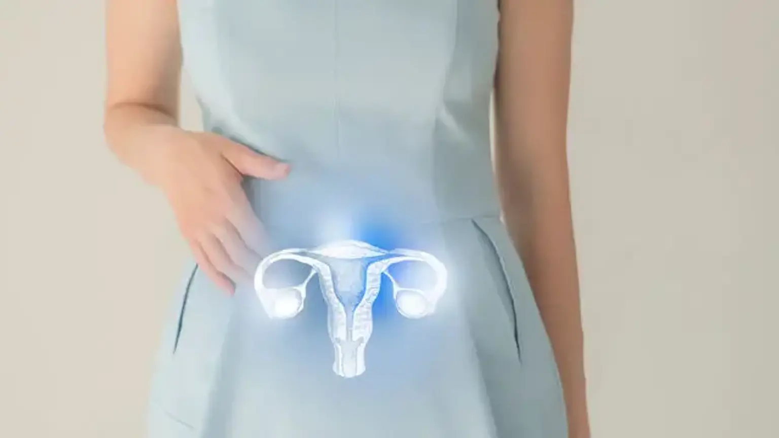
Hysteroscopy is a minimally invasive procedure used to examine the inside of the uterus. It involves the use of a thin, lighted tube called a hysteroscope, which is inserted through the cervix to view the uterine cavity. This diagnostic tool allows doctors to identify uterine abnormalities that could affect a woman's reproductive health.
In addition to diagnosing conditions like fibroids, polyps, and cancer, hysteroscopy can also be used to treat certain uterine conditions. It plays a crucial role in diagnosing the cause of abnormal bleeding, infertility, and recurrent miscarriages. As one of the most effective and precise diagnostic procedures, hysteroscopy has become increasingly popular worldwide for uterine health management.
How Hysteroscopy is Used for Uterine Diagnosis
Hysteroscopy is a highly effective procedure for diagnosing a variety of uterine conditions. When women experience abnormal bleeding, pain, infertility, or recurrent pregnancy loss, hysteroscopy can provide valuable insights into the underlying causes. By directly visualizing the uterine lining and other internal structures, the procedure helps doctors identify a wide range of uterine issues that may not be detected through traditional imaging techniques like ultrasounds.
Conditions such as uterine fibroids, polyps, endometrial cancer, and infections can be diagnosed with great precision using hysteroscopy. During the procedure, a hysteroscope is inserted into the uterus through the cervix, allowing the doctor to view the uterus in real-time on a monitor. This method is more accurate than imaging tests, enabling a comprehensive diagnosis that often includes biopsy samples for further analysis.
Common Uterine Conditions Diagnosed Through Hysteroscopy
Hysteroscopy is particularly valuable in diagnosing uterine conditions that can affect a woman’s fertility or overall health. Some of the most common conditions that can be diagnosed and treated with hysteroscopy include:
Fibroids: These benign tumors can cause heavy bleeding, pelvic pain, and fertility issues. Hysteroscopy helps doctors locate submucosal fibroids (those growing inside the uterine cavity) that may be causing these symptoms.
Polyps: Endometrial polyps, which are growths on the uterine lining, can lead to irregular bleeding and affect fertility. Hysteroscopy allows doctors to see and remove these polyps.
Uterine Septum: A congenital condition where the uterus is divided into two parts, which can lead to miscarriages or infertility.
Cancer: Hysteroscopy is instrumental in detecting uterine cancer, especially when abnormal cells are suspected in the uterine lining. It can provide biopsy samples for further examination.
By diagnosing these conditions early, hysteroscopy allows for timely treatment, which can improve reproductive outcomes and overall health.
The Procedure: What Happens During a Hysteroscopy?
The hysteroscopy procedure typically takes place in an outpatient setting, meaning patients can go home the same day. The procedure is done under local or general anesthesia, depending on the complexity of the case and the patient's comfort level.
To begin, the doctor gently inserts a speculum into the vagina to hold it open. The hysteroscope, a thin tube with a camera and light, is then inserted through the cervix and into the uterus. The camera allows the doctor to view the uterine cavity on a monitor in real-time. In some cases, the doctor may also inject a saline solution into the uterus to help improve visibility.
During diagnostic hysteroscopy, the doctor will assess the condition of the uterine lining, check for abnormalities such as fibroids, polyps, or infections, and take biopsies if needed. In therapeutic hysteroscopy, the doctor may perform minor surgical procedures, such as removing polyps or fibroids, to treat the condition.
The procedure usually lasts between 15 to 30 minutes, depending on the complexity of the case. Afterward, the patient may experience some cramping or spotting, but recovery is typically quick, with most women resuming normal activities within a day or two.
Why is Hysteroscopy Done?
Hysteroscopy is performed to diagnose and sometimes treat a variety of uterine conditions. It is particularly useful when a woman experiences unexplained symptoms like abnormal bleeding, infertility, or recurrent miscarriages. These conditions can often be traced to uterine abnormalities such as fibroids, polyps, or congenital issues like a uterine septum.
When doctors cannot pinpoint the cause through other diagnostic methods like ultrasounds or blood tests, hysteroscopy becomes the gold standard. It allows for a direct, clear view of the uterus, ensuring that doctors can diagnose conditions accurately and plan the appropriate treatment. Whether for women experiencing heavy bleeding, pelvic pain, or difficulties with conception, hysteroscopy provides essential insights into the health of the uterine lining and cavity.
Additionally, in cases of suspected uterine cancer or precancerous changes in the uterine lining, hysteroscopy offers the opportunity to take biopsies for histological examination, helping to identify malignant or abnormal growths.
Hysteroscopy for Abnormal Bleeding
One of the most common reasons for undergoing hysteroscopy is to investigate the cause of abnormal bleeding, such as heavy periods, bleeding between periods, or postmenopausal bleeding. These symptoms can often point to uterine conditions like fibroids, polyps, or endometrial hyperplasia (thickening of the uterine lining), which may require treatment.
During the procedure, the doctor uses the hysteroscope to examine the uterine cavity and look for any abnormalities in the endometrium (the uterine lining). If polyps or fibroids are found, they can often be removed during the same procedure. This provides immediate relief from the bleeding, and the patient typically experiences a reduction in symptoms afterward. In some cases, a biopsy may be taken to rule out cancer or other serious conditions.
By identifying the underlying cause of abnormal bleeding, hysteroscopy allows for targeted treatments that can improve quality of life and reproductive health.
Hysteroscopy for Uterine Fibroids
Uterine fibroids are non-cancerous tumors that grow in or on the uterus. They can range in size and number, and while many women have fibroids without experiencing symptoms, others suffer from heavy menstrual bleeding, pelvic pain, and infertility.
Hysteroscopy is an excellent tool for diagnosing fibroids, especially those located inside the uterine cavity (submucosal fibroids). These types of fibroids are more likely to interfere with a woman's ability to conceive or cause significant bleeding. During the hysteroscopy procedure, the doctor can directly visualize the fibroids, assess their size and location, and, if necessary, remove them in the same procedure.
The ability to treat fibroids during the diagnostic process is one of the key benefits of hysteroscopy. For women struggling with fibroid-related symptoms, hysteroscopic myomectomy (removal of fibroids) can provide significant relief, improve fertility outcomes, and restore normal menstrual cycles.
Hysteroscopy for Uterine Polyps
Uterine polyps are growths that develop on the inner lining of the uterus. These benign growths can cause irregular bleeding, and in some cases, they may interfere with fertility. Women who experience abnormal bleeding or difficulty conceiving may have uterine polyps, which can be detected and removed through hysteroscopy.
The hysteroscope provides a clear view of the uterine lining, allowing doctors to identify and biopsy any polyps present. In many cases, the polyps can be removed during the procedure, leading to a significant reduction in bleeding and an improved chance of conception. By diagnosing uterine polyps early, hysteroscopy helps women avoid unnecessary treatments and offers a minimally invasive solution for both diagnosis and treatment.
Furthermore, polyps can sometimes be a sign of endometrial hyperplasia, which can lead to more serious conditions like endometrial cancer. In such cases, hysteroscopy provides crucial diagnostic information that guides treatment decisions.
Role of Hysteroscopy in Diagnosing Uterine Cancer
Uterine cancer, especially endometrial cancer, is one of the most common cancers affecting women. It typically presents with abnormal vaginal bleeding, particularly in postmenopausal women, but can sometimes be overlooked or misdiagnosed. Hysteroscopy plays a critical role in detecting uterine cancer or precancerous changes in the uterine lining, often in cases where the cause of bleeding cannot be identified through other diagnostic methods.
During a hysteroscopic procedure, the doctor can directly view the endometrium and assess any suspicious areas that might be indicative of cancerous or pre-cancerous changes. If any abnormalities are detected, a biopsy can be taken for further analysis, allowing for a definitive diagnosis. Early detection of uterine cancer is crucial because it significantly improves the chances of successful treatment.
In addition to its diagnostic role, hysteroscopy also helps guide surgical procedures, such as the removal of cancerous tissue, ensuring that the procedure is as precise as possible.
Infertility and Hysteroscopy
Infertility can be caused by many factors, including structural issues in the uterus. Conditions like uterine fibroids, polyps, or congenital uterine abnormalities can prevent a woman from becoming pregnant or carrying a pregnancy to term. Hysteroscopy is an invaluable tool in diagnosing these conditions, as it allows for direct visualization of the uterine cavity.
For women who have experienced recurrent miscarriages or are unable to conceive, hysteroscopy can help uncover underlying uterine issues that might be hindering their fertility. Conditions like a uterine septum, which is a congenital abnormality, can be diagnosed and treated during the procedure. Additionally, fibroids or polyps that affect the uterine lining can be removed to improve the chances of a successful pregnancy.
The ability to perform both diagnosis and treatment during the same procedure makes hysteroscopy an effective and efficient option for women struggling with infertility.
Hysteroscopic Treatment Options
Hysteroscopy is not only used for diagnosing uterine conditions but also for treating many of them. One of its main advantages is its minimally invasive nature, allowing doctors to treat conditions like fibroids, polyps, and septums without the need for large incisions or lengthy hospital stays.
Fibroid Removal: For submucosal fibroids that are causing symptoms like heavy bleeding or infertility, hysteroscopy offers a method for removing the fibroids using specialized instruments.
Polyp Removal: Polyps can be removed directly during the procedure, preventing further bleeding and improving fertility outcomes.
Septum Resection: A uterine septum, a condition where the uterus is divided by a wall of tissue, can be surgically corrected during hysteroscopy, improving the chances of pregnancy.
These treatments can often be performed in an outpatient setting, meaning that women can go home the same day. This significantly reduces recovery time compared to more invasive surgeries. Hysteroscopy offers a quicker, less painful solution with minimal downtime, making it a highly appealing option for many women.
Recovery After Hysteroscopy
The recovery time following hysteroscopy is generally short, as it is a minimally invasive procedure. Most women can resume their normal activities within a day or two, though some may experience mild cramping, spotting, or light bleeding for a few days after the procedure. These symptoms are typically mild and should subside within a few days.
For women who underwent a more extensive procedure, such as the removal of large fibroids or polyps, recovery may take a bit longer. However, the majority of patients are able to return to work and other routine activities within a few days. It's important to avoid strenuous activities, including heavy exercise and sexual intercourse, for at least one to two weeks to ensure proper healing.
Your doctor will provide specific aftercare instructions and will schedule a follow-up appointment to ensure that healing is progressing as expected. In most cases, any discomfort after the procedure is easily managed with over-the-counter pain medications.
Risks and Safety Considerations
Like any medical procedure, hysteroscopy carries some risks, although they are relatively rare. Most women tolerate the procedure well, but it’s essential to understand the potential complications to make an informed decision.
Some of the possible risks include:
Infection: While infections are uncommon, they can occur after any surgical procedure. Following proper hygiene and aftercare instructions significantly reduces the risk.
Perforation: In rare cases, the hysteroscope may accidentally puncture the uterine wall. This can often be managed quickly, but it may require additional procedures to correct.
Heavy Bleeding: Some bleeding after the procedure is normal, but excessive bleeding may require further treatment.
Anesthesia Risks: As with any procedure requiring anesthesia, there are risks associated with sedation, particularly for women with certain health conditions.
To minimize these risks, it’s important to have the procedure done by an experienced doctor in a medical facility that is equipped to handle any potential complications. Additionally, women should discuss their medical history with their healthcare provider to ensure that they are good candidates for the procedure.
Success Rates of Hysteroscopy
Hysteroscopy is considered highly successful in diagnosing and treating a variety of uterine conditions. For conditions like fibroids, polyps, and septums, hysteroscopy offers an effective method for both diagnosis and treatment, with high success rates for improving symptoms and fertility outcomes.
For women suffering from uterine fibroids, a study has shown that hysteroscopic myomectomy (the removal of fibroids) can significantly reduce symptoms like heavy bleeding and pelvic pain. In cases of infertility, removing uterine fibroids or correcting anatomical abnormalities, such as a uterine septum, can improve pregnancy rates.
The success rate of hysteroscopy varies depending on the specific condition being treated, but overall, the procedure is associated with positive outcomes, minimal recovery time, and reduced complications compared to more invasive surgeries.
Global Popularity and Accessibility of Hysteroscopy
Hysteroscopy has gained significant popularity worldwide due to its effectiveness, minimal invasiveness, and ability to treat various uterine conditions in a single procedure. It is now routinely used in many countries as a first-line approach for diagnosing and treating conditions like abnormal bleeding, fibroids, polyps, and infertility-related issues.
In developed countries, hysteroscopy is widely accessible, with many outpatient clinics offering the procedure as a safe and efficient alternative to traditional surgeries. As awareness of the procedure grows and technology advances, it is becoming more accessible in developing nations as well, offering women worldwide the chance to benefit from its diagnostic and therapeutic potential.
Hysteroscopy’s ability to provide accurate, real-time diagnosis while treating conditions in a single session makes it a globally popular choice for both physicians and patients.
Frequently Asked Questions (FAQs)
1. Is hysteroscopy painful?
Most women experience only mild discomfort during a hysteroscopy, especially if local anesthesia is used. The procedure is typically well-tolerated, and any discomfort usually subsides after a few hours. In some cases, general anesthesia may be used, in which case the patient will be asleep during the procedure.
2. How long does the procedure take?
The actual procedure usually lasts between 15 to 30 minutes, depending on whether additional treatments, like fibroid or polyp removal, are performed.
3. How soon can I get pregnant after a hysteroscopy?
If you had a diagnostic hysteroscopy with no treatment, you can usually try to conceive as soon as your doctor clears you, which is typically within a few days. For women who have undergone a therapeutic hysteroscopy, such as the removal of fibroids or polyps, it’s best to wait for a few months before trying to conceive to allow the uterus to heal fully.
4. Are there any risks associated with hysteroscopy?
While hysteroscopy is generally safe, there are some risks, including infection, bleeding, and uterine perforation. These risks are low, and the procedure is usually well-tolerated. Your doctor will discuss these risks with you beforehand to ensure you are fully informed.
5. Will my insurance cover hysteroscopy?
Hysteroscopy is typically covered by most insurance plans when performed for diagnostic purposes or to treat medical conditions like fibroids, polyps, or infertility. However, coverage may vary, so it’s important to check with your insurance provider to understand your specific benefits.
Advances in Hysteroscopy Technology
Hysteroscopy has evolved significantly over the years, with advancements in technology making the procedure even more precise and less invasive. Modern hysteroscopes are equipped with high-definition cameras, allowing for a clearer view of the uterine cavity. Some newer models are also more flexible, enabling easier navigation through the cervix without causing trauma.
In addition to better imaging, innovations in instrumentation have made it possible to perform complex procedures, such as the removal of large fibroids or the resection of a uterine septum, with minimal discomfort and faster recovery times. The use of saline solution to expand the uterus (hysteroscopic distension) also allows for clearer visualization, ensuring that abnormalities are detected more easily.
These technological improvements have made hysteroscopy a preferred choice for both diagnosis and treatment, offering safer, more efficient outcomes for patients.
Emotional and Psychological Impact of Hysteroscopy
While hysteroscopy is a relatively quick and straightforward procedure, the emotional and psychological impact on patients should not be overlooked. For many women, undergoing a uterine procedure can be anxiety-inducing, especially if they are experiencing symptoms such as abnormal bleeding or infertility, which can already cause stress.
It’s important for healthcare providers to offer emotional support, address any concerns, and explain the procedure thoroughly before the patient undergoes it. Reassurance about the minimal invasiveness and short recovery time can help ease anxiety. After the procedure, most women experience relief from their symptoms, leading to improved emotional well-being. In cases where fertility is a concern, successful outcomes from hysteroscopic treatments can also have a profound positive impact on mental health.
Cost Considerations of Hysteroscopy
The cost of hysteroscopy can vary depending on factors such as location, the complexity of the procedure, and whether additional treatments are performed. On average, hysteroscopy can range from $1,500 to $5,000 in many countries, though this figure may be higher for complex cases that require surgical intervention.
Insurance typically covers hysteroscopy when performed for medical reasons such as the treatment of fibroids, polyps, or infertility. However, it's important for patients to check with their insurance provider to confirm coverage details. Some healthcare facilities also offer payment plans or financing options to help manage costs.
While the upfront cost may seem high, hysteroscopy is a minimally invasive procedure that can save money in the long term by reducing the need for more invasive surgeries or lengthy hospital stays.
Conclusion
Hysteroscopy has proven to be a valuable tool in diagnosing and treating a range of uterine conditions, from fibroids and polyps to infertility and abnormal bleeding. With its minimally invasive nature, high success rates, and ability to treat multiple conditions in one procedure, it continues to grow in popularity and accessibility worldwide.
As technology advances, hysteroscopy is becoming even more refined, allowing for more complex surgeries with quicker recovery times. This makes it an attractive option for women seeking effective treatment with minimal disruption to their lives.
Looking forward, ongoing research and development in hysteroscopy technology are likely to improve its precision, further enhancing patient outcomes and broadening its global reach. With increasing awareness and accessibility, hysteroscopy is set to remain a cornerstone of uterine care, offering hope and relief for many women across the globe.