Image guided radiotherapy (IGRT)
Overview
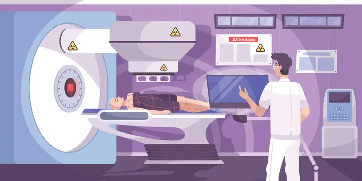
IGRT, or image guided radiation therapy, is a procedure that uses regular imaging to improve accuracy during a patient's radiation treatment. Imaging equipment can be integrated within the machine that generates high-energy radiation beams (linear accelerator) or installed nearby in the treatment room. Modern technologies, such as IGRT, are critical to generating ongoing improvements in patient outcomes and quality of life.
What is Image-guided Radiation Therapy (IGRT)?
The use of imaging during radiation therapy to increase the precision and accuracy of treatment delivery is known as image-guided radiation therapy (IGRT). IGRT is used to treat malignancies in moving parts of the body, such as the lungs. Radiation therapy devices include imaging technology that allows your doctor to see the tumor before and during treatment.
When these pictures are compared to the reference images obtained during simulation, the patient's posture and/or the radiation beams can be modified to more accurately target the radiation dosage to the tumor. Some IGRT techniques may include fiducial markers, ultrasound, MRI, x-ray imaging of bone structure, CT scan, 3-D body surface mapping, electromagnetic transponders, or colored ink tattoos on the skin to help orient and target the radiation equipment.
If you are going to get IGRT, your doctor will most likely utilize CT scanning to simulate the therapy and provide reference pictures. Other imaging methods, such as an MRI or PET scan, may be used to help pinpoint the precise form and location of your tumor, and a specific gadget may be developed to assist you in maintaining the same exact position throughout each treatment. Depending on the sort of exam, your doctor will give you precise instructions.
In IGRT, radiation-delivery machines, such as a linear accelerator (for x-ray or photon) or cyclotron/synchrotron (for proton), are outfitted with special imaging technology that allows the physician to image the tumor immediately before or even during the delivery of radiation, while the patient is positioned on the treatment table. These photos are compared to the reference photographs captured during simulation using specialist computer software. The patient's posture and/or radiation beams are adjusted as needed to more accurately target radiation at the tumor while avoiding healthy surrounding tissue.
IGRT may be performed using computed tomography (CT), magnetic resonance imaging (MRI), ultrasound (US), and x-ray imaging to visualize bone or soft-tissue architecture. Other IGRT approaches involve placing markers on the patient's body surface or implanting markers into the patient's body.
IGRT is used to treat tumors that are prone to movement, such as the lungs (influenced by breathing), liver, pancreas, and prostate gland, as well as cancers that are adjacent to vital organs and tissues. It is frequently used in conjunction with advanced modes of high-precision radiotherapy such as intensity-modulated radiation therapy (IMRT), proton beam therapy, stereotactic radiosurgery, or stereotactic body radiotherapy (SBRT), which use computer-controlled x-ray accelerators to deliver precise radiation doses to a malignant tumor or specific areas within the tumor.
Who will be involved in this procedure?
Radiation therapy is delivered by a treatment team that includes a radiation oncologist, therapeutic medical physicist, dosimetrist, and radiation therapists. The radiation oncologist is a doctor that assesses the patient and decides on the best therapy or combination of therapies, as well as the kind of IGRT. The doctor decides which location to treat and what dose to provide. The radiation oncologist, in collaboration with the therapeutic medical physicist and the dosimetrist, selects which procedures will be used to give the recommended dose.
Following that, the physicist and dosimetrist do thorough therapy calculations. Radiation therapists are highly skilled techs who collect pictures and provide daily treatments. The radiation oncology nurse evaluates the patient and offers more information about the therapy and any side effects. In consultation with the physician, the radiation oncology nurse also manages any responses or side effects from therapy that may arise.
How does IGRT help?
Cancers may shift somewhat in some places of the body during or between treatments. For example, the prostate gland changes position depending on whether or not the bladder is full. You may also sleep in a little different position every day. As a result, there is a chance that some of the cancer will be beyond the treatment region (radiotherapy field) during certain treatment sessions.
Having scans and x-rays taken right before your radiographers turn on the radiation treatment beam ensures that you are in a position very near to your planned scan.
If your radiographers see that your position is quite similar each day after the first few photographs, they may not need to take images every day.
- 4D-RT
Images are taken throughout your treatment with 4D-RT. Using the information from these photos, the radiotherapy machine may then alter the position of the couch.
If the tumour travels out of the radiation field, the machine in some versions of 4D-RT may turn off. Cancers in regions of the body that move while we breathe, such as the lungs, benefit from this form of therapy. When the cancer returns to the proper place, the radiation beam is turned back on. The radiation machine is constantly under the direction of your radiographers.
When do doctors recommend IGRT?
IGRT is frequently utilized to treat tumors that are adjacent to delicate tissues or organs in the body. It's particularly beneficial for tumors that are prone to moving during or between treatments, such as lung tumors, which might be influenced by breathing.
IGRT is most commonly used to treat lung cancer, breast cancer, prostate cancer, brain cancer, spine cancer, bladder cancer, esophageal cancer, liver cancer, bone cancer, and lymphoma.
What equipment is used?
Imaging equipment, such as a linear accelerator, is installed on or incorporated into the unit that administers radiation in IGRT. Imaging equipment may be installed in the treatment room as well. X-rays, computed tomography (CT), 3-D body surface mapping, magnetic resonance imaging (MRI), and ultrasound (US) are among the imaging modalities employed in IGRT. IGRT is sometimes accomplished via a detector in the room that measures motion by localizing markers on a patient's surface, or by electromagnetic transponders inserted within the patient.
Is there any special preparation needed for the procedure?
Women should always notify their doctor or technician if they suspect they are pregnant or if they are nursing their child. If MRI is utilized for simulation or IGRT, patients with pacemakers or loose metal in their body should notify their treatment team.
For some IGRT treatments, extremely small markers known as fiducial markers or, in rare situations, electromagnetic transponders may be implanted within the body near or in the tumor to assist the treatment team in identifying the region. They are typically implanted at least one week before the first radiation therapy session. To assist orient and target the radiation equipment, the patient's skin may be tagged or tattooed with colored ink. Patients with prostate cancer who receive IGRT with ultrasound must drink enough water approximately an hour before each treatment to maintain their bladder full so that the ultrasound equipment can image or "see" the prostate.
How is the procedure performed?
The patient is carefully positioned at the start of each radiation therapy session, guided by the markings on the skin indicating the treatment region. Devices may be utilized to assist the patient in maintaining the correct posture. Imaging equipment incorporated within the radiation delivery unit or installed in the treatment room is then used to capture images.
Some IGRT treatments necessitate patients holding their breath for 30 to 60 seconds. If IGRT requires fiducial markers or electromagnetic transponders to be implanted within the body, they will be placed with a needle about a week before the simulation procedure.
Depending on the kind of IGRT employed, an x-ray, CT scan, or ultrasound will be acquired prior to therapy on each treatment day. To make position modifications, the physicians or radiation therapist evaluate the photos and compare them to the reference images produced during simulation. The patient could be moved, and further imaging might be done. Radiation therapy is administered after any required changes are made to match the patient's reference placement.
Each radiation treatment session is likely to take longer due to the image-guidance procedure.
What will I feel during and after this procedure?
Prior to or during radiation therapy sessions, imaging is painless. During the radiation treatment session, you may notice or hear equipment moving about you as the imaging technique is being performed. During treatment, patients may notice an unusual odor created by the ozone produced by the linear accelerator. When receiving therapy, some patients may perceive a colorful light; this is especially true for people having their brain treated.
Radiation therapy can have unintended consequences. These issues might be caused by the treatment itself or by radiation damage to healthy cells in the treated region. The frequency and severity of adverse effects will vary depending on the type of radiation used, the dose, and the body area being treated. Inform your doctor and/or nurse so that they can assist you in managing them.
Radiation can have both immediate and delayed negative effects. Early side effects occur during or immediately following therapy. They usually vanish within a few weeks. Fatigue and skin issues are common early adverse effects. The treatment area's skin may become sensitive, red, irritated, or swollen. Dryness, itching, peeling, and blistering are some of the other alterations.
Depending on the area being treated, other early side effects may include:
- Hair loss in the treatment area
- Mouth problems and difficulty swallowing
- Eating and digestion problems
- Diarrhea
- Nausea and vomiting
- Headaches
- Soreness and swelling in the treatment area
- Urinary and bladder changes
Late side effects may occur months or years following treatment. While they are often permanent, they are rare. They include:
- Brain changes
- Spinal cord changes
- Lung changes
- Kidney changes
- Colon and rectal changes
- Infertility
- Joint changes
- Lymphedema
- Mouth changes
- Secondary cancer
Radiation treatment carries a small chance of acquiring cancer. After treatment, your radiation oncologist will monitor for problems and recurring or new malignancies on a regular basis.
Imaging specialists, using techniques like as IGRT, are increasing radiation treatment's cancer-destroying powers while limiting its influence on healthy tissues and organs, as well as the treatment's own negative effects.
Conclusion
Image guided radiation therapy (IGRT) is one of the most cutting-edge advances in cancer treatment. It also allows for the use of specialized radiotherapeutic procedures with small safety margins near radiosensitive organs. For these reasons, IGRT may be advised for treating tumors that are adjacent to essential and sensitive organs like the heart and kidneys. Image guided radiation treatment can also be used to treat malignancies in regions of the body that move, such as the lungs, prostate, and liver.
Proper patient selection for IGRT must include treatment goals, planning features, and available technical and human resources.