Implantable cardioverter-defibrillator (ICD)
Overview
What is the Implantable Cardioverter Defibrillator?
The implantable cardioverter defibrillator (ICD) is a life-saving device that prevents sudden cardiac death in high-risk patients such as those with severe left ventricular systolic dysfunction (LVSD) and those with specific structural heart diseases such as hypertrophic obstructive cardiomyopathy (HOCM), sarcoidosis, and others. ICDs have been implanted in millions of people worldwide since the first human implant in 1980.
Clinical trials have contributed to the development of guidelines for implantable cardioverter defibrillator insertion in the primary and secondary prevention of sudden cardiac death. Recent trials have also explored and compared different programming ways to reduce unwanted shocks and increase ICD patient survival.
Mechanisms of ICD shock
How the implantable cardioverter defibrillator works?
Sudden cardiac death is frequently caused by ventricular arrhythmias, which are more common in LVSD patients. Implantable cardioverter-defibrillators are used to detect and treat these dangerous ventricular arrhythmias, frequently using high-energy shocks for defibrillation.
The patient gets a sudden intracardiac shock, similar to external defibrillation. An ICD shock has been described by patients as "an earthquake," "being hit by a vehicle," or "being kicked by a mule."
Given the traumatic nature of ICD shocks, it would be ideal if the implantable cardioverter defibrillator could always discriminate between ventricular arrhythmias and non–life-threatening tachyarrhythmias and only deliver shocks for VT or ventricular fibrillation (VF).
Unfortunately, the algorithms that distinguish VT or VF from less dangerous arrhythmias have not been developed in practice. Furthermore, many ICD patients—as many as one in three in some studies—receive incorrect shocks. When the device provides a high-voltage discharge for a reason other than a ventricular arrhythmia, inappropriate shocks occur. As a result, while the primary function of the implantable cardioverter defibrillator is to detect ventricular arrhythmias and give treatments to restore normal sinus rhythm, this advantage comes at a cost to patients who get unnecessary shocks for non-life-threatening rhythms and other causes.
Periprocedural workup
What are the requirements of the implantable cardioverter defibrillator surgery?
Laboratory studies
Platelet counts (preferred: >50×103 per μL) and the international normalized ratio (INR) is generally used to determine the risk of bleeding. However, continuous warfarin usage has been proved to be safe and has become standard practice.
Some professionals would evaluate a diabetic patient's hemoglobin A1c level prior to implantation to verify their condition is adequately managed and to limit the risk of infection.
Anesthesia
ICD implantation is often performed under a mix of local anesthetic and conscious sedation. Intravenous (IV) midazolam and fentanyl can be used to induce conscious sedation. In severely uncooperative or high-risk patients, general anesthesia may be required. The operation typically takes 30 to 90 minutes to perform.
Local anesthetic with lidocaine and conscious sedation are sufficient for the vast majority of patients. The skin and subcutaneous tissue should be anesthetized using a local anesthetic. With one or two injections and needle repositioning, the entire region can be properly anesthetized. Repeated skin penetration with the anesthetic needle should be avoided since it raises the danger of introducing microorganisms.
Consideration should be given to general anesthesia in certain patient populations, such as pediatric patients, patients undergoing subpectoral implantable cardioverter defibrillator (ICD) insertion, and those requiring lead tunneling.
Positioning
Because of the close location of the incision, the patient's hair should be restrained with a surgical hat. To avoid skin irritation and the creation of a route for pathogens, the chest should be shaved using a clipper rather than a razor.
The patient should be lying down. In rare circumstances, left arm extension may be required to allow for the installation of a submammary device. Patches of external defibrillator should be put anteriorly and posteriorly.
The implantable cardioverter defibrillator insertion site should be washed with detergent solution, dried, and then treated with chlorhexidine-based solution; this may improve faster healing following ICD implantation and reduce bacterial activity. Patients should also wash their entire body (including their hair) with a cleaning solution 24 hours before implantation.
Implantation
An incision is made below the collarbone for the surgery. The leads will be inserted into the heart via a vein. One lead will be inserted into the right ventricle and another into the right atrium.
After the procedure
A chest radiograph is required to ensure that the leads are in the appropriate location and that the lung has not been harmed. Patients frequently spend the night in the hospital after the implantable cardioverter-defibrillator surgery.
Equipment
What are the tools of implantable cardioverter-defibrillator surgery?
The following equipment is required in the insertion of an implantable cardioverter-defibrillator (ICD):
- Surgical tray
- Fluoroscope
- Peelable hemostatic sheath(s)
- External defibrillator
- ICD pulse generator and lead(s)
- Pacing cable(s)
- Pacing system analyzer (PSA)
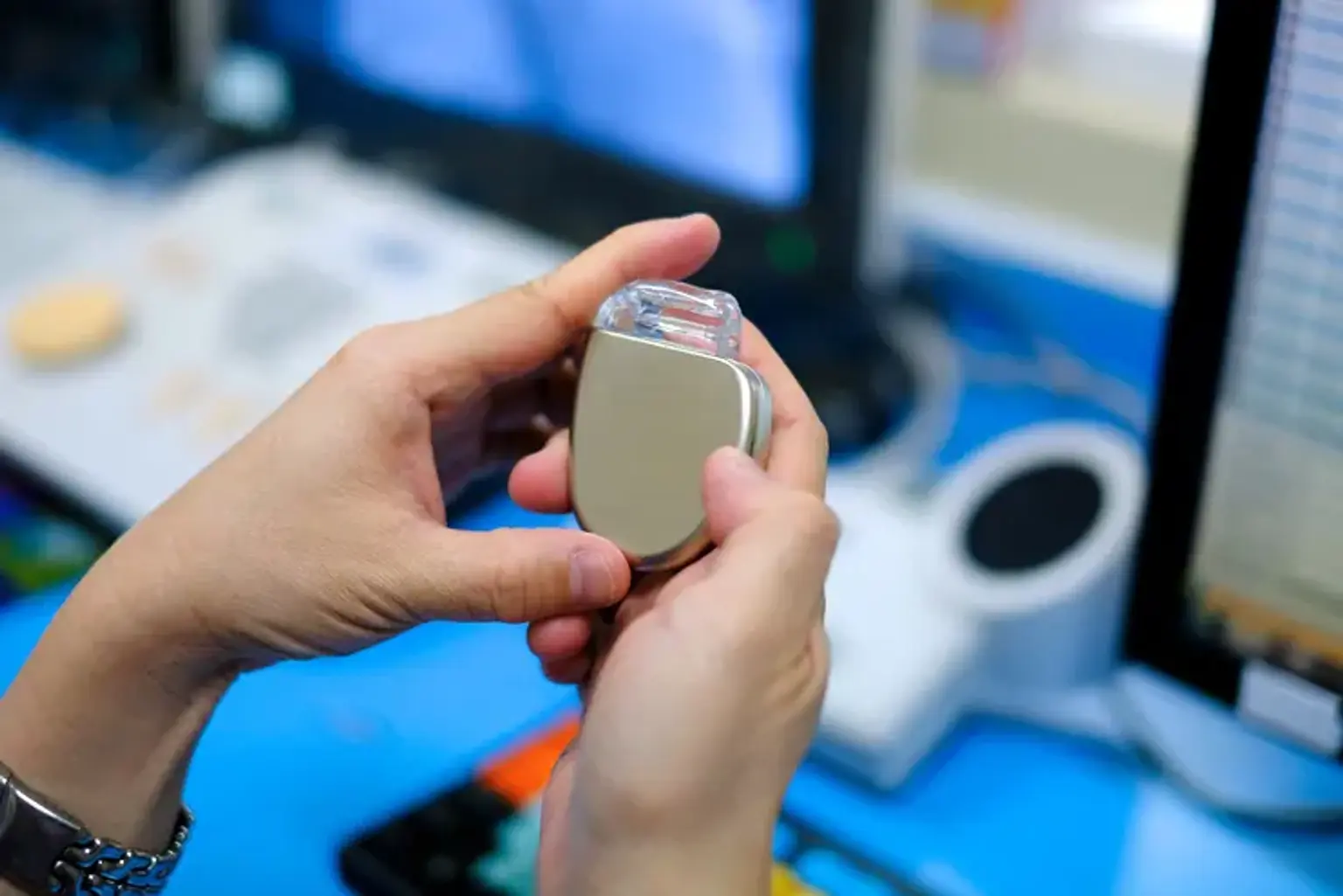
ICD systems consist of a pulse generator and pacing leads. Endocardial leads are inserted transvenously and advanced to the right ventricle, where they are implanted into the myocardial tissue. The pulse generator is implanted in the chest wall, either subcutaneously or submuscularly.
On September 28, 2012, the US Food and Drug Administration approved the first subcutaneous implantable cardioverter defibrillator for ventricular tachyarrhythmias, which allowed the lead to be placed under the skin rather than through a vein into the heart.
With no leads inside the heart, a subcutaneous implantable cardioverter defibrillator (S-ICD) can be implanted beneath the skin. It is attached to the rib cage just below the armpit or the left axilla. The lead is inserted through a tube beneath the skin. Because the lead is not directly in the bloodstream, this approach has a lesser chance of infection. However, the device is bigger than a transvenous device.
Technique
What are the steps of implantable cardioverter defibrillator surgery?
The insertion of an implantable cardioverter defibrillator (ICD) is a minimally invasive surgery. Transvenous access to the ventricle and atrium is normally conducted under local anesthetic. Access is usually gained by the subclavian vein, cephalic vein, femoral vein, or, in rare cases, the internal jugular vein. The procedure might take place in either the cardiac catheterization facility or the operating room.
Incision
Depending on the patient's handedness, the skin incision is commonly done in the right or left infraclavicular region. In general, the implantable cardioverter defibrillator (ICD) should be placed on the side opposite the patient's dominant hand. Other factors to consider while deciding where to place the implant are as follows:
- Previous mastectomy or lymph node resection: Such sites should be avoided.
- Recreational activities: In hunters, for example, the site on which the gun butt rests should be avoided for implantation.
- Existing cardiac devices: When an upgrade to a defibrillator is planned, the site where the previous device was implanted is preferred, provided that the venous circulation is sufficiently patent to allow lead placement.
The location of the incision is heavily influenced by the anticipated vascular access. If a cephalic vein cutdown is planned, an incision in the deltopectoral groove may be preferable for easier vein visibility and the formation of a subpectoral pocket with minimum hemorrhage. If axillary vein access is desired, the incision should be guided fluoroscopically by an examination of the first and second rib positions.
In patients who already have a cardiac device in place, it is critical to ensure that the incision allows simple access to vascular structures and is not too far away from the initial system.
Pocket creation:
Subcutaneous
A 5-7 cm long incision is created and continued down to the subcutaneous tissue to create a subcutaneous pocket. Electrocauterization is used to produce hemostasis, with care given to avoid any skin burns that might impair the healing process. Electrocauterization, blunt dissection, or both are used to deepen the dissection to the prepectoral fascia. It should not extend beyond the prepectoral fascia; doing so frequently leads in pectoralis muscle hemorrhage.
Electrocautery is utilized to generate a new plane in the inferior portion of the incision once the incision has been carried down to the prepectoral fascia. The pocket for the device is then formed using a mix of electrodissection and blunt dissection with the fingers. To prevent device migration on the lateral side of the chest, the pocket should be oriented obliquely medially. The size of the pocket is determined mostly by the size of the device to be used. Once the pocket has been created and hemostasis achieved, attention is turned toward obtaining vascular access.
Subpectoral
If feasible, cephalic vein access is favored for lead insertion in a subpectoral pocket; the incision is performed in the deltopectoral groove, and the cephalic vein is clearly visible. To avoid harm to the cephalic vein, the incision is continued down to the prepectoral fascia and the deltopectoral groove, generally first with cautery and then by blunt dissection. After that, the cephalic vein is separated and secured.
Following that, the lateral edge of the deltopectoral muscle is pulled and gently separated from the pectoralis minor using blunt dissecting scissors. At this time, blunt dissection with fingers is also an option. hemostasis must be carefully monitored.
After the subpectoral pocket has been formed, the focus shifts to establishing vascular access.
Insertion of Pacing and Defibrillation Lead(s):
There are several types of defibrillation leads available. Active fixation leads, which allow for controlled insertion in the intraventricular septum, are favored. Passive fixation leads, on the other hand, need a more apical location, increasing the risk of ventricular perforation and the problems that come with it. Other components of the lead design, such as the sensing circuit and the presence or absence of a proximal defibrillation coil, must also be addressed.
The pace-sensed portion of the lead either permits sensing from the lead tip to the dedicated ring in close proximity to the distal defibrillation coil or offers a larger sensing vector between the lead tip and the distal (right ventricular) defibrillation coil.
The defibrillation leads may have two defibrillation coils, with the distal coil inserted at the right ventricular apex and the more proximal coil commonly running from the junction of the high right atrium to the superior vena cava (SVC), or they may simply have a single distal coil.
The single-coil defibrillation device offers several advantages, including improved future extractability due to decreased adhesion and scarring in the area of contact between the proximal coil and the right atrium/SVC junction. Single-coil defibrillation leads also take up less space in the SVC (thus reducing the risk of venous obstruction) and are easier to implant in patients who already have many pacing leads. For these reasons, single-coil leads are preferable.
Finally, one of the defibrillation lead types has a polytetrafluoroethylene (PTFE) covering on the defibrillation coils, which may reduce tissue ingrowth and collision artifact when compared to conventional pacing and defibrillation leads.
Placement of the lead(s)
The form and thickness of the stylet determine the steerability of the lead. At the same time, keep in mind that inserting even the softest stylet into the lead greatly increases the force transferred via the lead; hence, extreme caution is required at all times during lead manipulation to avoid heart perforation.
The stylet must be bent to allow passage through the tricuspid valve. Such curve is typically not difficult to navigate via the venous system and the right atrium. If there is a problem, a straight stylet can be implanted and the lead progressed to the right atrium. If the venous system is tortuous or if many leads are already present, a hydrophilic wire should be put first, followed by a lengthy peel-away sheath to allow for simple lead mobility.
To confirm that the lead is in the right ventricle, it is advanced to the right ventricle and then to the right ventricular outflow tract (RVOT). The lead will be placed most anteriorly above the heart silhouette in the right anterior oblique (RAO) projection in this case. This guarantees that the lead did not mistakenly pass a patent foramen ovale (PFO) or a ventricular septal defect (VSD) and, eventually, into the left ventricle.
At this point, the stylet is withdrawn and a soft stylet with 135° angulation at the distal 2-3 cm is inserted into the lead. As the lead is gently removed from the RVOT, counterclockwise torque is maintained on the stylet, enabling it to sink to the bottom region of the right ventricle. The lead is then slowly advanced while maintaining counterclockwise rotation to facilitate septal placement.
The lead location is checked in the left anterior oblique (LAO) projection after the full length of the distal defibrillation coil has passed the tricuspid valve. In this projection, the tip of the lead should be pointed to the right of the screen, perpendicular to the interventricular septum.
In patients with existing leads, pay close attention to new lead placement to ensure that any new lead is placed far enough away from the previously placed leads because collision artifacts might be formed by the new lead hitting the old lead ("chattering"). If chattering happens, the device can sense/detect it as ventricular arrhythmia, resulting in incorrect shocks. Any lead position on the intraventricular septum from the RVOT to the apical septum may be suitable; no specific sites have been shown to be especially beneficial.
Once proper sensing is established, the lead is secured by extending the fixing screw. Following lead fixation, the stylet is drawn to one-third of its length and the lead position is evaluated in the anteroposterior (AP) view to check that the complete defibrillation coil has crossed the tricuspid valve and that there is enough slack on the lead. At this point, the electrical waveform should be carefully examined on an analyzer.
Evaluation of electrical parameters
It is critical to search for the "current of injury," which is represented by ST elevation on the Pacing system analyzer (PSA); higher ST elevation has been linked to decreased dislodgment rates. The lead impedance is examined to ensure that it is within the acceptable range (as specified by the lead manufacturer), keeping in mind that excessive fixation might result in perforation. Depending on the lead manufacturer, an acceptable impedance range is typically 300-1100 ohms. It is allowed to have a capture threshold of less than 1 V.
Securing of the lead(s)
If the electrical parameters are satisfactory, the hemostatic sheath is divided and pulled, and a suture sleeve is advanced over the lead to the level of the muscle to establish hemostasis at the access site, anchoring it to the pocket's floor with nonabsorbable sutures.
A sufficient amount of slack should be supplied to allow the lead to move freely without putting traction on the myocardium. The slack is frequently large, especially in obese people, since the diaphragm and heart are greatly displaced superiorly in the recumbent posture. Pay close attention to the amount of laxity in the right atrium in pediatric patients. If substantial more growth is anticipated, a wide loop in the right atrium may be left to allow the lead to follow the heart's growth.
The lead is typically attached to the pectoralis muscle using 0 to 2-0 silk sutures, with the first ties placed directly beneath the lead. To reduce the danger of muscular necrosis, several knots should be tied for secure placement, but avoid a particularly tight ligation on the pectoralis muscle. If muscular necrosis occurs, the necrotic section of the pectoralis muscle might separate from the remainder of the muscle, causing the lead to get dislodged. Once attached, the lead should be checked for enough slack and corrected as needed.
Following that, the suture is wrapped around the suture sleeve and many additional knots are securely made while maintaining continual strain on the silk suture to keep the lead from shifting. For secure lead installation, a minimum of two sutures should be put over the suture sleeve. Always double-check that the lead cannot be moved within the suture sleeve. When this is accomplished, the stylet is completely removed from the lead.
Connection to the device
To guarantee that there is no blood contamination, the lead connections are cleaned using wet and dry gauze. If the lead has separate connections for the pacing-sensing section and the defibrillation coils, ensure that these connections are not swapped. In the future, such a mistake might lead to inappropriate therapy from the device. This is especially important for defibrillation devices that use prolonged bipolar sensor leads.
Insertion of Generator
An anchor suture is put in the superior and medial side of the pocket after the pacing and defibrillator leads have been implanted. An antibiotic solution is irrigated into the pocket. Antibiotics of several types may be utilized; the decision is normally governed by the facility's infection control policy. Cephalexin is commonly utilized; however, bacitracin or vancomycin may be used in individuals with allergy to penicillin and cephalosporins. Although there is no clear proof that this method is beneficial, the orthopedic experience suggests that the mechanical force of the washing may remove certain pollutants.
After pocket lavage, the device is inserted in the pocket. It is critical to insert the lead(s) at the bottom of the pocket before positioning the device so that it covers the lead (s).
Interrupted sutures with absorbable material are utilized to connect the pectoralis major to the deltopectoral muscle for subpectoral devices. To guarantee pocket integrity, an initial layer of 0 to 2-0 absorbable suture (applied in a continuous pattern) is required for devices put subcutaneously. To prevent accidentally injuring the lead(s) or the device, suture in an inferior-to-superior orientation.
The bodily habitus dictates the subsequent suture layers. Only an intradermal layer is generally necessary in patients with a very thin layer of adipose tissue. Another continuous layer of absorbable 2-0 suture material may be required in obese individuals. Suturing should be done horizontally. For the intradermal layer, absorbable 4-0 monofilament material is required for effective incision edge apposition.
Once the incision is completely closed, cover it with cyanoacrylate glue or adhesive strips (skin closure strips). If cyanoacrylate adhesive is utilized, no additional treatment is required. When using skin closure strips, an occlusive sterile dressing should be put over the incision.
Testing of the Defibrillation Threshold
In some patients, defibrillation threshold (DFT) testing should be undertaken after the lead is secured and attached to the defibrillator and the defibrillator is put in the pocket but before the pocket is closed. Although there is significant debate in the literature about the value of DFT testing, with some studies demonstrating that it has little therapeutic significance, some practitioners continue to utilize it in this scenario. The most prevalent reasons for not completing DFT testing are very advanced heart failure, recent heart failure exacerbation, borderline hemodynamic condition, the presence or suspicion of intracardiac thrombi, and patients with atrial fibrillation who had anticoagulation discontinued before to surgery.
The pocket may be closed once DFT testing is done.
Postoperative Care
What care required after implantable cardioverter defibrillator surgery?
Following the implantation operation, appropriate pain medication is required. Patients who had the device implanted subpectorally reported much higher discomfort than those who had the device implanted subcutaneously.
Typically, an overnight stay is required. The patient can be safely released home the next morning if the device parameters are within the acceptable range, the pain is managed, and no local problems in the pocket region are evident. Instructions on how to care for an incision should be supplied. If cyanoacrylate adhesive was used, patients can shower within 24 hours as long as they do not irritate the incision site. Patients should avoid bathing if skin closure strips, were used until postoperative days 5-7 when the occlusive dressing is removed. The patient can shower on the day after discharge if a sterile Aquacel dressing is utilized.
The evidence for postprocedural antibiotic treatment is ambiguous and, for the most part, lacking in randomized controlled studies. Nonetheless, most doctors prescribe oral antibiotics for a limited time. Cephalexin (500 mg every 8 hours for 5 days) is normally used for patients who do not have a penicillin or cephalosporin allergy, while clindamycin (300 mg every 8 hours for 5 days) is often used for patients who are allergic to penicillin.
Patients should also be advised to avoid excessive arm motions on the implant side for up to 6 weeks. Within a few days of the implantation, the patient should be examined for a wound check to ensure that the wound is healing properly.
Anticoagulant therapy is provided as warranted after the procedure. In the BRUISE CONTROL (Bridge or Continue Coumadin for Device Surgery Randomized Controlled) trial, patients at high risk for thromboembolism who remained on uninterrupted warfarin therapy before, during, and after the implantation of a pacemaker or ICD had a significantly lower device-pocket hematoma rate than did similar pacemaker and ICD patients whose antithrombotic treatment was bridged with heparin.
Complications
What are the possible complications after implantable cardioverter defibrillator surgery?
Complications of placing an implantable cardioverter-defibrillator (ICD) placement include the following:
- Inadvertent access to the axillary/subclavian artery rather than the axillary/subclavian vein.
- Arteriovenous (AV) fistula formation if both vessels (artery and vein) are accessed.
- Thrombosis of the axillary vein or subclavian vein (incidence, 1%-3%).
- Injury to the lung parenchyma, or pneumothorax or hemothorax.
- Perforation of any vascular structures, including perforation of the right atrium/ventricle and cardiac tamponade.
- Infection of the system, including intravascular hardware and endocarditis (incidence, 1%-7%).
- Local pocket hematoma.
- Obstruction of the superior vena cava by lead bulk.
- High defibrillation thresholds (DFTs) and failure to defibrillate.
- Death (incidence, 0.2%).
- Rejection phenomena.
- Erosion through the skin/muscle.
- Oversensing, causing inappropriate shocks or undersensing/failure to detect and/or terminate arrhythmia episodes.
- Surgical complications, such as hematoma, infection, inflammation, and thrombosis.
- An additional complication for ICDs is the acceleration of ventricular tachycardia (VT) to a faster VT either by antitachycardia pacing (ATP) therapy or shock.
Monitoring and Follow-up
Assessment of the implantable cardioverter defibrillator (ICD)/leads system is usually necessary every 3 months. A programmer is used to control the ICD pulse generator (PG). Manufacturers have also created a remote monitoring system that allows patients to interrogate their device from home using the internet or a phone. Remote monitoring happens every three months, and patients' devices should be personally checked once a year. The device's data may be examined to determine the remaining battery life, the stability of the lead(s), the program settings, shock and pacing parameters, and rhythm disruptions.
Conclusion
- The implantable cardioverter defibrillator (ICD) is a life-saving device that prevents sudden cardiac death in high-risk patients.
- It would be ideal if the implantable cardioverter defibrillator could always discriminate between ventricular arrhythmias and non–life-threatening tachyarrhythmias and only deliver shocks for VT or ventricular fibrillation (VF), but unfortunately inappropriate shocks may occur.
- Before implantable cardioverter defibrillator surgery, patients must have some routine laboratory studies (e.g., platelet count, INR, and others).
- Local anesthetic with lidocaine and conscious sedation are sufficient for the vast majority of patients. Consideration should be given to general anesthesia in certain uncooperative patients.
- The patient’s hair must be restrained with surgical hat, chest hair should be shaved with a clipper rather than a razor and the patient must wash out his/her body 24 hours before surgery.
- Implantable cardioverter defibrillator surgery involves the following steps:
-
- Incision
- Creation of the device pocket
- Insertion of Pacing and Defibrillation Lead(s)
- Placement of the lead(s)
- Evaluation of electrical parameters
- Securing of the lead(s)
- Connection to the device
- Insertion of Generator
- Testing of the Defibrillation Threshold
-
- Postoperatively, the patient must receive good analgesia for pain control and monitored for 1 day.
- If the device parameters are within the accepted range and there are no complications, the patient can be discharged safely the next day.
- Patients should also be advised to avoid excessive arm motions on the implant side for up to 6 weeks
- Assessment of the implantable cardioverter defibrillator (ICD)/leads system is usually necessary every 3 months.