Intravascular Ultrasound (IVUS)
Overview
Intravascular ultrasonography (IVUS) generates sound waves and pictures of blood vessels using a transducer or probe. When used to assess the coronary arteries, IVUS may reveal the complete artery wall and offer critical information regarding the quantity and kind of plaque deposition, assisting in determining if you are at risk for a heart attack. Ultrasound does not utilize ionizing radiation, has no known side effects, and may offer clear views of soft tissues that x-ray scans cannot.
This method does not need any extra preparation. However, surgeons frequently combine it with another operation. As a result, consult with your doctor on how to prepare and whether you'll be admitted to the hospital. If you will be sedated during your treatment, your doctor may advise you not to eat or drink anything for up to eight hours before your exam.
What Heart Consist of?
The parts of your heart are like the parts of a house. Your heart has:
- Walls.
- Chambers (rooms).
- Valves (doors).
- Blood vessels (plumbing).
- Heart walls
The muscles that contract (squeeze) and relax to send blood throughout your body are the walls of your heart. The septum is a layer of muscle tissue that separates the left and right sides of your heart.
Your heart walls have three layers:
- Endocardium: Inner layer.
- Myocardium: Muscular middle layer.
- Epicardium: Protective outer layer.
One layer of your pericardium is the epicardium. The pericardium is a protective sac that surrounds your heart. It generates fluid to keep your heart lubricated and from rubbing against other organs.
- Heart chambers
Your heart is made up of four chambers. The heart has two chambers on top (atrium, plural atria) and two on the bottom (ventricles), one on each side.
- Right atrium: Two major veins carry oxygen-depleted blood to the right atrium. The superior vena cava transports blood from your upper body to your heart. The inferior vena cava transports blood from the lower body to the heart. The blood is then pumped to your right ventricle by the right atrium.
- Right ventricle: The lower right chamber delivers oxygen-depleted blood to your lungs via the pulmonary artery. The lungs replenish the oxygen in the blood.
- Left atrium: The pulmonary veins transport blood to the left atrium after the lungs have filled it with oxygen. This upper chamber is responsible for pumping blood to your left ventricle.
- Left ventricle: The left ventricle is somewhat bigger than the right ventricle. It circulates oxygen-rich blood throughout your body.
- Heart valves:
Your heart valves function as gates between your heart chambers. They open and close to enable blood to circulate.
The atrioventricular (AV) valves connect the upper and lower chambers of your heart. They are as follows:
- Tricuspid valve: A valve that connects your right atrium to your right ventricle.
- Mitral valve: The valve that connects your left atrium to your left ventricle.
When blood rushes out of your ventricles, semilunar (SL) valves open. They are as follows:
- Aortic valve: When blood travels from your left ventricle to your aorta, the aortic valve opens (artery that carries oxygen-rich blood to your body).
- Pulmonary valve: When blood travels from your right ventricle to your pulmonary arteries, the pulmonary valve opens (the only arteries that carry oxygen-poor blood to your lungs).
- Blood vessels:
Your heart circulates blood through three types of vessels:
- Arteries transport oxygen-rich blood from your heart to the tissues of your body. The pulmonary arteries, which provide blood to the lungs, are an exception.
- Veins transport oxygen-depleted blood back to your heart.
- Capillaries are tiny blood arteries that interchange oxygen-rich and oxygen-depleted blood.
A network of coronary arteries transports nourishment to your heart. These arteries travel along the surface of your heart. They are there to service the heart.
What is Intravascular Ultrasound?
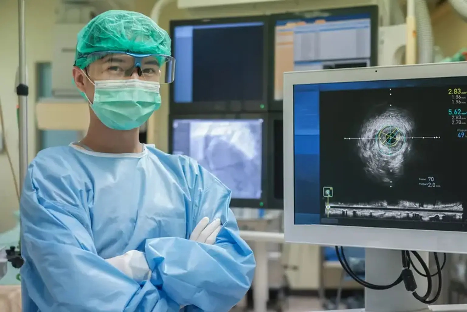
Endovascular ultrasound or intravascular echocardiography are other terms for intravascular ultrasonography (IVUS). It generates sound waves using a transducer or probe and produces images of the insides of blood vessels. A special catheter with a tiny ultrasonic transducer on one end is used for IVUS. The catheter is threaded through an artery or vein to the desired place by the doctor. Once there, the transducer emits sound waves that form pictures of the blood arteries and aid in the diagnosis of various conditions.
How Does It Work?
IVUS employs high-frequency sound waves (also known as ultrasound) to create a moving image of your heart. The images are transmitted from within the heart rather than via the chest wall. The sound waves are transmitted via a device known as a transducer. The transducer is connected to the end of a catheter, which is inserted into your heart via an artery. The sound waves bounce off the artery walls and return to the transducer as echoes. The echoes are transformed into pictures on a television monitor, creating a picture of your coronary arteries and other body vessels.
Common Uses of IVUS
IVUS is used by doctors to aid in the diagnosis and treatment of both arteries and veins. In the veins, doctors most commonly utilize IVUS for acute and chronic blood clots, especially if constriction of the veins is suspected. IVUS can assist in locating regions of constriction or blockage deep within the body. It also aids in the measurement of veins for the proper size of stents to keep the vessel open.
IVUS may be used to evaluate every artery in the body. It is very beneficial in assisting doctors in seeing peripheral arteries in the legs and coronary arteries. IVUS is frequently used in combination with catheter angiography to aid in the diagnosis of peripheral artery disease (PAD). PAD is best detected by IVUS, which is also valuable in determining the size of stents needed to keep the artery open.
IVUS can be used to see the coronary arteries in combination with or to assist in the planning of catheter angiography, angioplasty, and vascular stenting. Unlike angiography, IVUS can disclose more information regarding plaque accumulation (atherosclerosis), which increases your risk of a myocardial infarction. IVUS information frequently influences treatment decisions, such as stent size and placement. Doctors frequently utilize it after angioplasty and vascular stenting to ensure the stent is in the proper position and has repaired the issue.
IVUS is also used to examine abdominal aortic aneurysms before, during, and after vessel repair procedures.
Benefits vs. Risks
IVUS has many benefits, including:
- Showing the existence and extent of plaque in arteries.
- Determining the extent to which the vessel has gotten constricted due to plaque.
- Giving details on the plaque's building.
- Restenosis detection.
- Increased stent accuracy and decreased stent thrombosis in arteries and veins.
- Detecting stenosis or constriction that is not seen with angiography.
- Identifying locations of external vein compression that may be prone to blood clots.
- No ionizing radiation exposure.
Any operation that involves inserting a catheter into a blood artery entails some risk. These dangers include blood vessel injury, bruising or bleeding at the puncture site, and infection. To reduce these dangers, the doctor will take steps.
Other risks may include:
- Irregular heart rhythms (arrhythmia).
- Blood clot.
- An allergic reaction to the medications used during the procedure.
- In very rare cases, a heart attack, stroke, or blood clot in the lung.
IVUS itself adds little additional risk to angioplasty and catheter angiography.
Who Performs an Intravascular Ultrasound (IVUS)?
Interventional cardiologists perform intravascular ultrasounds. An interventional cardiologist specializes in diagnosing and treating conditions and diseases of the heart and blood vessels. They use nonsurgical, catheter-based procedures and specialized imaging techniques.
Is IVUS Painful?
Both you and your care staff value your comfort and relaxation. During the IV insertion and local anesthetic injection, you may feel a pinch and brief stinging. You may also feel pressure during the incision, but you should not experience any discomfort. There will be enough sedative drugs to keep you comfortable. If you have chest discomfort or other symptoms during the procedure, notify your doctor or a member of your healthcare team.
How Should I Prepare?
While intravascular ultrasound does not use any contrast material, the procedures associated with it (catheter angiography, angioplasty, etc.) do. You should follow the below instructions:
- Inform your doctor about all of the drugs you are taking. Include any allergies, particularly to iodine contrast materials. Inform your doctor about any recent illnesses or medical issues (e.g. asthma).
- Make sure to tell your doctor about your renal condition and diabetes mellitus.
- For the test, you may need to remove some clothing and/or change into a gown. Remove any jewelry, detachable dental appliances, eyeglasses, other metal items or clothes that might interfere with the x-ray pictures.
- If a woman is pregnant, she should always notify her doctor and a gynecologist. To prevent exposing the fetus to radiation, doctors will not do many tests during pregnancy. If an x-ray is required, the doctor will take care to limit the baby's exposure to radiation.
- If you are breastfeeding at the time of the exam, see your doctor. It may be beneficial to pump breast milk ahead of time. Keep it on hand until all contrast material has been removed from your body (about 24 hours after the test). However, according to the most recent American College of Radiology (ACR) Manual on Contrast Media, studies reveal that the quantity of contrast absorbed by the newborn while nursing is exceedingly minimal.
- Some women may be concerned about passing on contrast to their child. They may decide to stop breastfeeding for 12 to 24 hours.
- Ask your doctor how long you should stop breastfeeding if the surgery requires anesthesia. Furthermore, if sedation is used, your doctor may advise you not to eat or drink anything for four to eight hours before your exam.
- If your exam includes sedation, do not drive for 24 hours; arrange for someone to take you home. Because IVUS requires an observation period and is frequently used in conjunction with another treatment, ask your doctor whether you will need to stay in the hospital overnight.
- Be sure that you have clear instructions from your health care facility.
What Happens During IVUS?
Here’s what happens during the procedure:
- You lie down on a table and are offered sedative drugs to help you relax. You will be awake and cognizant during the process, but you will not be in any pain.
- A medical professional cleans the skin at the incision site. They next provide an anesthetic injection to numb the region.
- A plastic sheath is inserted into the incision by the interventional cardiologist or vascular surgeon. This makes inserting and advancing the catheter easier.
- They utilize the ultrasound probe to acquire pictures after the catheter has reached the precise location.
- When the operation is finished, the catheter and sheath are removed by the healthcare professional.
- Stitches are usually not required to close the wound. To avoid infection, a surgical dressing is used.
What Will I Experience During & After IVUS?
- When the nurse puts the needle into your vein for the IV line and injects the local anesthetic, you will feel a tiny pinch. The majority of the feeling is felt at the location of the skin incision. The doctor will use local anesthetic to numb this region. When the doctor puts the catheter into the vein or artery, you may feel pressure. However, you will not experience severe discomfort.
- If sedation is used, you will feel peaceful, drowsy, and at ease. Depending on how profoundly you are drugged, you may or may not remain conscious.
- When the doctor inserts the catheter, you may feel little pressure, but no major discomfort.
- During ultrasound scanning, you will not feel the catheter in your artery or vein, nor will you experience any pain.
- To prevent bleeding, you may need to rest flat on your back for a few hours following the test and apply pressure to the catheter insertion site. Your doctor may use a "closure device" to plug the tiny hole in the artery in some circumstances. This will allow you to move about more quickly.
- The doctor or nurse will examine your catheter site for bleeding or swelling and will monitor your blood pressure and heart rate for many hours.
- You may feel tired until the sedative wears off.
- The duration of your hospital stay will depend on whether IVUS was performed in combination with another surgery, such as catheter angiography or angioplasty. While IVUS does not increase to your recovery time, catheter angiography recovery will require you to stay in the hospital for up to six hours for observation. Recovery time for angioplasty and vascular stenting might range from 12 to 24 hours. Recovery from vein procedures is largely influenced by the complexity of your surgery.
- You should relax and drink lots of water after you get home. Lifting large things and intense activity should be avoided for at least 24 hours, if not longer. It is strongly advised that you stop smoking because it is a key contributor to artery and vein disease.
- The catheter insertion site may be bruised and sore. If bleeding begins where the catheter was inserted, you should lie down, apply pressure to the site and call your doctor.
- Call your doctor immediately if you notice any change in the color of your leg, pain, swelling or warm feeling in the area where the catheter was inserted.
Advantages of IVUS over Angiography
The most beneficial application of IVUS is to visualize plaque that angiography cannot see. This approach has grown over time into a highly helpful research tool for modern invasive cardiology, and it is increasingly being employed in research to better understand the behaviour of the atherosclerosis process in living people.
IVUS may see not only the lumen of the coronary arteries, but also the atheroma (membrane/cholesterol laden white blood cells) "hidden" within the wall. Thus, IVUS has enabled advances in clinical research by offering a more comprehensive view and greater comprehension.
Furthermore, because IVUS examinations were performed more frequently, they served to reveal and confirm the autopsy research findings, demonstrating that atheromatous plaque tends to cause expansion of the internal elastic lamina, causing angiography to greatly underestimate the degree of plaque burden. Angiography only shows the atheroma's edge that protrudes into the lumen.
Current clinical applications of IVUS technology include determining how to treat difficult lesions prior to angioplasty and determining how well an intracoronary stent was implanted into a coronary artery following angioplasty. If a stent is not extended flush against the vessel wall, turbulent flow may develop between the stent and the vessel wall, which some believe may produce a nidus for acute thrombosis of the artery.
When Should I Call My Doctor?
Following an intravascular ultrasound, it is critical to keep your follow-up appointments. In between checkups, contact your doctor if you have any questions or concerns. Call your doctor immediately away or seek emergency medical attention if you have:
- Bleeding.
- Chest pain, chest tightness, chest pressure, or palpitations.
- Dizziness or passing out.
- Fever.
- Numbness, a feeling of coolness, or change in color in the arm or leg that had the catheter.
- Pain that is not controlled by your pain medication.
- Cellulitis, unexpected drainage, pus, redness or swelling of your incision.
Conclusion
Intravascular ultrasound (IVUS) or intravascular echocardiography is a medical imaging technique that employs a specially constructed catheter with a small ultrasound probe attached to the distal end. The catheter's proximal end is connected to computerized ultrasound equipment.
The most common imaging focus for IVUS is the arteries of the heart (the coronary arteries). IVUS is utilized in the coronary arteries to assess the degree of atheromatous plaque that has accumulated at any given site in the epicardial coronary artery. Intravascular ultrasonography is a novel technique for studying the development or reversal of atherosclerotic lesions in vivo.