Isotope therapy

    Last updated date: 10-Jul-2023

    Originally Written in English

    Isotope therapy

    Isotope therapy

    Overview

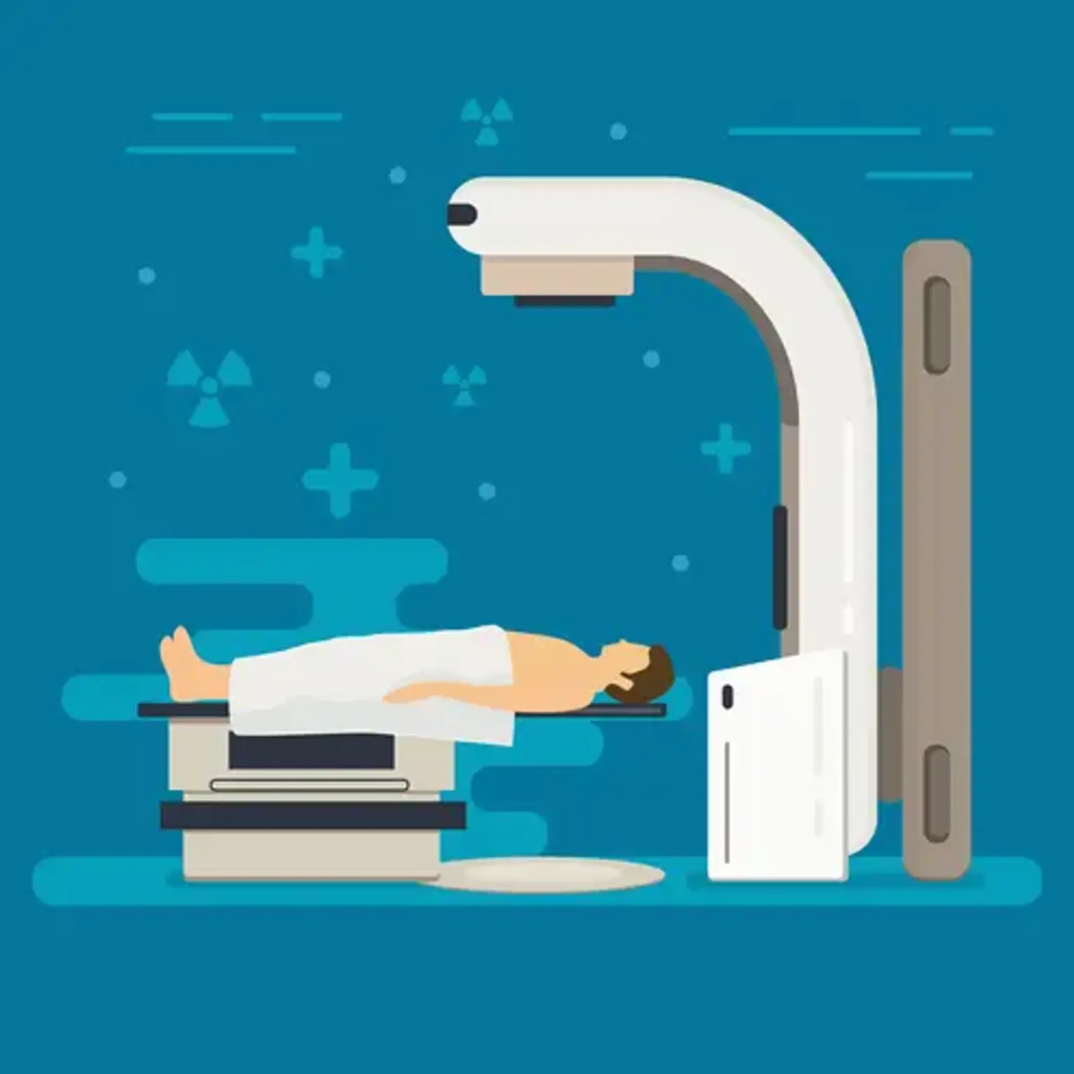
    The two most well-known cancer therapies are chemotherapy and radiation therapy. There are several forms of radiation treatment, just as there are numerous types of chemotherapy. Internal radiation is a cancer treatment that involves inserting a radioactive substance into your body to treat cancer. Internal radiotherapy includes radioisotope treatment.
     

    What are Radioisotopes?

    Radioisotopes

    Radioisotopes are radioactive isotopes of a chemical element found on the periodic table. The varying amount of neutrons in the nuclei of isotopes leads to their instability. An unstable combination of neutrons and protons exists in radioisotopes of a certain atom, which may result in an excess of energy (and instability)

    Every chemical element has at least one isotope, and radioisotopes are identifiable by that chemical and its atomic weight. Iodine isotopes, for example, are written as Iodine-131, indicating that they have an atomic weight of 131 neutrons. The average weight of an iodine atom is 127.

    Radioisotopes are employed in industry and medicine. A radiopharmaceutical is formed when radioisotopes are linked to a tiny molecule (such as a peptide).


    What is Radioisotope therapy?

    Radioisotope therapy

    Radioisotope treatment use radioisotopes to kill cancer cells. Thyroid cancer, bile duct cancer, liver cancer, bone metastases, and neuroblastoma are among the cancers that can be treated using radioisotope treatment. Different radioactive isotopes will be utilized depending on the type of cancer present. I-131 radiation, for example, is used to treat thyroid cancer, whereas radium Ra 223 dichloride may be utilized to treat prostate cancer. Radioisotope therapy can be used in conjunction with other cancer therapies.

    One such medication is Xofigo (Radium-223), which is used to treat individuals with advanced prostate cancer that has migrated to the bones. Because Xofigo is preferentially absorbed by bone, radiation can be delivered directly to the site of your prostate cancer's painful bone metastases.

    Radioisotope therapy is a noninvasive treatment. The radiation is administered through a beverage in one of two ways: by mouth (as a drink or pills) or by injection into a vein. We provide radioisotope therapy for a variety of cancers, including thyroid cancer, non-lymphoma, Hodgkin's prostate cancer, and osteoblastic metastatic bone lesions.

    You will meet with a radiation oncologist prior to the administration of radioisotope therapy to discuss if you are a candidate for the treatment, what other tests or scans you may need prior to the therapy, and what the risks and advantages of the treatment may be.

    PET-CT and SPECT-CT imaging are essential components of radionuclide treatment. It provides an estimate of the needed therapeutic dosage and its effects by determining the amount to which radioactive material accumulates in the tissues. Imaging confirms that an adequate dosage of radiation is delivered to the tumor tissue during treatment. It may require multiple treatment sessions to acquire an adequate radiation dosage.


    Type of cancers treated with Radionuclide Therapy

    Neuroendocrine Tumors (NETs)

         1. Neuroendocrine Tumors (NETs)

    Neuroendocrine tumors of the pancreas, colon, and lung are treated using radionuclide treatment. To target these tumors, somatostatin, a chemical that targets a particular receptor on the cell surface, is labeled with a radioactive particle. This is referred to as peptide receptor radionuclide treatment (PRRT). Lutetium-177 or Yttrium-90 is the radioactive particle. Peter Mac has a big neuroendocrine multidisciplinary team that treats for all aspects of patients with NETs and has great expertise with PRRT.

    Furthermore, this method can be used to treat a variety of uncommon tumors such as pheochromocytoma, parangalioma, and neuroblastoma. Another treatment option for these malignancies is Iodine-131 MIBG.

         2. Prostate cancers

    Many prostate tumors, particularly those that have progressed or grown resistant to hormonal treatments, display a unique molecule termed prostate-specific membrane antigen on their cell surface (PSMA). Lutetium-177 PSMA is a developing treatment that is now being tested in clinical studies to provide large doses of targeted radiation to prostate cancer spots while preserving most normal tissues. The Prostate Imaging Center of Excellence is supported by a grant from the Prostate Cancer Foundation (PCF) and is made up of a multi-disciplinary team of nuclear medicine, medical oncology, radiation oncology, urology, and laboratory-based doctors and researchers who have a strong patient-centered philosophy.

         3. Bone metastases

    Tumors that have progressed to the bone (most typically prostate or breast cancer) can be treated with radioactive particles that are absorbed in places with significant bone turnover. Radium-223 (Xofigo), Strontium-90, and Samarium-153 EDTMP are among the radioactive particles available for this sort of therapy.

         4. Thyroid cancers

    For almost 80 years, radioactive iodine (Iodine-131) has been used to treat thyroid cancer. This is used to treat certain people following thyroidectomy in order to eliminate any leftover cancer cells and prevent the thyroid cancer from returning. It's also used to treat those who have advanced thyroid cancer.

         5. Lymphoma

    Radioimmunotherapy (RIT) is a cancer treatment that employs monoclonal antibodies to deliver targeted radiation directly to lymphoma cells. To treat lymphoma, an antibody called rituximab, which targets a specific antigen (CD20) on the cell surface of lymphocytes, is labeled with radioactive iodine (Iodine-131 rituximab). In general, your haematologist will decide if this therapy is appropriate for you.

    Radionuclide therapy has long been recognized to have the potential to cure some uncommon solid tumors (e.g., feocromocytomas and neuroblastomas) and haematological disorders (e.g., leukemia), as well as in intracavitary and intravascular therapies, but the number of patients treated has been limited. The bulk of these therapy procedures need collaboration among physicians from other specialties, and only a few centers worldwide have been able to carry out such therapies.


    How does radioisotope therapy work?

    Because radiopharmaceuticals have a radioisotope and a tracer that is linked to the pharmaceutical, the radioisotope can target a specific tissue or portion of the body. Because cancer cells absorb more radioisotope than noncancer cells, when the radioisotope decays, it damages the targeted tissue or tumor. The higher the dosage of radiation, the more cancer cells are destroyed. The amount of radiopharmaceutical used is precisely calculated depending on your unique tumors, their location, and their size.


    What happens after radioisotope therapy?

    after radioisotope therapy

    Following your therapy, you will be given precise advice on how to prevent radiation exposure to people in your household. Small quantities of radiation are expelled by your body through urine and bowel movements, saliva, and perspiration. As a result, patients must take special care to avoid exposing others to radiation. For these reasons, you could be told to:

    • Avoid sharing cups, food, towels, or sleeping spaces 
    • Wash your linens, towels, and clothing separately for a few days
    • Avoid physical contact with others for a week
    • Maintain a space of at least three feet between you and others 
    • Avoid caring for small children for a week 
    • Avoid physical contact with pregnant women for at least a week after your treatment


    Side effects of Radionuclide Therapy 

    Side effects of Radionuclide Therapy 

    The type of therapy chosen for your tumor type determines the adverse effects. Because the therapy delivers far more radiation to tumors than to normal tissues, side effects are usually minor or moderate. However, a variety of side effects are possible and will be addressed with you before to therapy.


    Radioisotope in Diagnosis 

    Radioisotope in Diagnosis 

    Radioisotopes are critical components of medical diagnostic techniques. They may be utilized for imaging to examine the dynamic processes taking place in various sections of the body when combined with imaging equipment that detect the gamma rays released from within.

    When radiopharmaceuticals are used for diagnosis, a radioactive dosage is given to the patient, and the activity in the organ may then be analyzed as a two-dimensional picture or as a three-dimensional picture using tomography. Nuclear medicine diagnostic procedures employ radioactive tracers that generate gamma rays from within the body. These tracers are typically short-lived isotopes attached to chemical compounds that allow for the examination of certain physiological processes. They can be administered through injection, inhalation, or oral administration.

    The first technology devised makes use of single photons detected by a gamma camera, which can examine organs from a variety of angles. The camera creates a picture from the locations where radiation is released; this image is improved by a computer and shown on a monitor to look for signs of abnormal conditions. The current principal scanning method for diagnosing and monitoring a wide variety of medical problems is single photon emission computerized tomography (SPECT).

    Positron emission tomography (PET), a more accurate and complex technology that uses isotopes generated in a cyclotron, is a more recent invention. A positron-emitting radionuclide is injected into the target tissue and accumulates there. It generates a positron as it decays, which quickly interacts with a neighboring electron, resulting in the simultaneous emission of two distinct gamma rays in opposing directions. A PET camera detects them and provides highly specific indicators of their origin. PET's most significant clinical application, with fluorine-18 as the tracer, is in oncology, where it has shown to be the most accurate non-invasive means of identifying and assessing most malignancies. It's also useful for heart and brain imaging.

    New technologies combine PET scans with computed tomography (CT) scans to provide co-registration of the two images (PET-CT), allowing for 30% better diagnosis than a typical gamma camera alone. It is an important and strong instrument that delivers unique information on a wide range of disorders ranging from dementia to cardiovascular disease and cancer.

    PET-MRI offers diffusion-weighted imaging in soft tissue with dynamic contrast and magnetic resonance spectroscopy, particularly for brain imaging.

    The main distinction between nuclear medicine imaging and other imaging modalities such as X-rays is the placement of the radiation source within (rather than outside) the body. Gamma imaging, using any approach outlined, offers a picture of the radioisotope's position and concentration within the body. Organ dysfunction can be suggested if the isotope is either partially or completely taken up in the organ (cold spot) (hot spot). If a sequence of photos is collected over time, an unusual pattern or pace of isotope transport might suggest an organ problem.

    Nuclear imaging has a major advantage over X-ray methods in that it can examine both bone and soft tissue. This has led to its widespread adoption in industrialized nations, where the likelihood of having such a test is one in two and growing.


    Diagnostic Radiopharmaceuticals

    Diagnostic Radiopharmaceuticals

    From a chemical standpoint, each organ in our body behaves differently. A variety of substances that are absorbed by certain organs have been identified by doctors and scientists. The thyroid, for example, absorbs iodine, but the brain needs large amounts of glucose. Radiopharmacists can use this information to attach different radioisotopes to physiologically active drugs. When a radioactive form of one of these compounds enters the body, it is absorbed by regular biological processes and eliminated normally.

    Diagnostic radiopharmaceuticals can be used to monitor bone development, blood flow to the brain, liver, lungs, heart, or kidney function, and to corroborate other diagnostic tests. Another key use is predicting the effects of surgery and assessing changes after therapy.

    The radiopharmaceutical dose given to a patient is just enough to collect the necessary information before it decays. The radiation dosage received is negligible from a medical standpoint. The patient feels no pain throughout the test, and after a short time, there is no indication that the test was ever performed. This method is a significant diagnostic tool because to its non-invasive nature and ability to view an organ operating from outside the body.

    A radioisotope used for diagnostics must generate enough gamma rays to escape from the body and have a short enough half-life to fade away shortly after imaging is concluded.

    Tc-99 is the most often used radioisotope in medicine, accounting for approximately 80% of all nuclear medicine treatments. It is an isotope of the man-made element technetium, and it possesses nearly excellent properties for nuclear medicine scans such as SPECT. They are as follows:

    • It has a half-life of six hours, which is long enough to investigate metabolic processes but short enough to keep the radiation exposure to the patient to a minimum.
    • It decays by an 'isomeric' mechanism that emits gamma rays and low energy electrons. The radiation exposure to the patient is modest since there is no high-energy beta emission.
    • Its low-energy gamma rays easily leave the human body and are detected properly by a gamma camera.
    • Technetium's chemistry is so diverse that it can construct tracers by incorporating it into a variety of physiologically active chemicals that ensure it concentrates in the tissue or organ of interest.

    Its logistics also make it useful. From the nuclear plant where the isotopes are created, hospitals get technetium generators (a lead pot enclosing a glass tube carrying the radioisotope). They contain molybdenum-99, which has a half-life of 66 hours and gradually decays to Tc-99. When necessary, saline solution is used to wash the Tc-99 out of the lead pot. The generator is returned for recharging after two weeks or less.

    A similar generator technique is used to generate rubidium-82 from strontium-82, which has a half-life of 25 days, for PET imaging. Myocardial perfusion imaging (MPI) is a technique that employs thallium-201 chloride or Tc-99 to identify and predict coronary artery disease.

    Fluoro-deoxy glucose (FDG) with a half-life of little under two hours is the primary radiopharmaceutical used in PET imaging. FDG is easily absorbed into cells without being broken down, making it an excellent indication of cell metabolism.

    As PET and CT/PET become more widely available, there is a strong tendency toward employing more cyclotron-produced isotopes such as F-18 in diagnostic medicine. However, the operation must be performed within two hours of a cyclotron, limiting its value in comparison to Mo/Tc-99.


    Conclusion

    Radionuclide therapy is a systemic treatment that employs a radionuclide-labeled molecule to provide a high amount of radiation to treat some tumors. Radioisotopes are administered either intravenously or orally by consumption. Iodine-131 and Strontium-89 are two radioisotopes often utilized in Radiation department. Thyroid cancer is treated with iodine-131.