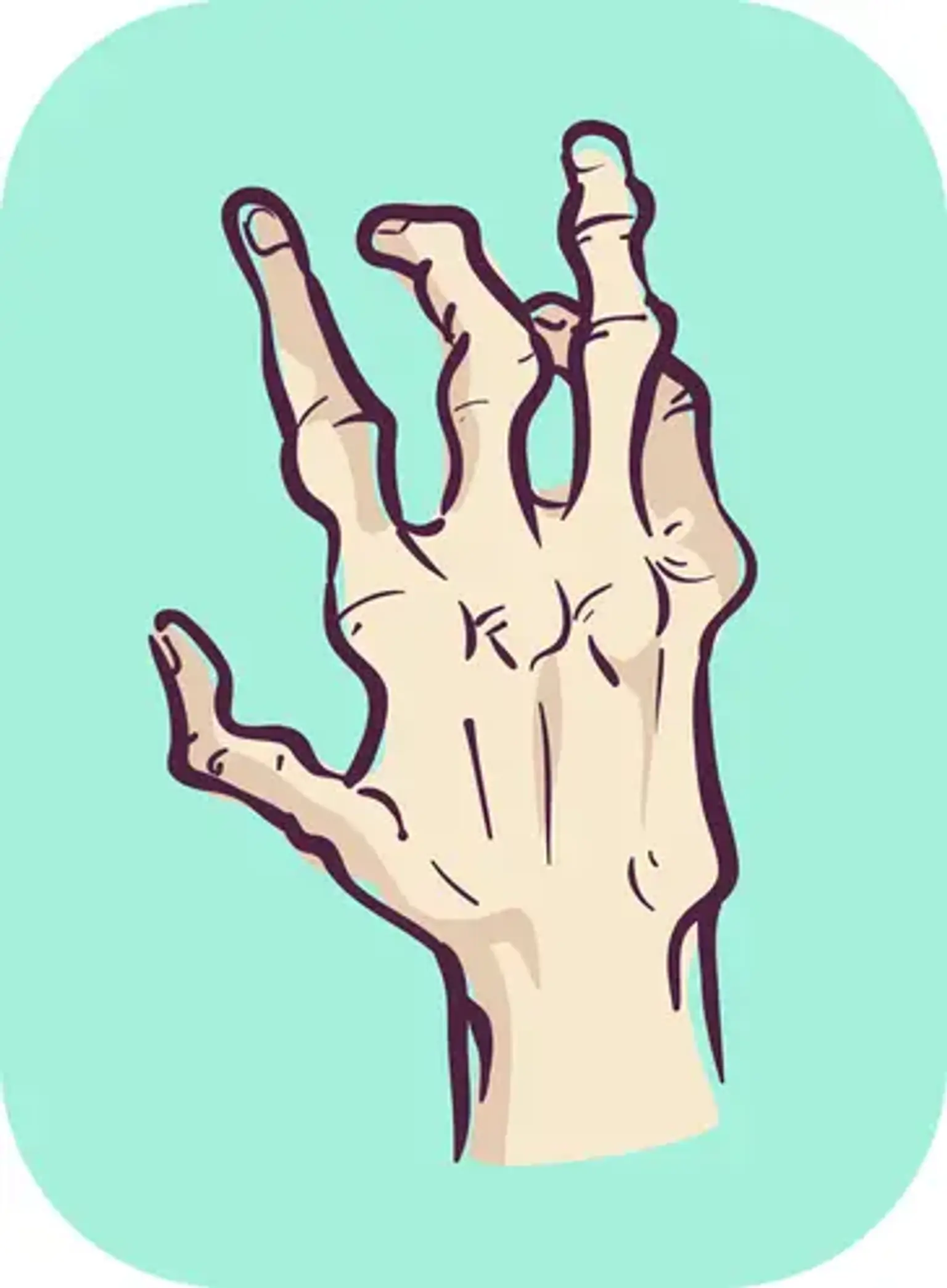
Joint deformity
Overview
Rheumatoid arthritis (RA) used to wreak havoc on the hands and feet. Joints began to deform. The fingers and toes were twisted out of form. Because to earlier detection and better treatment, these alterations are becoming less prevalent and less severe. Crooked fingers, for example, can be caused by osteoarthritis. Bunions can be caused by wearing shoes that are too small. However, if you have RA, joint abnormalities indicate that your condition is not under control.
What is Joint deformity?
Joint abnormalities can occur in every joint in the body, but they are most typically encountered in the hands, which have a high number of joints that are used almost constantly. There are several reasons of joint deformities. Rheumatoid arthritis and osteoarthritis are two of the most frequent causes.
Hand Deformities
- Mallet Finger – End joint is flexed, cannot actively straighten – due to extensor tendon injury or small fracture
- Ulnar drift – When one or more finger leans towards the pinky – common with rheumatoid arthritis.
- Knuckle subluxation – The joint becomes unstable from injured or stretched ligament
- Swan neck finger – The joint at the base of the finger is bent, the middle joint is straight, and the outermost joint is bent.
- Boutonnière finger – The middle joint in the finger is flexed and end joint hyperextended
- Z-shaped thumb – The base joint of the thumb is bent.
- Bouchard’s nodes – Bony bumps/nodules on the middle joint of the finger from arthritis
- Heberden’s nodes – Bony bumps on the end joint of the finger from arthritis
- Wrist subluxation – The wrist joint becomes unstable from arthritis or injury
Foot Deformities
- Hammertoe – A bending of the middle joint, most commonly the second toe.
- Bunion – Causes a bony bump on the inner (medial) side of the big toe.
- Claw toe – The base joint of the toe is straight, the middle joint is bent, and the third joint may be curled.
- Mallet toe – Causes the last joint of the toe bend downward; most common in the second toe.
- Valgus deformity – Deformity where the joint bends outward, away the midline. Found mostly in the knees, though any joint can have this deformity.
- Varus deformity – Deformity when the joint bends inward, toward the midline. Found mostly in the knees, though other joints can also have this.
Bouchard’s and Heberden’s Nodes
A bony growth of the distal interphalangeal finger joint is referred to as a Heberden node. It is a symptom of osteoarthritis, which is a degenerative joint condition. A Bouchard node is a swelling that affects the proximal interphalangeal finger joint in a similar way.
Who gets Heberden and Bouchard nodes?
Heberden and Bouchard nodes are equally common in males and females of all races.
- They are frequent in the elderly. However, more than half of Heberden node and osteoarthritis patients are diagnosed before the age of 65.
- Bouchard nodes are rarer than Heberden nodes and are linked to more severe arthritis.
- The existence of the nodes is closely related to the family.
- Heberden nodes are present in more than 60% of patients with knee osteoarthritis.
What causes Heberden and Bouchard nodes?
Exostosis is a bone growth caused by osteoarthritis that results in a Heberden or Bouchard node. The exact etiology of osteoarthritis is unknown. There is a hereditary propensity for node formation.
Osteoarthritis is said to be an indication of joint 'wear and tear.' The ligaments that support the joints show the earliest structural abnormalities in osteoarthritis. The cartilage in the joints deteriorates, osteophytes (bone spurs) form, and the ends of the bones rub together. Friction-induced capsular rupture and synovial leaking cause inflammation in the nodes.
What are the clinical features of Heberden and Bouchard nodes?
A Heberden node is a bony swelling of a distal interphalangeal joint and a Bouchard node is a bony swelling of the proximal interphalangeal joint.
- Bony swelling can affect either the lateral or midline aspects of the joint or both.
- Multiple nodes can be present on one digit.
- The nodes can affect one or many digits.
- Nodes most often affect the middle finger or thumb of both hands.
- They may grow slowly or rapidly.
- The nodes may become inflamed and painful but are often painless and unnoticed.
- A Heberden node may be associated with a digital myxoid pseudocyst.
In addition to damaging the fingers, osteoarthritis causes discomfort, pain, and stiffness in the hands, knees, hips, and spine.
How are Heberden and Bouchard nodes diagnosed?
The diagnosis of Heberden or Bouchard node is usually made clinically due to their characteristic appearance. Imaging may be performed on the affected digit.
- Ultrasound scanning reveals osteophytes, synovitis, and bony erosions of osteoarthritis.
- A simple X-ray of the afflicted joint may reveal interphalangeal osteophytes, although association with clinical Heberden nodes is weak. Osteophytes are more frequent in inflammatory arthritis patients.
- Magnetic resonance imaging (MRI) of the afflicted digit may confirm the diagnosis of arthritis. Soft tissue bulging through the capsule between the dorsal tendons and collateral ligaments is related with Heberden and Bouchard node development. Ligamentous changes might be noticed in neighboring but clinically normal joints.
Histological investigation of the Heberden or Bouchard node is uncommon. It may reveal the existence of osteophytes as well as dorsal contractures.
How are Heberden and Bouchard nodes treated?
There is no particular therapy for Heberden or Bouchard node repair. Lifestyle adjustments including as exercise, weight loss, a low-inflammatory diet, heat packs, and cold compresses may be used to treat osteoarthritis.
Pain relievers and nonsteroidal anti-inflammatory medications are among the medical therapies available. Some osteoarthritic individuals may require surgery to repair or replace one or more joints.
Swan neck finger
Swan-neck deformity is characterized by a bending in (flexion) of the finger's base, a straightening out (extension) of the middle joint, and a bending in (flexion) of the finger's outermost joint.
Rheumatoid arthritis is the most prevalent cause of swan-neck deformity. Untreated mallet finger, looseness (laxity) of the fibrous plate inside the hand at the base of the fingers or of the finger ligaments, chronic muscle spasm affecting the hands caused by nerve damage (called muscle spasticity), other types of arthritis, a ruptured finger tendon, and misalignment in the healing of a fracture of the middle bone of the finger are all possible causes.
Normal finger bending may become impossible. As a result, the malformation might cause significant handicap. The thumb, which has one fewer joint than the other fingers, is not affected by true swan-neck malformation. However, in a swan-neck deformity variant known as duck-bill, Z (zigzag), or 90°-angle deformity, the top joint of the thumb is substantially overstraightened, with a bending in of the joint at the base of the thumb to form a 90° angle. When duck-bill and swan-neck deformities of one or more fingers coexist, the ability to pinch is severely impaired.
Treatment of Swan-Neck Deformity
When feasible, treatment for swan-neck deformity aims to repair the underlying disease. Mild abnormalities that have not yet resulted in scarring can be treated using finger splints (ring splints), which correct the deformity while enabling the client to use the hand.
Problems with pinching ability can be considerably addressed by surgically realigning the joints or fusing the thumb or finger joints together (called interphalangeal arthrodesis) into optimal function positions.
Claw Toe
People sometimes attribute the common foot deformity claw toe to wearing shoes that compress your toes, such as overly short or high heels. However, claw toe is frequently caused by nerve damage caused by disorders such as diabetes or alcoholism, both of which can weaken the muscles in your foot. Claw toe occurs when your toes "claw," digging into the bottoms of your shoes and causing painful calluses. Without treatment, claw toe worsens and may become a lifelong deformity.
Claw Toe Symptoms
- Your toes are bent upward (extension) from the ball of your foot joints.
- The middle joints of your toes are bent downward (flexion) toward the sole of your shoe.
- Your toes may also bend downward at the upper joints, curling beneath the foot at times.
- Corns can form on top of the toes or under the ball of the foot.
Evaluation of Claw Toe
If you experience symptoms of claw toe, consult your doctor. Certain tests may be required to rule out neurological problems that might weaken your foot muscles, causing imbalances that cause your toes to bend. Claw toe deformity can also be caused by trauma or inflammation.
Claw Toe Treatment
Claw toe malformations are often flexible at initially, but stiffen with time. If you have claw toe in its early stages, your doctor may advise you to use a splint or tape to keep your toes in the proper position. Additional suggestions:
- Wear shoes with soft, roomy toe boxes and avoid tight shoes and high-heels.
- Use your hands to stretch your toes and toe joints toward their normal positions.
- Exercise your toes by using them to pick up marbles or crumple a towel laid flat on the floor.
If you have claw toe in later stages and your toes are fixed in position:
- A customized cushion can disperse your weight and ease strain on your foot's ball.
- Try special "in-depth" shoes with an additional 3/8" depth in the toe box.
- To fit the deformity, have a shoe repair business expand a small pocket in the toe box.
If these treatments do not help, you may need surgery to correct the problem.
Mallet Finger
In routine clinical practice, mallet finger injuries are widespread. In the 1800s, the name mallet, which meaning hammer, was used to describe the hammer-like deformity that developed in sports-related injuries. Because other people do not recognize the hammer similarity, some have suggested modifying the name to remove the term "finger" because of its look.
It is sometimes referred to as the "baseball" finger. Although it is the most frequent closed tendon injury observed in athletes as a result of high velocity and contact sports, it may also be caused by relatively modest trauma such as performing housework (tucking in a shirt, tucking in sheets) or work-related tasks. Though some players and coaches feel mallet injuries are minor, each case should be evaluated thoroughly.
Mallet Finger Causes
The bones that make up the digits are known as phalanxes or phalanges. They consist of two bones in the thumb or three bones in the fingers, as well as two to three phalangeal joints. The distal interphalangeal joint (DIPJ); the metacarpophalangeal joint (MCPJ), which links the digit to the carpal or hand bones; and the proximal interphalangeal joint (PIPJ), which joins the DIP and MCP joints. To promote stability, the joints are supported by volar plates, which are collateral ligaments linked to thick fibrous connective tissue. Tendons are the connective tissues that link muscles to bone.
Long tendons connect the muscles that move the digits (fingers and thumbs) to the bones of the digits in the forearm. Extensor tendons on the top of the hand stretch or straighten the digits, whereas flexor tendons on the palm side of the hand flex or bend the fingers. The digit tendons stretch across three joints. When the extensor tendon is torn, mallet finger injuries occur.
Diagnosis of Mallet Finger
Mallet finger is a clinical diagnosis that necessitates a detailed medical history and physical examination. Imaging investigations are used as a supplement to assess bone damage. To identify the mallet finger category - to separate a bony injury vs a tendinous mallet injury - an anterior-posterior (AP), lateral, and oblique view X-ray focused at the DIPJ of the afflicted finger should be taken. The lateral view is the most effective for detecting avulsion fractures and volar (palmar) distal phalanx dislocation. Some have proposed that ultrasonography be used for diagnostics as well.
Management of Mallet Finger
A variety of therapies, ranging from reassurance to conservative splint implantation to medically corrected operations, have been explored. Although somewhat contentious, there is considerable agreement in the research that, in the absence of significant articular surface disruption or subluxation, non-operative therapy with a splint is beneficial for both soft tissue and bone mallet. Splints have been reported to be both safe and beneficial for acute and chronic soft tissue mallet finger. However, the type of splint, the length of full-time wear, and the requirement for supplementary night orthotic wear are usually determined by the provider.
Conservative treatments include the Stack splint, thermoplastic splint, or aluminum foam splint, all of which have the same goal of extending or slightly hyperextending the DIP joint. Perforated splints are more conforming than typical solid splints. The finger should be splinted until it is examined by a hand specialist. The standard length for extension splinting is 6-8 weeks, with increasing flexion exercises starting at six weeks.
The PIPJ should be permitted to move freely, with the DIPJ splinted solely in extension or modest hyperextension. The splint should be worn at all times since removal and bending of the joint reset the 6 to 8-week clock back to zero. Patient education on skin hygiene care without permitting DIPJ bending is an important aspect of the therapy.
Indications for surgical intervention:
- Open injuries.
- Bony mallet with a large fragment and subluxation of the DIP joint.
- Unstable fractures (30–50 % of the joint surface involved).
- Intolerance to splints.
- Chronic injuries (older than 12 weeks).
- Painful arthritic DIPJ.
- Swan neck deformity.
Boutonnière Deformity
Boutonnière deformity is most commonly caused by rheumatoid arthritis, however it can also be caused by trauma (such as severe cuts, joint dislocations, or fractures) or osteoarthritis. Rheumatoid arthritis can occur in people who have long-standing inflammation of the middle joint of a finger.
If the deformity is the result of an accident, the damage is commonly near the base of a tendon (called the middle phalanx extensor tendon). As a result, the middle joint (called the proximal interphalangeal joint) becomes "buttonholed" between the outer bands of the tendon that runs from the center of the finger to the end of the finger. In other words, the bones of the joint protrude through the tendon bands like a button through a buttonhole. The malformation may impair hand function.
Treatment of Boutonnière Deformity
A boutonnière deformity caused by an extensor tendon (a tendon that pulls the finger up) injury is generally treated with a splint that maintains the middle joint completely extended for 6 weeks. However, if scarring and irreversible abnormalities have already formed, the splint will be ineffective (usually after many weeks)
Surgery may be required to enhance function when splinting is inadequate or when boutonnière deformity is caused by rheumatoid arthritis.
Conclusion
Hand arthritis is most commonly found in the base of your thumb, knuckles, second joint, and top joint of your fingers. Common forms include osteoarthritis, rheumatoid arthritis, and psoriatic arthritis. Splints/braces, medicines, steroid injections, and healthy lifestyle choices are among the treatments. Joint fusion, joint replacement, and tendon transfer are all surgical procedures.