Keloid Scars

Last updated date: 23-Mar-2023

Originally Written in English

Keloid Scars

Overview

Keloids are caused by scar tissue overgrowth. Keloid scars are often larger than the original wound. Keloid development is influenced by both hereditary and environmental factors. Darker-skinned people of African, Asian, and Hispanic heritage have a higher prevalence. Overactive fibroblasts that produce a lot of collagen and growth factors are thought to be involved in the etiology of keloids.

As a result, characteristic histologic findings include huge, aberrant, hyalinized bundles of collagen known as keloidal collagen and a significant number of fibroblasts. Keloids appear clinically as stiff, rubbery nodules in areas of previous skin damage. Keloidal tissue, unlike normal or hypertrophic scars, expands beyond the primary site of damage.

Patients may experience discomfort, itchiness, or burning. There are several therapeutic options available, but none are universally effective. Intralesional or topical steroids, cryotherapy, surgical excision, radiation, and laser therapy are the most often used therapies.
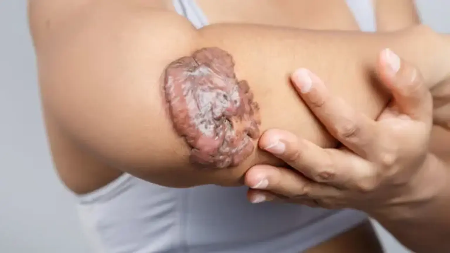
 

What are Keloids scars?

Keloids scars

Keloids are caused by an expansion of thick fibrous tissue, which normally forms after skin damage heals. The tissue expands beyond the initial wound's boundaries, does not normally retreat on its own, and tends to return following removal. In 1700 BCE, the first description of keloids (written on papyrus) involved surgical procedures employed in Egypt.

The fibroblasts in keloids continue to grow long after the lesion has healed. Keloids protrude above the skin's surface, forming huge mounds of scar tissue.

Keloids can form on any area of the body, but the upper chest, shoulders, and upper back are most vulnerable. Skin pigmentation, itching, redness, unusual sensations, and pain are all symptoms.Keloids are thought to affect roughly 10% of the population. While most individuals never develop keloids, others do after small lesions such as bug bites or pimples. People with dark skin appear to be more prone to the formation of keloids. Both men and women are impacted equally.


Epidemiology

Dark-skinned people of African, Asian, and Hispanic ancestry have greater rates of keloid formation than Caucasians. Keloids are more common among Polynesians and Chinese people than in Indians and Malaysians. Keloids were reported by as much as 16% of respondents in a random sample of black Africans. White people are the least likely to be harmed.

The prevalence has been observed to be higher in young females than in young men, most likely due to the higher incidence of earlobe piercing in females. In other age groups, keloids and hypertrophic scars affect both sexes equally.

In these darker-pigmented groups, the prevalence ranges from 4.5 percent to 16 percent. During pregnancy and adolescence, the incidence is significantly greater. Although no unique gene has been found, a positive family history increases the likelihood of developing keloids. 

The most prevalent age of onset is between the ages of 10 and 30. Keloids are less common at the extremes of age, however, a rising number of presternal keloids have developed from coronary artery bypass surgeries and other comparable treatments currently performed on people in their later years.


Etiology 

Keloid development is influenced by both hereditary and environmental factors. Predisposed individuals may develop a keloid as a result of any amount of skin damage, such as surgery, piercings, acne, tattooing, bug bites, burns, lacerations, abrasions, immunizations, and any other event that causes cutaneous inflammation. Increased wound tension may also lead to keloid development.


Pathophysiology

Keloids are a result of aberrant wound healing. Standard wound healing consists of three phases:

  1. inflammatory,
  2. fibroblastic, and 
  3. maturation.

The fibroblastic phase continues unabated in keloids, resulting in clinical and histological features.

When compared to usual wound healing, keloidal fibroblasts exhibit higher proliferative activity, remain for longer periods of time, and have lower rates of apoptosis. As a result, collagen and cytokines are overproduced. Keloids have 20 times the collagen production of healthy skin and three times the collagen synthesis of a hypertrophic scar.

The key drivers of this process are assumed to be transforming growth factor-beta and platelet-derived growth factor. TGF-beta, an essential component of wound healing, drives chemotaxis of fibroblasts to the site of inflammation and collagen production. Fibrosis and aberrant scar response result from dysregulation of this system.


Clinical manifestations of Keloids

Clinical manifestations of Keloids

Keloids are dermal growths that are caused by past skin damage or inflammation. Lesions can appear as early as 1 to 3 months after damage or as late as a year afterwards. There have been reports of spontaneous lesions. However, it is more possible that the injury was not reported because it was minor, or that the keloid growth was delayed by months or even years. Keloids can appear everywhere, although the deltoid, pre-sternal chest, upper back, and ear are the most typically afflicted areas.

These growths can be found in unusual places such as the eyelids, genitalia, palms, and soles. Keloids appear as hard, rubbery nodules that commonly protrude from the underlying skin. They might have a small base and evolve into pedunculated lesions, or they can develop into a more broad-based plaque.

As previously indicated, an essential differential diagnosis is hypertrophic scar, which will never spread beyond the initial location of damage and will never project beyond the underlying skin more than 4 millimeters. Keloids, on the other hand, invariably spread beyond the site of the original damage. Color might range from erythematous to flesh-colored to hyperpigmented and can alter as the lesion progresses. Although these tumors are benign, they usually cause symptoms.

In one research, 86 percent of patients reported pruritus and 46 percent reported discomfort. Tenderness and burning have also been mentioned as symptoms. Finally, because keloids can develop to be huge and disfiguring, they may have major aesthetic consequences for afflicted people.


Diagnosis

Clinical evaluation is the primary focus. Unless the diagnosis is in doubt, a biopsy is not necessary. In that circumstance, a biopsy is necessary. There is no need for any more testing.


Management

Corticosteroids

Keloids continue to be a therapeutic conundrum. They are not only difficult to cure, but insufficient therapy can cause a keloid to develop and expand. As a result, primary prevention is critical. Predisposed persons should avoid elective treatments, particularly ear piercing and tattoos, if at all feasible. If an accident happens or surgical intervention is necessary, there are strategies to reduce and maybe prevent the formation of keloid scars.

It is critical to lessen wound tension in order to reduce the likelihood of keloid formation. The recognized critical features in tension-free wound closure are rapid primary closure and sufficient hemostasis. Prolonged wound stabilization with silicone sheeting may also assist minimize wound tension. Compression treatment has been shown in the literature to minimize keloid formation, with pressures ranging from 15 to 45 mmHg advised for more than 23 hours per day for at least 6 months.

Several modalities alleviate symptoms of existing keloids:

  • Corticosteroids- Intralesional steroids are the first-line therapy for keloid scars. Multiple injections at 4 to 6 week intervals are necessary. The recommended dosages of triamcinolone vary from 10 to 40 mg/cc. This can be used on its own or in conjunction with other modalities. Topical ointments and steroid-impregnated tapes have also been found to alleviate itching and burning symptoms.
  • Cryotherapy- Delivery methods such as spray, contact, and intralesional-needle cryoprobe have all been documented. Several treatments are necessary. To produce scar tissue necrosis, freeze-thaw cycles of 10 to 20 seconds are advised. This approach is less suitable in darker skin types due to post-treatment pigmentary modification.
  • Surgical excision- Because of the high recurrence rate (45-100%), this approach should always be combined with adjuvant therapy such as post-surgical radiotherapy or intralesional steroid injections.
  • Radiotherapy is best used as an adjuvant treatment 24 to 28 hours after excision. Caution should be used in patients under the age of 18, as well as in susceptible locations such as the head, neck, and breast, due to the inherent danger of carcinogenesis.
  • Laser-Successive sessions with a 585 nanometer (nn) pulse-dye laser and a 1065 nanometer (nm) neodymium-doped yttrium aluminum garnet (ndYAG) laser have been proven to cause keloidal flattening and regression.
  • Other therapies, such as topical imiquimod after excision, intralesional botox, intralesional bleomycin, intralesional 5-fluorouracil, and silicone gel sheeting, have been shown to be effective in treating keloid scars, either alone or in conjunction with the modalities listed above. 

It is critical to control patient expectations because all treatments take numerous sessions and may not always result in complete keloid regression. The utilization of several modalities and adapting therapy to match the needs of the patient results in the most successful treatment.


Prognosis

Prognosis

Keloids seldom go away on their own; but, with therapy, they can become softer, less sensitive, less painful, and less pruritic. Keloids typically recur (>50%) after excision therapy alone.

Most keloids and hypertrophic scars are aesthetic in nature; however, certain keloids and hypertrophic scars can produce contractures, which can result in loss of function if covering a joint or substantial deformity if positioned on the face.


Differential Diagnosis

Differential Diagnosis

The differential diagnosis is restricted when assessing a patient with suspected keloids. However, critical characteristics must be noted in order to decide whether lesions require a skin biopsy. As previously noted, hypertrophic scars can resemble keloids and form as a result of skin damage. In contrast to keloids, which can continue to expand and include the surrounding skin, hypertrophic scars are often smaller and isolated to the region of damage.

Dermatofibroma is another type of abnormal scar response that manifests as a flesh-colored or hyperpigmented papule or nodule. The "dimple sign" is the characteristic clinical hallmark of these lesions, which is central depression while applying lateral pressure. Dermatofibrosarcoma protuberans is a rare, locally aggressive spindle cell tumor that develops on the trunk and proximal extremities of young people. These lesions, unlike keloids, have no prior trauma and have more uneven boundaries. Morphea and scleroderma are keloidal variations.

In the absence of a recognized inciting event, these individuals would have progressive illness and may also exhibit additional indications of connective tissue disease. Xanthoma disseminatum is an uncommon histiocytic growth with keloidal-like skin lesions. Acute lesions manifest in a diffuse symmetric pattern, and systemic involvement may lead to the development of diabetes insipidus.

Finally, lobomycosis is a deep fungal infection produced by the Lacazia loboi organism. It manifests as a slow-growing keloid-like nodule on the distal extremities and is linked to dolphin exposure or rural soil in Central and South America.


Hypertrophic scar

Hypertrophic scar

A hypertrophic scar is an enlarged or ugly scar that does not extend beyond the original wound borders. Unlike keloids, the hypertrophic scar grows to a specific size before stabilizing or regressing. Hypertrophic scars, like keloids, are connected with negative wound healing variables.

Unlike keloid development, hypertrophic scarring has no racial or family predominance. SEM imaging reveals flattened collagen bundles that are parallel in orientation. These scars, like keloids, have a larger amount of type III collagen than typical wounds. Hypertrophic scars are more prone to form in the same anatomic regions as keloids (e.g., the mandibular border), owing to increased strain in these places. 


Keloid VS Hypertrophic scar

A hypertrophic scar resembles a keloid. Scars that have hypertrophy are more prevalent. They do not grow as large as keloids and may diminish with time. They can be found in all racial groupings. Keloids are called benign tumors, however they are mostly aesthetic in nature and never turn malignant. Because operating on a keloid frequently spurs the formation of additional scar tissue, persons with keloids may have been informed that there is nothing they can do to get rid of them.

Both represent abnormal responses to dermal injury, with exuberant deposition of collagen developing over three basic stages: 

  1. inflammation (first three to 10 days); 
  2. proliferation (next 10 to 14 days); and
  3. maturation or remodeling (two weeks to years).

Keloids and hypertrophic scars are treated similarly, however hypertrophic scars have a better prognosis.


Keloids Prevention

Patients should be asked if they have ever experienced scarring difficulties before undergoing any surgical operation. As part of informed consent, discuss the possibility of keloids, and discourage ear piercing and other elective treatments in those with dark skin.

If your ears are pierced notwithstanding this recommendation, pressure earrings are commercially available to help reduce the likelihood of keloid formation. In a high-risk patient, if surgery cannot be avoided, urgent silicone elastomer sheeting or corticosteroid injections should be started. Anything that speeds up wound healing and reduces skin tension (for example, postsurgical taping for 12 weeks) reduces risk.

Wounds repaired using normal suture procedures appear to have a comparable aesthetic effect to those closed with 2-octyl cyanoacrylate dermal glue (Dermabond). One small research found that hypertrophic scars appeared in five out of 24 Dermabond repairs vs three out of 28 standard suture repairs.


Conclusion 

Keloid

Keloid, also known as keloid disease and keloidal scar, is the creation of a kind of scar that is mostly formed of either type III (early) or type I (late) collagen depending on its maturity. It is caused by an excess of granulation tissue (collagen type 3) at the site of a healed skin injury, which is gradually replaced by collagen type 1.

Keloids are hard, rubbery lesions or glossy, fibrous nodules that can range in color from pink to the color of the person's skin or from red to dark brown. A keloid scar is harmless and not contagious, although it can cause significant itching, discomfort, and textural changes. In extreme circumstances, it can impair skin mobility.

Keloid scars are observed 15 times more common in persons of Sub-Saharan African heritage than in those of European descent in the United States. These elevated scars can form in men and women of African, Asian, or Hispanic origin all throughout the world. Some people, however, are more likely to develop a keloid when they scar, such as those with a family history of keloids and those aged 10 to 30 years.

Keloids should not be confused with hypertrophic scars, which are elevated scars that do not extend beyond the original wound's limits.