Keratoconus

Last updated date: 06-Nov-2023

Originally Written in English

Keratoconus

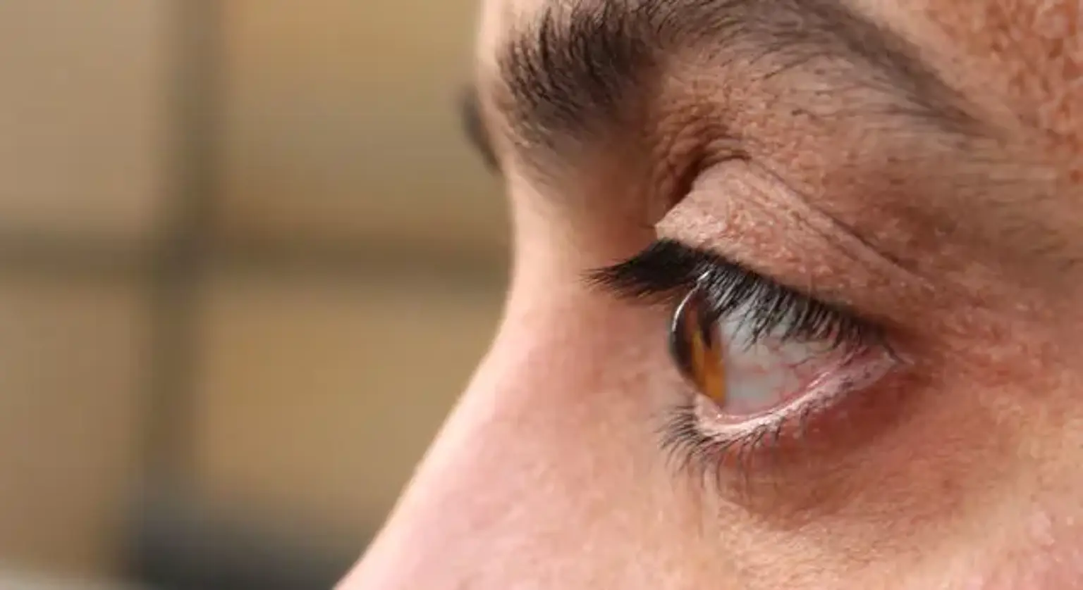
Keratoconus is a chronic corneal ectatic condition that affects both eyes. It causes a cone-like steepening of the cornea, which is accompanied by uneven stromal thinning, leading to a cone-like protrusion (hump) and substantial visual impairment.

A severe and varying loss in visual function, picture distortion, and greater sensitivity to glare and light are all optical impacts. Because of the substantial asymmetry, spherocylindrical contact lenses are less effective in correcting vision. Keratoconus can be characterized as mildly asymmetrical oblique astigmatism if it continues subclinical (i.e., undiagnosed).

Keratoconus may appear clinically during adolescence (late teenagers for males and young adulthood for females) and continue (continuous stromal thinning and corneal steepening) till the third to fourth decade. It is quite rare for anyone to advance beyond this age. In rare situations, keratoconus can develop later in life as a result of a change in endocrinologic conditions, such as pregnancy. The disease's presentation and course are exceedingly diverse, and most cases are asymmetrical between the two eyes of the same patient. It is commonly acknowledged that there is no unilateral keratoconus, in the sense of a disease that affects only one eye; even if there are no medical indications of the disease in the other eye, it is assumed that the disease is simply not present in that eye. Acute hydrops can lead to severe keratoconus.
 

Epidemiology

The prevalence of the disease in the general population differs, possibly due to discrepancies in diagnostic criteria. It is most often an isolated eye disorder, although it can also occur in the presence of other ocular and systemic conditions.

Seasonal keratoconjunctivitis, retinitis pigmentosa, and Leber congenital amaurosis are all well-known ocular connections. Many connective tissue diseases (e.g., Ehlers-Danlos and Marfan syndromes), mitral valve prolapse, atopic dermatitis, and Down syndrome have been suggested as systemic possible correlations, however, none of these were observed in the Collaborative Longitudinal Evaluation of Keratoconus study.

Atopic history, particularly ocular sensitivities, and maybe either or both stiff contact lens usage and forceful eye rubbing are also risk factors. The majority of keratoconus cases develop spontaneously, although roughly 15% of cases show evidence of hereditary inheritance.

Keratoconus prevalence varies with race. With odds ratios of 1.5 and 1.4, keratoconus is more prevalent in blacks and Latinos than in Europeans. The evidence on whether Asians are at a higher or decreased risk of keratoconus is equivocal. Considering that anecdotal accounts imply an increase in keratoconus prevalence in various places of the glob, including Arabia, the Indian subcontinent, and New Zealand, the variance might be due to the great heterogeneity of Asians (e.g., subcontinental Indian vs Middle Eastern vs Chinese).

Keratoconus usually develops during puberty and continues through the third and fourth decades of life, though it can develop at any age. Keratoconus evolves at varying paces, but it usually advances faster in young people and remains stable after about 20 years of disease onset.


Keratoconus Etiology

Keratoconus has an unknown origin and pathophysiology. Rigid gas permeable contact lens uses, persistent eye rubbing, Down syndrome, atopic disease, Leber congenital amaurosis, tapetoretinal degeneration, and genetics have all been linked. Keratoconus is frequently linked with no other systemic or ocular diseases. A chromosomal rearrangement, altered enzyme function, and depletion of collagen and/or ground material have all been linked to some unusual relationships. Down syndrome, Leber congenital amaurosis, and mitral valve prolapse are all strong keratoconus correlations.

Keratoconus appears to be caused or exaggerated by persistent eye rubbing. In people who are genetically susceptible to keratoconus, persistent eye rubbing and wearing firm contact lenses can cause mechanical damage that can lead to keratoconus advancement. The mechanical change could be linked to keratocytes changing to a repair phenotype in reaction to rubbing-induced stress.


Pathophysiology

Keratoconus Pathophysiology

Keratoconus is thought to affect all layers of the cornea. Epithelial basement membrane fragmentation and scar formation, a breach in the anterior limiting lamina (i.e., Bowman membrane), and axial stromal thinning and scar tissue are all common structural alterations. Fleischer rings are formed when the iron is deposited in the basal epithelial cells. Striae and acute hydrops are common striae and folds near the Descemet membrane.

Regarding the involvement of disruption of collagen fibers, lamellae, and proteoglycans in keratoconus, the research is extensive and inconsistent. Antioxidants, cytotoxic oxygen/nitrogen molecules accumulations, activated caspase pathways, and mitochondrial DNA damage have all been seen in keratoconic corneas. Keratoconus corneal cells have been revealed to have abnormal oxidative stress-related characteristics. Degradative enzymes are activated by oxidative stress, and tissue inhibitors of metal-low proteolytic enzymes are degraded. The disease has also been linked to a chromosomal deletion in the superoxide dismutase 1 gene.

Despite the fact that it is commonly thought to be noninflammatory, some evidence suggests that it has an inflammatory component.


Keratoconus Symptoms

Keratoconus Symptoms

Medical History

Keratoconus patients frequently experience deteriorating vision (distortions, glare, and monocular diplopia or ghost pictures), as well as many unsuccessful attempts to get optimal spectacle correction.

Soft contact lenses and spectacles may provide adequate vision at first, but vision deteriorates with time, necessitating the use of rigid gas-permeable contact lenses for correction. Piggyback lens devices (typically a gas-permeable lens over a soft, maybe silicone hydrogel) are another alternative, as are hybrid lenses (e.g., Synergeyes TM) and, more recently, gas-permeable scleral lenses.


Physical Examination

  • Mild cases: External and corneal manifestations are frequently absent or minor. Numerous unsatisfactory spectacle corrections of one or both eyes, including oblique astigmatism on refraction and moderate-to-high myopia, may be noticed. Keratometry results that are irregularly astigmatic (egg-shaped) but not necessarily on the steep side of normal (about 45 D) are compatible with the diagnosis. Corneal topography or tomography can make the diagnosis, revealing inferior corneal steepening (about 82% of keratoconus patients), central corneal astigmatic steepening (about 14% of keratoconus case scenarios), or even bilateral temporal steepening (about 14% of keratoconus case scenarios). Paraxial corneal thinning is visible on corneal tomography. Corneal sensitivity is reduced, as is tear secretion.


  • Moderate cases: The following are some of the most common keratoconus corneal signs:
  1. The corneal nerves have a more prominent appearance.
  2. In patients with mild keratoconus, Vogt striae (fine-stress lines) form in the deep stroma in around 42% of cases.
  3. Fleischer ring, a buildup of iron in the basal epithelial cells at the base of the conical projection in a ring (often partial) shape, affects about half of the patients.
  4. About 21% of people acquire corneal scars.

Scarring on the surface of the cornea might be fibular, nebular, or nodular. Deep stromal scarring may develop, possibly indicating the resolution of mini-hydrops episodes. Scarring at the Descemet membrane (posterior limiting lamina) is seen in certain cases, which is compatible with posterior polymorphous corneal dystrophy. This could be a type of posterior polymorphous corneal dystrophy. The keratometry value usually rises to 45-52 D. The oil drops sign or "scissoring" of the retinoscopy and direct ophthalmoscope red pupillary reflex can be seen when the retinoscopy and direct ophthalmoscope red pupillary reflex are distorted. When looking at downgaze, a "V" shape in the cornea's profile against the lower lid margin is evident. This is known as the Munson sign. The conical form of the cornea in moderate to advanced keratoconus is emphasized in this way.


  • Advanced cases: The keratometry value exceeds 52 D. All corneal indications, symptoms, and vision loss/distortion are improved, including Vogt striae, Fleischer ring, and scarring. Acute corneal hydrops is a possibility.


Keratoconus Diagnosis

Keratoconus Diagnosis

While considerable myopia is frequently associated with keratoconus, it is not a requirement in and of itself. Irregular corneal astigmatism and focal stromal thinning are the two most common manifestations. It's important to identify focused thinning from a thin cornea in general. Substantial astigmatism is therefore not a keratoconus requirement if it is symmetrical. It is important to remember that astigmatism seen in keratoconus is quite asymmetric. Corneal thinning and asymmetric astigmatism both occur in the inferotemporal portion of the corneal protrusion. In addition to biomicroscopic (slit-lamp) examination, topography, tomography, and pachymetry are the primary techniques employed in the diagnosis and assessment of keratoconus.

Marc Amsler provided the first use of Placido topography for the detection and categorization of keratoconus. Before other clinical or biomicroscopic indicators may be established, topography can detect small corneal surface irregularities. From early changes in cornea curvature to clinically apparent keratoconus, Amsler recorded a classification scheme. Then he divided keratoconus into two clinically visible stages and two latent (subclinical) stages that could only be detected with the Placido disk corneal topography: the first is forme-fruste and the second is early or mild keratoconus.

Several ocular imaging techniques, including corneal topography, tomography, and biomechanical evaluation techniques, have improved our ability to diagnose early keratoconus in a quantitative and repeatable way in recent years. The major diagnostic method for keratoconus diagnosis is corneal topography. The color-coded corneal curvature maps generated by corneal topography may allow visualization of anterior corneal surface irregularity, which is characterized by a typically infero-temporally positioned cone area of significantly high steepening, often as high as 65 D, and a supero-nasally positioned flat area of significantly low steepening, often as low as 35 D. Conversely, the corneal aberrations map given by these tools may reveal a significant degree of high-order aberrations, with coma as the primary aberration and spherical aberration following closely behind.

Pachymetry data from Scheimpflug imaging equipment like the Pentacam (in the past, scanning slit-scan systems like the Orbscan) is also used. These instruments provide precise pachymetry maps that show corneal thinning in conjunction with corneal curvature. The position of the largest corneal steepening coincides well with the thinnest cornea. These technologies can also be utilized to create corneal surface elevation maps, both anterior and posterior. A particular and sensitive sign for the condition is a distinctive asymmetry in posterior surface elevation.

Anterior surface irregularity indicators, such as the Index of Height Decentration, are other particular quantitative data derived by Placido topography or Scheimpflug imaging topometry that can be employed as disease progression determinants.

The use of anterior-segment optical coherence tomography as a diagnostic technique of keratoconus has been increasing in recent years. Optical coherence tomography can produce meridional cross-section pictures of the cornea, demonstrating asymmetric corneal thinning and asymmetry of the posterior curvature. More recently, spectral-domain optical coherence tomography has been used as a corneal pachymetry technique, demonstrating keratoconus-related corneal thickness asymmetry.

The epitheliums' thickness works as a cover for underlying stromal thickness abnormalities, which is an important aspect. As a result, it might be thinner where the cornea is most projecting and thicker where it is flatter. If the epithelial thickness distribution is not taken into account, a falsely homogeneous corneal thickness is presented, disguising early signs of corneal thickness irregularities measured with other devices, such as those relying on Scheimpflug imaging, and lowering the degree of corneal curvature irregularity evaluated by Placido topography. Tear fluid as a biomarker for keratoconus and corneal biomechanics are two more ocular clinical tests that can aid in the identification of keratoconus. The use of new Fourier-domain optical coherence tomography technologies to investigate corneal epithelial thickness distribution can be a very sensitive and specific diagnostic for keratoconus. 


Keratoconus Treatment

Keratoconus Treatment

The treatment choices for keratoconus are mainly dependent on the stage of the disease and how far it has progressed. When the condition has reached a plateau (no further advancement), the focus shifts to vision correction. If the disease is advancing, the focus should be on slowing (or stopping) the progression.

Vision repair with spectacles and spherical soft contact lenses is poor and only relevant to the early stages of keratoconus because the effects of keratoconus in cornea shape distortion and stromal thinning are very asymmetric. The principal aberrations related to keratoconus, such as coma and spherical aberration, may be reduced with custom-designed soft contact lenses that include aberration-controlled designs.


Rigid Gas Permeable Contact Lenses

The cornerstones of visual treatment for moderate-to-advanced keratoconus are rigid gas permeable contact lenses and scleral lenses. Their primary benefit is the formation of a tear pooling between the lens and the cornea, which naturally neutralizes the ocular aberrations associated with keratoconus ectasia, potentially resulting in near-perfect adjusted vision.  The usage of rigid gas permeable lenses has the disadvantage that they may not be accepted. The use of rigid gas permeable lenses in keratoconus is frequently problematic. Intolerance, allergic reactions (such as giant papillary conjunctivitis), corneal scratches, and neovascularization are the most common complaints. Hydrogel contact lenses, piggyback contact lenses, and scleral contact lenses are all options. The latter gives you better eyesight and more comfort.


Corneal Collagen Cross-linking

Corneal Collagen Cross-linking

Corneal collagen cross-linking is a minimally invasive outpatient technique that has been found to effectively stop keratoconus from progressing. It causes an increase in stromal stiffness, which slows and eventually stabilizes keratectasia advancement. Mechanical and biological tests have been used to show this in ex vivo research. When applied successfully, corneal collagen cross-linking reduces the requirement for penetrating keratoplasty.

When triggered by UVA light, riboflavin 5'-phosphate acts as a visual enhancer, producing singlet oxygen and reactive molecules that cause cross-linking. The classic, or Dresden, procedure includes epithelial debridement (to facilitate riboflavin penetration and a high level of UVA uptake in the stroma), soaking the vulnerable stroma in riboflavin for about 25 minutes (to allow adequate saturation in the stroma), and irradiation with UVA light for 30 minutes. 5.4 J/cm2 is the total energy released. The US Food and Drug Administration has licensed a drug and device combination product for cross-linking for the management of progressive keratoconus.

Other corneal collagen cross-linking procedures involving greater irradiation and shorter exposure times have been proposed and are in use outside the United States. In an attempt to integrate the benefits of ectasia arrest with enhanced visual acuity, corneal collagen cross-linking has been paired with topography-guided laser-ablation corneal surface normalization.

While corneal collagen cross-linking does not always enhance vision clarity, it is crucial to note that early recognition can make a major difference in keratoconus treatment success. If the condition is detected early on, both the irregularity and thinning of the cornea may be slightly affected. The effect of corneal collagen cross-biomechanical linking stabilization will avoid future blindness in these scenarios.


Other Treatment Options

Intracorneal ring segments are another therapy option. A little, bent PMMA ring (or collection of rings) is inserted in the cornea to assist straighten corneal curvature and improving eyesight.

When the cornea is too thin to undergo corneal collagen cross-linking and the symptoms are serious, a corneal transplant is regarded as the last option. Healthy donor cornea tissue is used to replace the cornea completely (penetrating keratoplasty) or partially (lamellar keratoplasty).


Keratoconus Prognosis

Keratoconus Prognosis

The prognosis of keratoconus varies depending on the patient. During puberty, it usually starts with modest astigmatism and myopia, and if the problem worsens, it can result in immediate vision loss or repeated prescription modifications. It is most typically detected in late adolescence or early adulthood, but it can manifest or progress at any age. Keratoconus usually does not cause blindness or serious vision loss. Because a patient's eyesight might vary dramatically in as little as a few months, prescription modifications may be necessary. Some people may be more stable, with a steady loss of vision. The illness normally progresses for 9 to 20 years before stopping in a person's thirties and forties. A corneal transplant will be required in about 10 percent of cases. Corneal cross-linking is a treatment that aims to enhance the long-term prognosis of a patient. It is a treatment in which riboflavin (vitamin B2) and UV radiation are combined to produce a structural alteration in the corneal cross-linking inside the stroma. The following are the long-term outcomes of this technique: a straightening or reduction in the steepness of the cornea; a strengthening of the cornea; and when paired with laser straightening, a reduction in corneal refractive astigmatism. The ensuing corneal haze, which can also reduce a patient's highest adjusted acuity, is a disadvantage of this operation.

For patients with health care plan coverage, corneal cross-linking costs roughly $190 per eye repaired. Only in extreme circumstances can the cornea get stretched to the point where vision loss is more significant. To address the significant eyesight loss in certain cases, a corneal transplant may be needed. This, however, is not normal for people with keratoconus. Vision swings and ghost visions are common symptoms of the disorder, which may usually be cured with contact lenses.


Conclusion

Much regarding the etiology and pathogenesis of keratoconus remains a mystery centuries after the disease was first described. Early identification of the disease, as well as providing effective optical and surgical repair for enhancing the quality of vision in affected patients, have made significant progress. In particular, in the last two decades, there have been exciting new advances that promise to change the natural history of keratoconus for the first time. For two fundamental causes, research interests in the disease are likely to stay considerable in the near future. The prospect of iatrogenic ectasia, or the unmasking of asymptomatic keratoconus, hangs over all refractive doctors like the sword of Damocles.