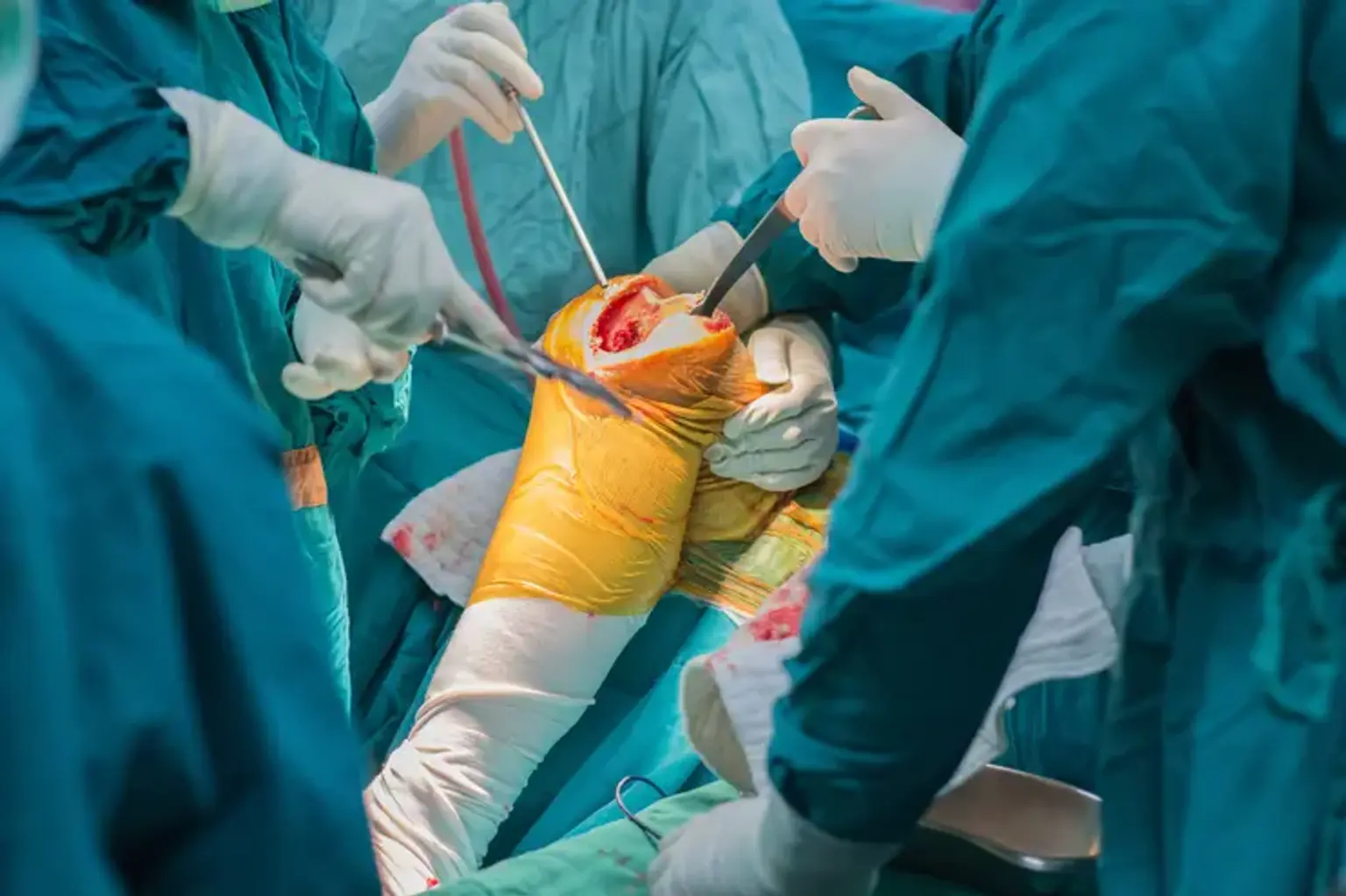
Knee osteotomy

Last updated date: 17-Jun-2023

Originally Written in English

Knee osteotomy

Overview

Knee osteoarthritis sometimes affects only one side of the joint, resulting in the well-known "bow-legged" or "knock-knee" abnormalities. When this happens, realigning the angle around the knee can change your body weight such that the healthy side of the knee joint bears more of the load. This alleviates discomfort and postpones the need for joint replacement surgery. This is referred to as an osteotomy.


What is Knee osteotomy?

Knee osteotomy

Knee osteotomy is the cutting of bone to adjust the alignment and function of the knee in order to relieve persistent arthritic knee discomfort.

Knee osteotomy is a possibility for individuals with unilateral knee arthritis, which means damage to only one side of the knee joint, or "compartment." The surgery's purpose is to redistribute some weight away from the painful, injured side of the knee joint and onto the healthy side. The surgeon does this by selectively cutting and reshaping a leg bone's alignment or form.

A knee osteotomy treatment, such as a high tibial osteotomy or a femoral osteotomy, restores the proper alignment of the knee joint by correcting the tibia or femur bone, reducing discomfort and eliminating pressure on the arthritic or damaged portion of the knee.

The technique includes splitting the bone and realigning the bones in the limb to straighten the leg by relieving pressure on the injured side of the knee joint. This realignment of the tibia or femur bone distributes body weight evenly across the knee joint, easing strain on injured tissue, enabling it to recover, and preserving the joint in the optimal position to help avoid future issues.

Patients' ability to resume regular activities promptly after surgery is typically due to the much reduced discomfort they experience. This can be especially advantageous for active younger and older individuals, including those who participate in sports on a regular basis. High tibial osteotomies, which reshape the shin bone, and femoral osteotomies, which reshape the thigh bone, are the two most prevalent forms of knee osteotomies.


High Tibial Osteotomy

High Tibial Osteotomy

When the joint deterioration is beyond repair, knee replacement surgery can help. A high tibial osteotomy, on the other hand, can realign the knee to relieve pressure on the afflicted side by wedging open the top section of the tibia to reorganize the knee joint in select individuals. Weightbearing is subsequently transferred from the injured or worn tissue to the better tissue.

Because these advantages often decrease after 8 to 10 years, this form of osteotomy is commonly seen as a technique to postpone the need for a knee replacement. This surgery is mainly reserved for younger patients suffering from discomfort caused from instability and malalignment. An osteotomy can also be used in combination with other joint preservation treatments to allow cartilage repair tissue to develop without being subjected to undue pressure.


Femoral osteotomy

Femoral osteotomy is a surgical treatment used to repair particular femoral - the long bone in the upper leg - and hip joint abnormalities. The procedure, which includes cutting the bone, is performed by orthopedic surgeons in attempt to realign it and restore a more normal structure, therefore resolving or preventing complications associated with the deformity. Damage to the articular cartilage in the hip joint, rips to the labrum (the crescent-shaped cartilage component that runs around the rim of the hip socket), and different kinds of hip impingement - improper contact between the two bones that meet in the hip joint - are examples of these issues.

Consider the typical hip to have a better understanding of various defects. This "ball and socket" joint is found where the femur (thigh bone) joins the pelvic bone. The femur is the biggest bone in the body and serves as the leg's primary joint connection as well as support and alignment. The femoral neck is the top part of the femur that curls and angles forward toward the pelvic.

The femoral head is located at the top of the femur and fits into the hollow in the pelvic bone that forms the socket, also known as the acetabulum, to create the hip joint. The surfaces of the joint are coated by articular cartilage, a smooth, cushioning layer. The articular cartilage is responsible for absorbing the load and allowing the bones to move smoothly. As depicted, the healthy hip has a perfect fit and the femoral head is securely positioned in the acetabulum.


Anatomy of knee joint

Anatomy of knee joint

Three bones make up the knee joint. The primary weight-bearing component of the knee is formed by the femur (thigh bone) meeting the tibia (shin bone). The patella (kneecap) is placed at the front of the knee and serves as a fulcrum to provide mechanical advantage to the thigh muscles when straightening the knee. These three bones are covered with cartilage, a white, highly polished surface that permits each bone to move freely against the others. Cartilage covers the femoral head, the top of the tibia, and the area behind the patella. When this surface cartilage goes down, osteoarthritis (OA) develops.


Why might I need knee osteotomy?

Knee osteotomy surgery

Knee osteotomy surgery is a less intrusive alternative to knee replacement surgery for people suffering from arthritis caused by misplaced bones in their legs. This misalignment places additional strain on the knee joint, producing discomfort and worsening knee injury.

Younger people, especially top athletes, and active older patients are two more groups of patients who may benefit from a knee osteotomy. An osteotomy allows these patients to return to their prior levels of activity, including competitive sports, more rapidly.

You might need this type of surgery if you are:

  • Bow-legged - this can cause arthritis on the inside side of the knee joint. Alignment surgery, also known as 'high tibial osteotomy,' involves realigning the top section of the tibia (shin bone). Bow-legged patients account for almost 80% of our realignment treatments.
  • Knock-kneed - this might result in arthritic damage to the outside side of the knee joint. The bottom section of the femur (thigh bone) is corrected during bone alignment surgery


Advantages and Disadvantages of Knee Osteotomy

Knee osteotomy is a viable surgical therapy option for a subset of active knee arthritis patients under the age of 60. The following are some of the potential benefits of knee osteotomy over knee replacement surgery:

  • The actual knee joint, including ligaments, is preserved.
  • Once healed, many patients can return to high-impact activities, such as jogging or playing basketball, which is not recommended after total knee replacements.

Compared to total knee replacement, knee osteotomy has potential disadvantages, including that it:

  • Requires more healing time
  • Is more prone to complications
  • During bone healing, the patient must wear crutches.
  • Depends on effective bone healing, because there are several elements that influence bone healing, not all of which are known or controllable.
  • Is less trustworthy in terms of pain alleviation (and then a partial or total knee replacement may be recommended)
  • Is not a treatment for arthritis
  • It does not restore or repair the cartilage that has already been gone.


Preoperative assessment

Knee osteotomy Preoperative assessment

Prior to any operation, your knee will be imaged to determine the extent of arthritis. This will involve x-rays that reveal narrowing of the afflicted compartment of the knee, as well as specialized x-rays (4-foot x-rays) that take a picture from the hip joint to the ankle joint 

Based on this, your surgeon will be able to anticipate where your knee now bears the majority of your body weight and how to correctly realign your leg to ensure a good conclusion. You will almost certainly undergo an MRI scan, which will provide an accurate assessment of the remaining cartilage throughout the knee joint as well as the related ligaments and meniscus. 


How is an Osteotomy Performed?

Osteotomy Procedure

  • Opening wedge osteotomy – In this procedure, a surgeon slices through the upper tibia on the medial side and opens a wedge, occasionally adding a piece of bone (auotgraft) taken from the pelvic area or utilizing cadaveric bone (allograft) to hold the wedge open and assist recovery. A plate is put across the osteotomy to stabilize it. This is the most often utilized approach since it does not shorten the leg and also simplifies future knee replacement operations. This causes your body weight to move from the inner part of the knee to the unaffected lateral compartment.
  • Closing wedge osteotomy – This entails cutting a wedge of bone from the top section of the tibia, generally right below the joint. The bone wedge is removed from the outside section of the tibia for patients with medial compartment arthritis (varus knees). After the bone wedge is removed, the two bone ends are joined and secured together with a metal plate or staples. This also causes your body weight to move from the inner part of the knee to the unaffected lateral compartment.

The surgeon has the choice of doing a closing wedge or opening wedge osteotomy, and the decision is made based on what would be the greatest option for you given your specific situation.

On the day of operation, you will be admitted to the hospital. Your anesthesiologist will discuss the best anesthesia for you and provide you with alternatives for postoperative pain treatment. Antibiotics will be given to you during surgery to reduce the chance of infection. Following the administration of your anesthesia, a tourniquet will be put to your upper thigh, and everything except your knee joint will be draped in sterile drapes.

The nerves and blood arteries that run behind the knee joint are protected throughout the treatment. A drain is often put into the incision within 24 hours following surgery. Dissolving sutures are used to seal all surgical wounds. Before you leave the operation room, the leg will be covered in a well-padded garment. After this sort of surgery, a brace may be necessary for a brief length of time.


After your operation

after Knee osteotomy Operation

In the recovery room, an x-ray of your knee will be taken. When you return to the ward, a variety of measurements (temperature, blood pressure, heart rate, circulation, and foot feeling) will be taken at regular intervals.

A physiotherapist will give you instructions on leg exercises you may do in bed and will help you walk with crutches while you're in the hospital. It is typical to be on crutches for up to six weeks after a closing wedge osteotomy and up to eight to ten weeks after an opening wedge osteotomy since this kind heals more slowly. The goal of your physiotherapy will be to recover knee movement, improve mobility, and maintain muscle strength surrounding the knee joint. You will also learn how to reduce the swelling in your knee.

Once you are comfortable and can move around safely, your surgeon will release you to go home. The majority of patients were released from the hospital within two days following their procedure. Your leg may be swollen and your knee may be stiff for the first month. It is typical to take pain medicine on a frequent basis throughout this time. To improve your outcome after surgery, it is critical that you execute your exercises on a regular basis at home.


  • Rehabilitation

It might take up to six months to recover completely after a knee osteotomy. Maintaining your strength and motion as the osteotomy heals is the most crucial element of your rehabilitation. It is critical that you follow the exercises prescribed by your physiotherapist during this time. Physiotherapy usually begins within the first week following your surgery.


How long does it take to recover from a knee osteotomy?

knee osteotomy recovery period

A knee osteotomy normally requires an overnight stay in the hospital, however you may be able to go home the same day in certain situations. Most patients can bear their own weight and walk around easily within 24 hours, whereas full recovery takes 6-12 weeks. Unless your job is physically demanding or requires manual labor, you should be able to return to work after six weeks.

Rehabilitation is an important aspect of a patient's rehabilitation from a knee osteotomy. Patients at rehabilitation facilities have access to a comprehensive team of specialized physiotherapists, occupational therapists, and rehabilitation gyms.

Sportsmen and women are often able to return to normal exercise and competitive sport relatively rapidly after a brief time of recovery.

Following an osteotomy, elite athletes have been able to return to normalcy and compete well, having restored their fitness levels. One example is an exceptional triathlon runner who, following his osteotomy treatment, went on to become the top-ranked athlete in his age group.


Complications

Knee Osteotomy Complications

Even when every attempt is made to avoid them, difficulties can emerge. All precautions are taken before, during, and after the procedure to avoid problems. If a problem occurs, you will be notified and counseled on the best treatment to manage the condition while also optimizing the outcome of the initial operation.

The most common complications following knee osteotomy surgery are:

  • Infection – The risk of infection after this operation is extremely minimal (1 in 100). This might be a superficial (mild) infection of the incisions or a severe infection of the knee joint and bone. Antibiotics will be necessary if this occurs. If the infection is severe, hospitalization and intravenous antibiotics are indicated. If the infection has spread to the knee joint, a knee arthroscopy may be necessary to remove the infection. Antibiotics are used before to surgery to lessen the risk of infection. Smokers are at a greater risk of infection, thus it is vital that you stop smoking two weeks before your operation. It is also critical to avoid any scrapes, rashes, or abrasions around the knee joint before to surgery since they increase the risk of infection and will force your operation to be postponed until they heal.
  • Blood clots “Deep Venous Thrombosis – A blood clot in a calf muscle vein occurs around once every 20 years. These blood clots seldom break free and migrate to your lungs. This is known as a "pulmonary embolus." A pulmonary embolus is extremely unlikely to occur. If you experience calf discomfort or soreness after your operation, you should consult your surgeon or family doctor. An ultrasound check of the calf will be conducted to look for a blood clot. To keep these blood clots from growing bigger, medications will be necessary. To reduce the chance of a blood clot, contact your surgeon if you have any inherited risk factors for blood clot development or if you have previously suffered a blood clot. You should also stop smoking since it raises your chances of developing this condition. Combining the oral contraceptive pill or HRT with smoking raises the risk of DVT. These aspects should be discussed with your surgeon before to the procedure.
  • Failure of the osteotomy to heal – Approximately 3-5 percent of persons have difficulty with bone repair. This may necessitate further surgery, such as bone grafts extracted from the pelvis and additional plate fixation. Smoking reduction and rigorous adherence to your post-op therapy lower the chance of this condition.
  • Fractures adjacent to the osteotomy – A crack (bone fracture) may form next to the osteotomy on rare occasions. This is frequently detected and addressed during the procedure. This is sometimes only shown on successive x-rays. If this occurs, more surgery may be necessary.
  • Nerve Injury – A extremely unusual consequence can include numbness or loss of power in the muscles that move the foot and ankle (foot-drop). In most situations, this is only transient and resolves itself within 6-12 months. It is crucial to note that this is not the same as the normal numbness that happens around surgical incisions with most types of surgery.
  • Compartment Syndrome – This is where swelling causes pressure in the leg, reducing blood supply to the muscles. It generally happens during the first 48 hours following surgery. If not treated with decompression (fasciotomy), it might cause irreversible muscular injury. The likelihood of this problem is less than one in a thousand.
  • Numbness around the wounds - Numbness around surgical incisions is frequent. The majority of these little patches are transient and do not disturb patients. On rare occasions, a persistent patch of numbness can be felt on the top section of the tibia's outer side.
  • Stiffness – Following surgery, there is a chance of developing knee stiffness. If this happens, you may need a second procedure in which the knee is manipulated or scar tissue from the interior of the knee joint is removed. To avoid stiffness after surgery, it is critical to have a decent range of motion before to surgery and to undertake frequent stretching exercises. Most essential, you must actively participate in your physiotherapist's post-operative rehabilitation program.
  • Ongoing swelling or pain – Because this surgery is conducted for arthritis, some patients may not experience considerable relief in arthritic symptoms despite the fact that the procedure is performed appropriately and with all required professional care. Furthermore, despite symptom reduction during typical activities, not all patients are able to return to high-impact activities. Your surgeon will advise you on whether it is safe for you to resume high-impact activities in the future.


Knee Osteotomy vs. Knee Replacement

Knee osteotomies have grown increasingly prevalent as surgery has advanced and surgical therapies for knee arthritis have become more popular. The two main reasons patients select a knee osteotomy are to maintain the natural tissue surrounding the knee and to postpone or avoid the necessity for a knee replacement.

A knee osteotomy preserves the actual knee joint—where the tibia, femur, and patella (kneecap) meet—as well as all of the knee's ligaments. Conserving the knee enhances joint function and keeps the knee feeling natural throughout its full range of motion. These considerations are critical for patients who desire to continue participating in activities that require greater degrees of knee function, such as jogging, squatting, kneeling, and climbing.

Knee osteotomies may be used to avoid or postpone the need for knee replacement surgery. Some patients prefer to postpone knee replacement surgery because, while the average prosthetic knee lasts 15 years or more, it may eventually necessitate a second procedure known as revision knee replacement surgery. Patients, logically, desire to make decisions that will help them avoid the need for a revision knee replacement.


Conclusion 

Knee osteotomy surgery is performed on individuals suffering from a variety of knee disorders, such as arthritis and sports-related discomfort and injury. A knee osteotomy can typically assist to postpone the need for knee arthroplasty (replacement) and, in many cases, eliminate the need for it entirely. New advancements in osteotomy procedures and anchoring systems have rekindled interest in knee osteotomies.