Laminectomy

Last updated date: 22-Aug-2023

Originally Written in English

Laminectomy

Overview

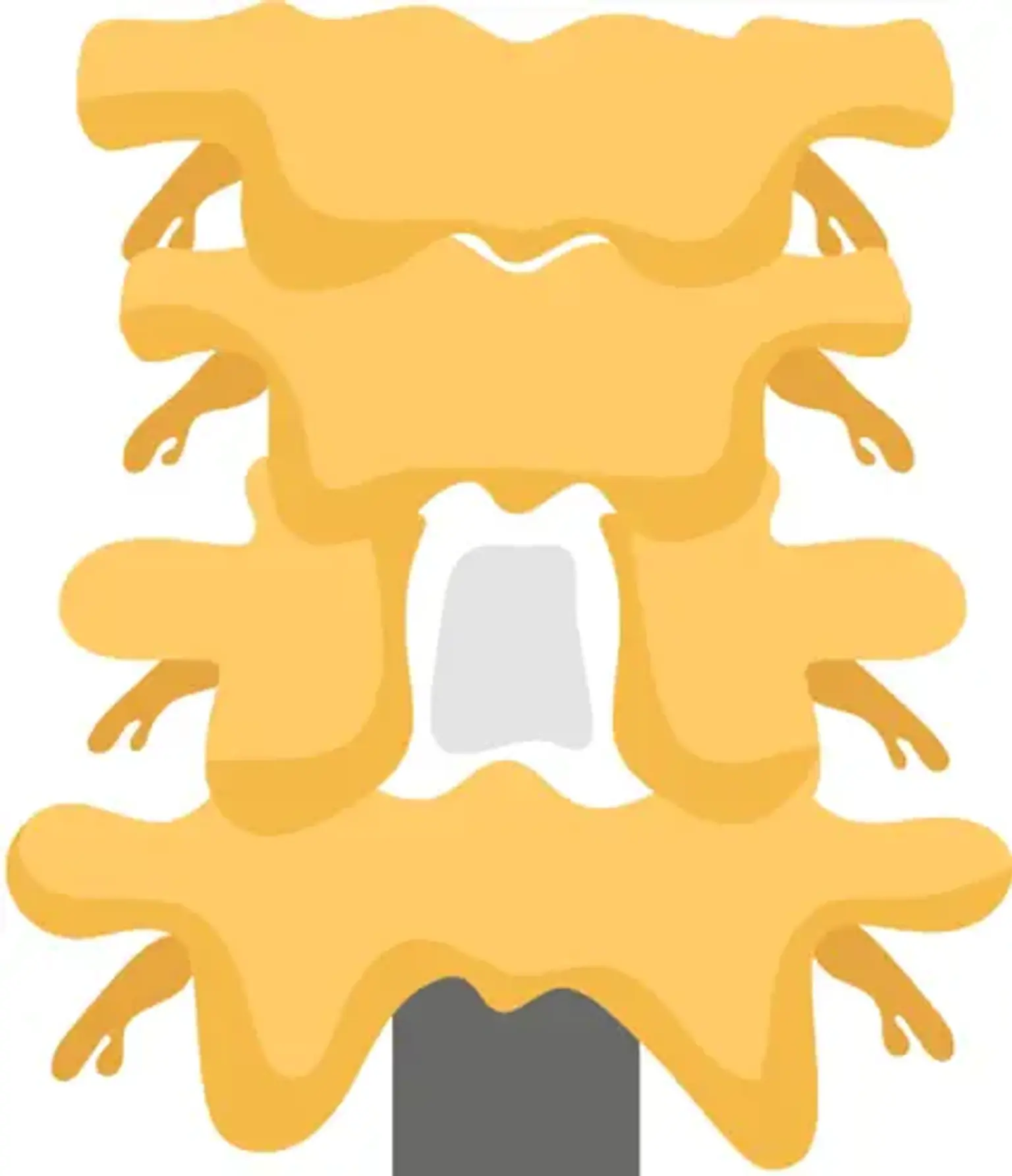
Laminectomy is one of the most popular treatments used to decompress the spinal canal in situations of restriction caused by degenerative stenosis, fracture, primary and secondary spinal tumors, abscess, and deformity. The removal of the spinous process and lamina is confined laterally to the medial region of the facet joints. For optimal clinical recovery and the avoidance of failed back surgery syndrome, the central canal, lateral recesses, and neural foramina must be decompressed.


What is Laminectomy?

Laminectomy surgery

Posterior spinal decompression is one of the most popular surgical techniques used to release neural structures after nonoperative therapy has failed. It is typically used to address degenerative disorders such as spinal stenosis, particularly in middle-aged and older patients. It is one of the most common spinal procedures among those over the age of 65.

The classical laminectomy is a facet joint sparing central laminectomy. The removal of the spinous process and lamina is confined laterally to the medial region of the facet joints. There was no reason to keep the midline structures. Decompression of the central canal, lateral recesses, and neural foramina is required for successful clinical recovery and avoidance of failed back surgery syndrome.

There are now various procedures available for doing posterior spinal decompression, including open or minimally invasive laminectomy, hemilaminectomy, laminotomies, and laminoplasty. There are two types of decompression techniques: direct and indirect. Direct methods, like as laminectomy, involve visualization of the dural sac during the surgery. In contrast, indirect decompression occurs in the absence of dural sac visibility. One of the most common operations performed by a spinal surgeon is laminectomy, either alone or in conjunction with fusion.

The goals of the surgery include:

  • Reduce neurological claudication
  • Halt clinical deficits and
  • Promote functional ambulation.


Anatomy and Physiology

Anatomy and Physiology

Grasp the concepts of laminectomy requires a thorough understanding of the posterior vertebral arch and laminae anatomy.

The laminae are part of the posterior vertebral arch and extend medially from the base of the spinous process to the junction of the superior and inferior facet joints, acting as a spine stabilization structure in conjunction with the facet joint as well as a spinal cord and nerve root protective layer. The laminae have a superior and inferior border, an anterior surface that contacts the medullary canal, and a posterior surface that serves as an attachment point for the erector spinae muscles. The form and thickness of the laminae differ depending on anatomical area.

Laminar height decreases from C2 to C4, then climbs to a peak at T8. From T9 to L4, laminae width reduces progressively and grows in length, with L5 having the lowest lumbar height; nevertheless, from cervical to lumbar, laminae width declines progressively up to the narrowest at T4 in the thoracic region and then expands steadily to reach the broadest at L5.

Regarding the thickness, it increases from the cervical to lumbar regions. 

A greater understanding of the architecture of the laminae in various spinal locations may enhance surgery success and help to minimize iatrogenic problems such nerve root or spinal cord damage.


Indications for Laminectomy

Indications for Laminectomy

The presence of spinal canal stenosis is the primary justification for laminectomy; spinal canal constriction has numerous etiologies, including congenital, metabolic, traumatic, or tumoral; nevertheless, degenerative stenosis is the most prevalent cause. Central stenosis, lateral recess, foraminal, and extraforaminal stenosis are all types of spinal stenosis. Laminectomy is very effective in treating central and lateral recess stenosis.

The most prevalent kind is central stenosis, and the major symptom is neurogenic claudication, which causes pain, tingling, or cramping in the lower extremities. Radiculopathy can be caused by lateral recess, foraminal, and extraforaminal stenosis, whereas individuals with central stenosis may feel greater discomfort in standing and during walking, and pain is generally reduced by leaning forward or reclining. Straight leg raising and femoral nerve stretching tests are frequently normal in situations with central stenosis.

When conservative therapy fails to relieve symptoms caused by stenosis, surgical treatments such as decompression with or without fusion is frequently considered.

When stenosis is coupled with spinal instability, degenerative or isthmic spondylolisthesis, kyphosis, or scoliosis, fusion methods are necessary, since laminectomy alone may increase the risk of spinal instability in these circumstances. However, the research shows mixed findings in cases of low-grade degenerative spondylolisthesis regarding the risk of instability following laminectomy alone; some studies recommend fusion in cases of degenerative spondylolisthesis. no higher risk of instability after laminectomy, particularly in individuals without significant complaints of mechanical back pain and after minimally invasive operations.

Primary or secondary malignancies, infection (peridural abscesses), trauma (fractures that damage the spinal canal), and stenosis associated with the deformity are all essential reasons for laminectomy. The radiological equipment required for the workup includes:

  • CT-criteria for central stenosis include anteroposterior diameter (< 10 mm) and cross-sectional area (< 70 mm2) of spinal canal. 
  • MRI –Gold standard imaging modality
  • Dynamic flexion /extension films- to rule out instability and spondylolisthesis
  • EMG- for differentiating distal neuropathies
  • Plan X-ray- hip and knee- confounding osteoarthritis


The causes of spinal stenosis can be categorized as:

  1. Congenital: Achindroplastis dwarfism
  2. Acquired:
  • Degeneration
  • Trauma
  • Space occupying lesions: Tumors, cysts
  • Osseous lesions: Paget and Ankylosing spondylitis.


Indications for laminectomy include:

  • Central or lateral canal stenosis refractory to 12 weeks of medication, physical therapy, and injections
  • Presence of intractable pain or progressive neurological deficits
  • Presentation with cauda equina syndrome


Candidates for Laminectomies and Laminotomies

Neurosurgeons executions

Neurosurgeons and orthopedic spine surgeons execute laminectomies and laminotomies on a regular basis. Laminectomies are one of the most successful treatments for spinal stenosis and spinal canal disorders such as tumors, some types of infections and abscesses, and spinal difficulties including bowel or bladder dysfunction. Patients who have not had satisfactory outcomes from non-surgical therapy may benefit from a laminectomy.

The most prevalent reason for laminectomy is spinal stenosis, which affects persons over the age of 60. However, this is not the only reason why someone may be a suitable candidate for laminectomy. Other factors and situations to consider include:

  • Failure of previous treatments, including medication, fusion surgery, physical therapy, and injections
  • The presence of tumors in or near the spine
  • Certain types of infection including peridural abscess
  • Neurologic defects
  • Bowel or bladder dysfunction, especially if in combination with cauda equina syndrome

It is critical to ask your surgeon questions before to your treatment to verify that you are comfortable proceeding with the process. Here are a few questions to consider:

  • What are the risks associated with this procedure?
  • Will this surgery relieve all my symptoms?
  • What are the risks if I don’t have surgery?
  • What will my recovery look like?


Surgical Technique

Surgical Technique

Laminectomy can be conducted using either an open method or a minimally invasive procedure.

The classic open method necessitates a posterior midline incision (3 to 4 cm in length for a single level) and subperiosteal dissection along spinous processes to separate and pull paraspinous muscles medially to the lateral laminar boundary while avoiding injury to the facet joint. Spinous processes can be resected with a bone cutting rongeur or a burr to expose the ligamentum flavum, ligamentum flavum can be removed with a Woodson elevator and spatula, and medial facetectomies can be done to decompress the lateral recess.

Kerrison rongeurs can reach the foraminal area. The use of a ball tip or an angled probe can aid in determining foraminal size. To reduce the danger of instability, great care must be taken to minimize injury to the pars interarticularis and more than 50% of the facet joint. The decompression operation is normally finished once the dural sac, exiting and descending nerve roots have been confirmed.

Laminotomy and microendoscopic laminotomy with tubular retractors are two minimally invasive surgical (MIS) methods. The current research supports these methods, which result in improved posterior muscular preservation, less intraoperative bleeding, and less postoperative discomfort.

MIS techniques may offer certain early-outcome benefits over open processes, but their economic worth and cost-effectiveness require additional examination.

A new comprehensive evaluation compares traditional laminectomy to three alternative methods that do not remove the spinous process (unilateral laminotomy, bilateral laminotomy, and split spinous process laminotomy). There was less postoperative back discomfort with bilateral laminotomy and split spinous laminotomy, but no clinically meaningful differences were detected. Furthermore, there was no difference between these procedures and standard laminectomy in terms of hospital length of stay, surgical time, or complications.

The salient surgical steps in classical decompressive laminectomy can be summarized as follows:

  • Prone positioning
  • The abdomen should be made free of any undue pressure
  • Anatomical localization aided with fluoroscopy
  • Superficial tissue and muscle dissection to reach the spinous process
  • Subperiosteal dissection of lamina-not exceeding axis of facet joints in classical laminectomy and not exceeding tip of the transverse process in others
  • Spinous process removal via large rongeur or Horsley bone cutter
  • Removal of the lamina via Leksell rongeur starting from the inter-laminar space
  • Removal of thickened ligament flavum via Kerrison rongeur
  • A high-speed drill to thin the lamina followed by its removal via Kerrison can also be undertaken
  • Undercutting of medial facet and decompression of respective foramina
  • Layered wound closure after ensuring hemostasis and placement of a drain

The minimally invasive and standard open laminectomy techniques both provide the same results. The minimally invasive version, on the other hand, includes one or more tiny incisions (or skin punctures) and small devices designed to separate muscles and soft tissues rather than cutting through them. Traditional open surgery necessitates a bigger skin incision as well as devices that retract, separate, and cut tissues.


Recovery

Laminectomy Surgery Recovery

Recovery following a laminectomy, whether open or micro, requires time. To properly heal, it is critical to carefully follow the doctor's advice. In general, patients should exercise caution for roughly a month after surgery. But this does not imply lying down or remaining motionless. In fact, mobility is essential for recuperation.

Things you can do to help with recovery include:

  • Using ice or cold packs to reduce swelling and pain.
  • Avoiding sitting for too long—, Usually sitting is the least comfortable position post-surgery.
  • Being mindful of body mechanics While you should avoid excessive bending or lifting, it is critical to practice proper body mechanics if you must bend or lift.
  • Post-operative ambulation Moving the body after surgery is critical for preventing post-surgical complications. "In simple circumstances, we want patients up and walking shortly after surgery."

There are also things you can do before surgery to make recovery easier:

  • No smoking – This is sound advice all the time, but it's especially important before surgery. Smoking cessation or abstinence for at least six weeks before to surgery will make recovery simpler.
  • Lose weight – Speak with your doctor about your weight; a loss of as little as five pounds can make a big difference for recovery.
  • Watch your blood sugar – if you have diabetes, having an A1C under eight prior to surgery is ideal.

Keep in mind that each patient's rehabilitation is unique, including yours. The amount of time required to recuperate is determined on the intricacy of the surgery, your particular medical history, and any health issues you may have. Your doctor will monitor your recuperation and provide advice as needed.

In average, people recover from the trauma of the procedure within two to four weeks following surgery, however this varies depending on the cause for the laminectomy.

If you suffer terrible nerve pain in your low back or legs caused by lumbar spinal stenosis, consult your doctor to see if spine surgery is correct for you. If this is the case, a decompressive lumbar laminectomy may help you regain your activity level and alleviate your symptoms.


Results

Result after Laminectomy surgery

Most patients who undergo lumbar and thoracic laminectomies recover slowly, with recurrence pain or spinal stenosis lasting up to 18 months following the treatment. According to a World Health Organization survey conducted in 2001, the majority of individuals who underwent lumbar laminectomy restored normal function after one year of the procedure.

Back surgery can alleviate spinal pressure, but it is not a cure-all for spinal stenosis. There may be significant discomfort soon following the procedure, and the agony may last for some time. Recovery can take weeks or months for some people, and they may require long-term occupational and physical treatment. The degenerative process is not stopped by surgery, and symptoms may recur after a few years.


Complications of Laminectomy

Complications of Laminectomy

Instability

There is a danger of damaging the pars interarticularis. Damage to more than 50% of the facets on both sides, or full facets on one side, necessitates fusion surgery. Because L1-3 have a restricted surgical corridor for decompression, pars preservation is critical at these levels.


Kyphosis

Due to disruption of posterior tension banding function of posterior Osseo-ligamentous complex. 


Spinal epidural hematoma

Maximum risk at the L2/3 level.


Dural tear 

The incidence of primary and revision surgery is 3.1% to 13% and 8.1% to 17.4%, respectively. It raises the likelihood of surgical site infections, postoperative impairments, and delirium. The lower surgical field at the nerve root is the most common site, followed by the dorsal sac. The dural closure approach has no effect on the rate of revision surgery or associated consequences. Primary repair is advised, followed by bed rest. Minimal access operations result in less dead space, which lowers the incidence of pseudomeningocele and CSF fistula.


What Other Spine Procedures are Performed with Laminectomy and Laminotomy?

Laminectomy and Laminotomy Performed

Laminectomies and laminotomies are seldom done together. If your operation affects numerous levels of your spine, your surgeon may do both. A laminectomy or laminotomy can be done with other spine procedures such as:

  • A foraminotomy, like a laminectomy or a laminotomy, is a decompression operation. Instead than removing a portion or all of the lamina, the surgeon gains access to the nerves through the foramen of the spine (the passageway that nerves pass through on both sides of each intervertebral disc).
  • Discectomy: This typical decompression operation for herniated discs includes the removal of all or part of the injured intervertebral disc.
  • Spinal fusion: Fusion with supporting spinal implants is frequently performed following decompression operations because the gap produced by removing a spinal structure(s) may induce instability to the spine.


FAQs

    1. What is post-laminectomy syndrome?

Post-laminectomy syndrome (PLS), often known as "failed back syndrome," is a disorder in which a patient has discomfort after surgery. However, there is significant disagreement over whether PLS or "failed back syndrome" are correct names. Patients who are still experiencing problems following a laminectomy should consult their doctor to discuss the next steps.

    2. How soon after laminectomy can I exercise?

It will take some time to return to the gym or get back into an exercise program following laminectomy surgery. It is critical to follow all doctor's directions and finish your whole course of physical therapy before beginning any extra fitness activities. Consult your doctor about your training intentions to find the optimum timetable for you and your spine.

    3. How long does it take to fully recover from a laminectomy?

Within four weeks of surgery, you should be able to resume some activities. Full recovery, on the other hand, takes longer, and each patient's recovery is unique. Depending on the intricacy of the operation, most patients are able to resume their normal activities within six to nine months.


Conclusion

Laminectomy is one of the most popular spine surgery techniques used to treat spinal stenosis. When executed appropriately, this approach corresponds with symptomatic improvement and early recovery with minimal complication rates. Even when the benefits of surgical vs nonsurgical lumbar stenosis therapy have not been proved. Laminectomy is still a successful surgical treatment for a variety of spinal canal disorders such as tumors, epidural abscess, and spondylotic myelopathy.