Laparoscopic Ureteroneocystostomy

    Last updated date: 13-May-2023

    Originally Written in English

    Laparoscopic Ureteroneocystostomy

    Laparoscopic Ureteroneocystostomy

    Overview

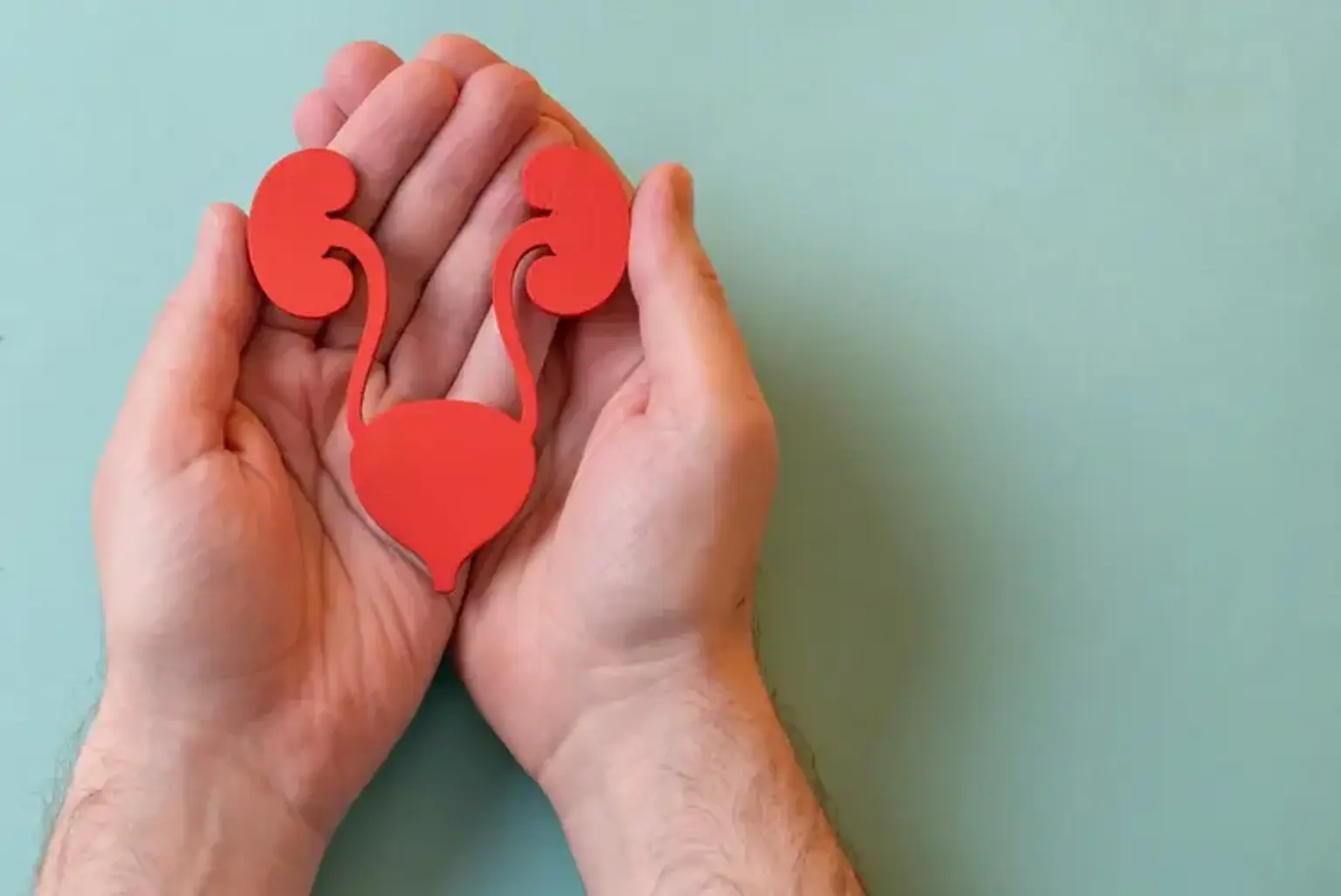
    Treatment for ureteral lesion, stricture, and blockage is often determined by the length of the defect, location, origin, and time of diagnosis. Laparoscopic ureteroneocystostomy is the surgery of choice for correcting distal ureteral injuries near the bladder without the need for major skin incisions. These injuries vary from more proximal injuries in that they are typically linked with disruption of the blood supply from the iliac arteries and so require a ureteroneocystostomy to be repaired.

    Correction of ureteral abnormalities longer than 5 cm is possible with modifications such as a psoas hitch (tacking the posterior bladder wall to the psoas muscle) and a Boari flap (tubularization of a bladder flap extending from the bladder to the ureteral orifice).

    Principles for obtaining successful ureteroneocystostomy outcomes include lack of tension, debridement and spatulation of the ureter, and postoperative drainage. 


    Structures & Functions of Kidney and urinary system

    Two kidneys

    • Two kidneys. This pair of purplish-brown organs is located below the ribs toward the middle of the back. Their function is to:
      • Remove waste products and drugs from the body.
      • Balance the body's fluids.
      • Release hormones to regulate blood pressure.
      • Control production of red blood cells.

    Urea is removed from the blood by the kidneys via microscopic filtering units known as nephrons. Each nephron is made up of a glomerulus, which is a ball of tiny blood capillaries, and a renal tubule, which is a small tube. Urine is formed as urea, water, and other waste substances travel through the nephrons and through the renal tubules of the kidney.

    • Two ureters. These thin tubes carry urine from the kidneys to the bladder. Muscles in the ureter walls contract and relax constantly, driving urine downward and away from the kidneys. A kidney infection might develop if urine backs up or is allowed to stand still. The ureters empty little volumes of urine into the bladder every 10 to 15 seconds.

    • Bladder. This hollow, triangle-shaped organ is found in the lower abdomen. Ligaments linked to other organs and the pelvic bones hold it in place. The walls of the bladder relax and expand to hold urine and contract and flatten to discharge urine through the urethra. The bladder of a healthy adult can hold up to two cups of urine for two to five hours. Upon examination, specific "landmarks" are used to describe the location of any irregularities in the bladder. These are:
      • Trigone: a triangle-shaped region near the junction of the urethra and the bladder.
      • Right and left lateral walls: walls on either side of the trigone.
      • Posterior wall: back wall.
      • Dome: roof of the bladder.
    • Two sphincter muscles. These circular muscles help keep urine from leaking by closing tightly like a rubber band around the opening of the bladder.

    • Nerves in the bladder. The nerves alert a person when it is time to urinate, or empty the bladder.

    • Urethra. This tube helps urine to exit the body. The brain causes the bladder muscles to contract, pushing pee out of the bladder. At the same moment, the brain instructs the sphincter muscles to relax, allowing urine to escape the bladder via the urethra. Normal urination happens when all of the signals occur in the proper order. 


    How Does Urinary System Work?

    urinary system filters blood

    The urinary system filters blood and produces urine as a waste product. The urinary system organs include the kidneys, renal pelvis, ureters, bladder, and urethra.

    The body uses nutrients from food to create energy. After the body has absorbed the necessary meal components, waste products are excreted in the colon and blood.

    The kidney and urinary systems aid in the elimination of urea, as well as the balance of substances such as potassium and sodium and water. Urea is created in the body when protein-containing meals, such as meat, poultry, and some vegetables, are broken down. Urea travels via the circulation to the kidneys, where it is excreted together with water and other wastes as urine.

    Blood pressure management and the generation of erythropoietin, which governs red blood cell formation in the bone marrow, are two other key roles of the kidneys. Kidneys also maintain fluid balance and control acid-base balance.


    Indications for Laparoscopic Ureteroneocystostomy

    Indications for Laparoscopic Ureteroneocystostomy

    Injury, stricture, or blockage of the ureter's distal 3-4 cm is a reason for laparoscopic ureteroneocystostomy. A vesico-psoas hitch or a Boari bladder flap can be used to bridge a more severe ureter loss. The distal ureter is the site of approximately one-third of traumatic injuries and the majority of iatrogenic injuries during pelvic operations. Other indications for ureteroneocystostomy include distal ureteral tumors that cannot be removed endoscopically, pelvic ureter malignancies, and renal transplantation and problems from transplanted kidneys. In the pediatric population, VUR is the most common indication for ureteroneocystostomy. However, ureteroneocystostomy with or without ureteral tapering is also performed for obstructive megaureters.

    • Indications for psoas hitch
      The psoas hitch is an effective method for bridging a gap in the bottom portion of the ureter. Distal ureteral damage, ureteral fistulae caused by pelvic surgery, segmental excision of a distal ureteral tumor, and unsuccessful ureteroneocystostomy are all indications.

    • Indications for Boari flap
      When the diseased portion of the ureter is too lengthy or ureteral mobility is too limited to conduct a primary ureteroneocystostomy, the Boari flap is a suitable adjuvant. Boari flaps may be made to span a 10- to 15-cm gap. In rare cases, spiral bladder flaps can be formed to reach the renal pelvis.


    When Laparoscopic Ureteroneocystostomy is Contraindicated?

    neuropathic bladder

    Significant bladder pathology linked with high intravenous pressures and poor compliance (e.g, neuropathic bladder) precludes ureteroneocystostomy. Individuals with defective voiding are more likely to develop problems, such as chronic VUR and blockage. It is equally dangerous to do ureteroneocystostomy on an irradiated bladder. In individuals with a history of bladder cancer, ureteroneocystostomy is not indicated.

    The Politano-Leadbetter ureteral reimplantation is contraindicated after puberty or in patients with megaureters.


    • Contraindications to psoas hitch and bladder flap

    A small constricted bladder with limited mobilization is a contraindication to psoas hitch. Furthermore, ureteral abnormalities at the pelvic brim frequently need more than a simple psoas hitch.

    Small bladder capacity is most likely related to difficult or insufficient Boari flap formation, necessitating evaluation of other procedures. In addition, urodynamic investigations should be performed prior to surgery to establish detrusor capacity and compliance if bladder outlet blockage, small capacity/high pressure, or neurogenic bladder is a possibility. Preoperatively, any existing bladder outlet blockage or neurogenic dysfunction must be treated.


    Laparoscopic Ureteroneocystostomy Success Rates

    Laparoscopic Ureteroneocystostomy Success Rates

    • In both adults and children, the success rate of ureteral reimplantation with a psoas hitch approaches 85%.
    • The success rate of laparoscopic ureteroneocystostomy in patients with low-grade primary VUR approaches 100% in expert hands.
    • In the pediatric population, nerve-sparing robotic extravesical ureteral reimplantation had a reported success rate of 97.6%.
    • In individuals having different operations such as psoas hitch, psoas hitch + Boari flap, and extravesical ureteral reimplantation, laparoscopic ureteral reimplantation had a success rate of 95.8%.
    • Although earlier studies of transvesicoscopic ureteral reimplantation demonstrated lower success rates (62%-79%), compared with open techniques, later studies have demonstrated more comparable success rates, ranging from 92%-96%.


    Preparations Before the Procedure

    Laparoscopic Ureteroneocystostomy Procedure

    When general anesthesia is required, there are specific food and drinking recommendations that must be followed in the hours preceding operation. A surgical nurse will call you between the hours of 1 and 9 p.m. one working day before your child's operation. (These calls are not made by nurses on weekends or holidays.) Please have paper and a pen on hand to record these vital instructions.

    Based on your child's age, the nurse will offer you particular feeding and drinking instructions. The following are the standard eating and drinking instructions. Regardless of your child's age, you must follow the particular instructions given to you on the phone by the nurse.

    For children older than 12 months: 

    • After midnight the night before the surgery, do not give any solid food or non-clear liquids. That includes milk, formula, juices with pulp, chewing gum or candy.

    For infants under 12 months: 

    • Up to 6 hours before the scheduled arrival time, formula-fed babies may be given formula. 
    • Up to 4 hours before the scheduled arrival time, breastfed babies may nurse.

    For all children:

    • Give only clear liquids up to 2 hours before the anticipated arrival time. Clear drinks include water, Pedialyte®, Kool-Aid®, and see-through juices like apple or white grape juice. Milk is not a transparent liquid.
    • Give nothing to eat or drink for 2 hours before the anticipated arrival time.
    • You may bring a "comfort" object for your kid to hold before and after the surgery, such as a favorite stuffed animal or "blankie."


    What Happens During the Procedure?

    diagnostic cystoscopy

    1. The patient is put in supine decubitus Trendelenburg position for transperitoneal laparoscopy after a diagnostic cystoscopy and RGP under general anesthesia in lithotomy posture.
    2. Before the surgery, a Foley catheter is inserted using a sterile method.
    3. The pneumoperitoneum is formed using the open Hasson's approach, and a 10 mm trocar for the 0° lens laparoscope is inserted at the umbilicus. There are four 5 mm ports, two on each side of the umbilical port.
    4. Following the release of any adhesions in the abdominal cavity, the ureter is dissected. It is released from above the iliac vascular junction until its passage into the bladder.
    5. Ureteral dissection must be gentle so that no healthy tissue is devascularized. The ureter is dissected distally up to the point where it is surrounded by scar tissue. 
    6. The bladder is totally freed and a completely open space of Retzius is observed. This step provides sufficient bladder mobility. 
    7. The proximal end of the stricture is spatulated. 
    8. The detrusor muscle is opened lengthwise for approximately 3 cm to expose the vesical mucosa. The vesical mucosa is then opened, and posterior ureterovesical anastomosis is done. 
    9. Then a DJ stent is placed, and anterior layer of ureterovesical anastomosis is done with interrupted vicryl 4.0 sutures. 
    10. In cases of tension due to the high ureteral stenosis, the ureteroneocystostomy with a psoas hitch muscle or Boari Flap technique is carried out. 
    11. For a psoas hitch procedure, interrupted polydioxanone sutures are used to approximate the bladder dome to the psoas tendon to allow a tension-free vesicoureteral anastomosis. 
    12. Contralateral vesicle pedicle is sacrificed if needed (it was done in only 4 cases). 
    13. A flap was raised on the anterolateral bladder wall for the Boari method, with the base and tip being 4 and 3 cm wide, respectively. The flap's tip was located immediately proximal to the bladder neck, while its base was located at the dome. The spatulated ureter and bladder flap were subsequently anastomosed tension-free using 4-0 polyglactin sutures over a 6Fr/26 cm DJ stent.
    14. Sutures were used to close the bladder in a single layer. At the end of the treatment, a soft silastic tube drain was placed into the pelvis. The drain was removed after 48 hours, the urethral catheter was removed after 7 days, and the DJ stent was removed after 6 weeks.
    15. A laparoscopic ureteroneocystostomy renal scan and/or excretory urogram was carried out 3 months after DJ catheter removal to rule out any obstruction. Follow-up was done every 6 months for at least 2 years with a renal scan.


    Post-operative Care

    Post-operative Care

    • Activity
      • Allow your child to gradually increase his or her activity level. Allow your child to rest as much as he or she requires. Make certain that your kid receives adequate rest at night.
      • If your child is old enough to walk, encourage them to walk every day. Increase your child's walking time gradually. Your child may be able to climb stairs. Walking increases blood flow and aids in the prevention of pneumonia and constipation.
      • Unless your kid still has a urinary catheter or drain, he or she may take a shower or bath. If your child has a catheter or drain, follow your doctor's bathing recommendations.
      • If your kid attends school or daycare, he or she may return whenever they are ready. This normally takes 1 to 2 weeks.
      • Allow your child to rest for 4 to 6 weeks, or until your doctor says it is safe. This includes biking, running games, wrestling, and participating in gym class.
      • Allow your child to swim or use hot tubs only if the doctor advises so.
      • Your kid is free to travel in the car with the car seat straps in their normal position.
    • Diet
      • Your child is able to eat a normal diet. If your child's stomach is upset, consider bland, low-fat items such plain rice, broiled chicken, toast, and yogurt.
      • Your child should drink plenty of fluids.
      • You may notice a change in your child's bowel habits immediately following surgery. This is common. If your child has not had a bowel movement after a couple of days, call the doctor.
    • Medicines
      • Your doctor will tell you if and when your child can restart any medicines. The doctor will also give you instructions about your child taking any new medicines.
      • The doctor may give your child medicine for bladder spasms. 
      • Make sure that your child takes pain medicines exactly as directed.
        • If the doctor gave your child a prescription medicine for pain, give it as prescribed.
        • If your child is not taking a prescription pain medicine, ask the doctor if your child can take an over-the-counter medicine.
      • If you think pain medicine is making your child sick to their stomach:
        • Give your child the medicine after meals (unless the doctor has told you not to).
        • Ask your child's doctor for a different pain medicine.
      • Your doctor may prescribe antibiotics. Your child should take them as directed. Do not stop giving them just because your child feels better. Your child needs to take the full course of antibiotics.
    • Incision care
      • If your child has strips of tape on the incision, leave the tape on for a week or until it falls off.
      • Every day, wash the area with warm, soapy water and pat it dry. Avoid using hydrogen peroxide or alcohol, both of which can slow recovery. You may cover the area with a gauze bandage if it weeps or rubs against clothing. Change the bandage every day.
      • Keep the area clean and dry.
    • Other instructions
      • Near the incision, your child may have a urinary catheter or a drain. Your doctor will advise you on how to care for it.
      • Your child may not be able to control when they urinate for a few weeks. For the next 2 to 3 weeks, your kid may require a diaper or pad to absorb leaking urine. Assure your child that this is common after surgery and will pass in time.

    Follow-up care is an essential component of your child's treatment and safety. Make and keep all appointments, and if your kid is having issues, call your doctor or the nurse call line. It's also a good idea to be aware of your child's test findings and to keep a record of the medications he or she is taking.


    Conclusion

    Laparoscopic Ureteroneocystostomy

    Laparoscopic ureteroneocystostomy is a procedure that uses a laparoscope to change the way a ureter joins to the bladder. The ureter is the tube that connects the kidney to the bladder.

    When a ureter is not properly connected to the bladder, urine can flow backward from the bladder into the kidney. Infections and renal damage might result from this. These issues can be avoided with surgery.

    Your child will be asleep during the surgery. First, the doctor makes a cut in your child's lower belly. This cut is called an incision. Then the doctor takes the ureter out of the bladder. Next, he or she connects it in a different place so urine cannot go backwards into the kidneys. Then the doctor closes the incision with stitches. The incision leaves a scar that usually fades with time.

    Most children go home 2 to 4 days after surgery.