Laparotomy

Last updated date: 02-Mar-2023

Originally Written in English

Laparotomy

Overview

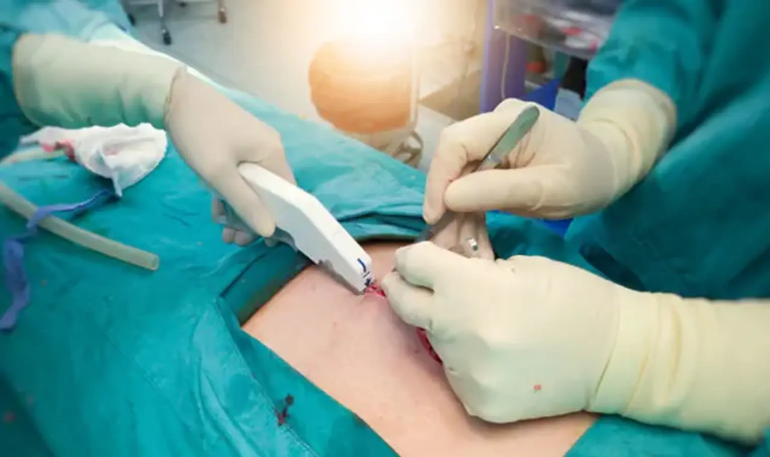
A laparotomy, also known as a celiotomy, is a surgical procedure that involves creating a wide incision in the belly to get access to the peritoneal cavity. 

A laparotomy is a surgical incision made into the abdomen. A laparotomy is used to examine the abdominal organs and help diagnose any issues. Infection and scar tissue development within the abdominal cavity is also possible risks.

A conventional laparotomy is performed by making a sagittal, midline incision along the linea alba.


Laparotomy definition

Laparotomy definition

The term laparotomy comes from the Greek terms lapara, which means flank, and tomy, which means cut. In surgical terms, this means making a large cut in the abdomen to gain access to the peritoneal cavity. A conventional laparotomy is often performed by making an incision in the midline along the linea alba. This is a popular treatment in the United Kingdom, with between 30,000 to 50,000 procedures performed each year.

Ephraim McDowell conducted the first successful laparotomy without anesthetic in Danville, Kentucky in 1809. George E. Goodfellow treated a miner outside Tombstone, Arizona Territory, who had been shot in the abdomen on July 13, 1881. When Goodfellow performed the first laparotomy to cure a gunshot wound, he was able to operate on the guy nine days after he was hit.


Anatomy and Physiology

Laparotomy Anatomy and Physiology

The abdominal wall protects the abdominal viscera by enclosing the abdominal cavity. Skin, subcutaneous fat, fascia of Camper, fascia of Scarpa, external oblique muscle, internal oblique muscle, rectus abdominis muscle, transverse abdominis muscle, pyramidalis muscle, transversalis fascia, and peritoneum are some of the layers of the anterior abdominal wall that may be encountered during a laparotomy.

The rectus abdominis muscles are two long vertical muscles in the abdominal wall on either side of the midline. The linea alba, a fibrous avascular plane that extends from the xiphoid process of the sternum superiorly to the pubic symphysis inferiorly, connects the two recti in the midline. The linea semilunaris is a surface feature formed by the lateral boundary of the two sides of this muscle. This muscle is separated by fibrous junctions that connect to the linea alba to provide the appearance of a six-pack.

The rectus abdominis connects to the pubic crest and inserts into the xiphoid process and costal cartilages of ribs 5–7. It is a flat muscle that compresses the abdominal viscera and stabilizes the pelvis during motions like walking. The thoracoabdominal nerve innervates it, which is supplied by nerve roots T7-T11.

The pyramidalis is a tiny triangular muscle, as the name implies. It is situated superficially and inferiorly to the rectus abdominis and is linked to the linea alba, with its base on the pubis bone.

The rectus sheath, which is created from the aponeuroses of the external oblique, internal oblique, and transversus abdominis, encloses the rectus abdominis and pyramidalis muscles. The rectus sheath is divided into two parts: anterior and posterior.

  • Anterior rectus sheath – made from the aponeuroses of the external oblique and half of the internal oblique.
  • Posterior rectus sheath – made from the aponeuroses of the external oblique and of half of the internal oblique.

Below the arcuate line, however, there is no posterior rectus sheath. This position is roughly halfway between the umbilicus and the pubic symphysis, putting the transversalis fascia in direct touch with the rectus abdominis.

The blood supply to the abdominal wall might be thought of as dual. The inferior and superior epigastric arteries combine to produce the deep epigastric arcade, which is located between the rectus abdominis muscle and the posterior rectus sheath.

Perforating veins supply the rectus muscle and branch out to supply the linea alba. The segmental arteries, which come from the aorta and supply the oblique and transverse muscles, are the second major supply. These connect the internal and transverse oblique muscles.

The abdominal wall is innervated by the thoracoabdominal nerve, the iliohypogastric and ilioinguinal nerves, and the ventral branches of the 5 to 12 thoracic nerves.


Indications

Laparotomy Indications

Since the introduction of minimum access surgery, the indications for a laparotomy have drastically decreased. It is crucial to emphasize, however, that access is essential for any surgical surgery. In many cases, keyhole surgery is exceedingly difficult, if not impossible, because the danger of injury to important structures outweighs the benefits of low access techniques. The more classic laparotomy remains the norm in these settings, whether emergency or elective.

Multiple thick adhesions from prior operations or inflammatory diseases, excessively inflated intestines in intestinal blockage, or enormous ascites in individuals with end-stage liver or heart illness are examples of these scenarios.

The most common causes for laparotomy include acute intraperitoneal bleeding, unmanageable gastrointestinal bleeding, traumatic or penetrating abdominal injuries, and widespread intraperitoneal infection owing to a ruptured gastrointestinal tract. Laparotomy is also indicated for elective surgeries involving a big specimen, such as pancreaticoduodenectomy, pancreatic or intestine transplants.


Contraindications

The most essential contraindication to be aware of is a patient who is unable to undergo general anesthesia owing to a combination of variables such as co-morbidities, sepsis, hemodynamic instability, and extensive metastatic malignant illness. The patient's desires are also highly essential, since it is their right to refuse a laparotomy if they have the mental ability to do so.

A thorough clinical evaluation and relevant radiological investigations are essential, as one should always maintain a high index of suspicion for medical conditions that may mimic acute abdomen, resulting in a false indication for laparotomy, such as acute pancreatitis, hyperglycemia, gout, gastritis, or urinary tract infection.


The surgical procedure

Laparotomy surgical procedure

Equipment

The size of the tools used in a laparotomy might vary depending on the reason for the procedure. However, the following tools are used to simply acquire access to the abdomen:

  • Scalpel 
  • Monopolar cautery
  • Tooth forceps
  • Abdominal packs/swabs
  • Fraser-kelly clips
  • McIndoe scissors
  • Retractor
  • Suction


Personnel

The surgeon, surgeon's assistant, scrub nurse, anesthetist, anesthesist technician, operating department practitioner, and some theatre workers to act as runners in case extra kit is necessary are normally present for a laparotomy.


Preparation

Depending on the goal of the surgery, the amount of the physical and emotional parts of preparation might vary. In the event of cancer, for example, it would be ideal to enlist cancer nurse experts to assist with pre-operative counseling. If the patient wishes, it is necessary to talk with the patient's next of kin (NOK)/loved ones in an emergency crisis to help enhance the patient's support network during the acute stressful period.

Informed consent is also important for educating the patient and his or her family for what to expect and the possible results. If the patient is asleep or lacks ability, the surgeon can complete a Consent Form 4 to act in the patient's best interests. 

To complete a Consent form 4, the patient must have a recorded mental ability evaluation and, if feasible, be discussed with a close family member. Children under the age of consent must have permission form 2 completed, with the parent or guardian providing legal consent.

Physical preparation differs depending on the nature of the surgery and whether it is an emergency procedure or a scheduled, elective procedure.

The points to consider include the following:

  • Hair removal from the abdominal wall
  • Patient BMI – Weight reduction may be suggested before an elective ileostomy to increase the patient's anaesthetic appropriateness and lower the distance the small bowel mesentery needs to traverse to be brought to the skin without stress.
  • Previous operative scars/deformities of the abdominal wall - A previous operation increases the extent of adhesions.
  • Hernia presence
  • In a planned setting, one would consider the effect of smoking and diabetic control on wound healing.
  • To consider the positioning of a stoma site if appropriate – This is usually at a level where the patient can see it, access it easily, and not have it interfering with belts or skin folds. It should also ideally avoid the costal margin and umbilicus.
  • A nasogastric tube in cases of obstruction/perforation or if anticipating a postoperative ileus
  • Adequate fluid and electrolyte resuscitation
  • Urinary catheter
  • Intravenous antibiotics
  • Crossmatching of units of blood products
  • Appropriate imaging, e.g. CT AP


Technique

The following incisions are used for access into the abdominal cavity:

Midline/Median Approach

The most usual method is a midline laparotomy, which involves making an incision through the middle of the abdomen along the linea alba. Depending on the location of the disease, the incision size may be restricted. An upper gastrointestinal condition, for example, may not necessitate a lower midline incision. However, if greater access is required, the choice may always be expanded lengthwise.

Some surgeons will curve their incision around the umbilicus, but a more visually acceptable procedure involves withdrawing the umbilicus away from the midline with a Littlewood's clamp to maintain the incision vertically straight. The incision is often performed with a scalpel, however cutting cautery is another option for the skin cut. The subcutaneous fat and superficial fascial layers are then dissected down to the rectus sheath using coagulative cautery.

The linea alba is an avascular plane where the aponeuroses converge, hence muscle should not be encountered. Once the rectus sheath  has been dissected, two Fraser-kelly clips can be attached to the peritoneum and pushed up.

McIndoe scissors are then used to cut between the clips, allowing access to the peritoneal cavity. The surgeon next inserts his or her fingers into the hole and expands the peritoneum incision, taking care not to hurt any underlying tissues such as the colon.

Paramedian Approach

The vertical incision is made lateral to the linea alb to provide access to lateral/retroperitoneal organs such as the kidneys and adrenal glands. The linea semilunaris, which is the rectus's lateral boundary, is commonly employed as a marker. Because the surgeon is more likely to come into contact with numerous veins and nerves feeding the muscles of the abdomen wall, the paramedian incision increases the risk of muscle atrophy, hematoma, and nerve damage.

Transverse Approach

This method, as the name implies, involves making a transverse incision lateral to the umbilicus (compared to the previous approaches which are vertical). This is a frequent strategy since it provides the least amount of harm to the nerve supply to the abdominal muscles because it follows a dermatome and heals effectively. The rectus abdominis incision heals, resulting in a new tendinous junction. An open right hemicolectomy is an example of where this is used.

Pfannenstiel Approach

To provide access to the pelvic cavity, Pfannenstiel incisions are performed 5 cm superior to the pubis symphysis in a curved transverse way. Because the fascia around the bladder is thin, care must be taken not to perforate it when executing this incision. Intestinal loops are very frequent in this area. This incision is frequently utilized in emergency cesarean sections and as a point of extraction for pathological specimens excised elsewhere inside the abdominal cavity.

Subcostal Approach

This incision begins inferior to the xiphoid process and continues inferiorly and parallel to the costal border. To lessen the risk of post-operative discomfort and poor wound healing, the incision should be at least two fingerbreadths below the costal margin. When performed on the right side, it is used to access the gallbladder and liver, while when conducted on the left side, it is utilized to access the spleen. A rooftop incision is formed when the left and right subcostal incisions are connected at the midline.

Other

Other common laparotomy incisions include:

  • Kocher (right subcostal) incision, suitable for some liver, gallbladder, and biliary tract surgeries The Kocher incision, which is used for thyroid surgery, is a transverse, slightly curved incision about 2 cm above the sternoclavicular joints.
  • Pfannenstiel incision, a transverse incision exactly under the umbilicus and above the pubic symphysis The skin and subcutaneous tissue are incised transversally in the typical Pfannenstiel incision, but the linea alba is opened vertically. It is the preferred incision for Cesarean sections and abdominal hysterectomy for benign illness. The Maylard incision is a version of this incision in which the rectus abdominis muscles are sectioned transversally to allow for more access to the pelvis. 
  • Lumbotomy involves making a lumbar incision that allows access to the kidneys (which are retroperitoneal) without entering the peritoneal cavity. It is usually reserved for benign renal lesions. It has also been proposed for upper urinary tract surgery.
  • Cherney Incision – developed in 1941 by the American uro-gynecologic surgeon Leonid Sergius Cherney.


Complications

Laparotomy Complications

Laparotomy complications might be site-specific or universal, although they are typically impacted by conditions present at the time of the procedure. As such, it can be categorised as patient-related or operator-dependent, and it should, of course, consider the surgery itself. The following is a comprehensive list of potential complications:

  • Bleeding
  • Infection
  • Bruising
  • Seroma/ hematoma
  • Wound dehiscence
  • Necrosis
  • Incisional hernia
  • Chronic pain
  • Skin numbness
  • Fistulation with underlying structures
  • Raised intra-abdominal compartment pressure
  • Damage to underlying structures
  • Poor cosmesis


There are some general risks inherent to all operations:

  • Small parts of the lungs may collapse, increasing the risk of infection in the chest. Antibiotics and physical therapy may be required.
  • Leg clots cause discomfort and edema. A portion of this clot may occasionally break off and go to the lungs, which can be deadly.
  • Because of the pressure on the heart, you might have a heart attack or a stroke. Death is an exceedingly unlikely outcome for anybody having surgery.


Some women are at an increased risk of complications, including:

  • Women who are extremely overweight are more likely to get wound infections, chest infections, heart and lung issues, and blood clots.
  • Smokers are more likely to get wound and chest infections, as well as heart and lung problems and blood clots.


Using a similar approach, a unique global study of 1,409 children undergoing emergency laparotomy from 253 centers in 43 countries revealed that adjusted mortality in children following surgery may be up to 7 times higher in low-HDI and middle-HDI countries compared to high-HDI countries, resulting in 40 extra deaths per 1000 procedures performed in these settings. Appendectomy, small bowel resection, pyloromyotomy, and intussusception treatment were the most frequent surgeries done globally.


Clinical Significance

In most surgical circles, the term laparotomy suggests a potentially large procedure; nevertheless, the terminology does not specify what operation the patient will undergo once the major open incision is completed. With advancements in minimally invasive surgery and robotics, an open cut is being used less frequently unless clinically necessary, such as in an emergency case when entry into the belly must be relatively quick or if the intra-abdominal environment is not receptive to alternative approaches.

As a result, it is critical that the patient be properly resuscitated, electrolytes and sepsis are treated, and that the surgical cut is made with care to limit the shock to the patient's physiology.


Discharge advice

  • It is important that you stay in the company of a responsible adult within the Brisbane region for 24 hours anddo not consume alcohol for the remainder of the day.
  1. Do not drive or operate any heavy machinery
  2. Do not consume alcohol for the remainder of the day
  3. Do not drive a car, motorbike or ride a bicycle until you can comfortably operate foot pedals and / or change gears
  4. Do not sign any legal documents or make any important decisions
  5. Do not engage in sports or heavy lifting.


  • Depending on your job and the severity of your sickness, you may need to take four to six weeks off.
  • A dressing may conceal staples or stitches in your abdominal incision. When they should be removed, your doctor will advise you.
  • If necessary, you will be given particular discharge medicine, although you may use paracetamol (Panadol) as needed (one to two tablets every four hours up to a maximum of eight tablets per day).
  • It is critical that you shower rather than bathe.
  • It is critical that you use sanitary pads rather than tampons.


Conclusion 

A laparotomy may be required to investigate abnormalities in the abdomen or internal organs, or to treat a specific ailment. It may be performed, for example, to determine the cause of abdominal (tummy) discomfort, if you have a damage to your abdomen, or to monitor the progression of certain disorders like as endometriosis and cancer. It is sometimes done in an emergency.

The surgery is often carried out under a general anaesthesia. A drip is placed in your arm. After you have been sedated, a catheter (a tube for urine drainage) is implanted. The incision is typically 15–20 cm long and located below the bikini line. In rare situations, rather of cutting across the abdomen, it may be required to cut down the abdomen from the belly button to the pubic area. Depending on the complexity of the operation, the process might take anywhere from one to several hours.