Laryngotomy
Last updated date: 10-May-2023
Originally Written in English
Laryngotomy
Whether blocked, traumatically damaged, or externally crushed, the acute airway necessitates immediate treatment. Acute airway treatment has been documented. Despite advances in our understanding of the structure, physiology, and disease causation, managing the acute airway remains difficult. Acute airway obstruction can occur in the prehospital context, as well as in the emergency department, intensive care units, and operating theaters. Acute airway management may be entrusted to clinicians at different stages of clinical development. Moreover, developments in technology and anesthesia have enabled a greater number of professionals to deal with acute airway obstructions.
In the treatment of the acute airway, the thoracic surgeon, particularly, has an extra responsibility. Post-intubation tracheal stenosis and cancerous airway lesions are the most prevalent tracheobronchial trunk diseases that can lead to life-threatening airway blockage if not managed and monitored adequately. As a result, thoracic surgeons are responsible for the proper and meticulous treatment and follow-up of these patients.
To be skilled at respiratory support, the practitioner must be familiar with the airway's important anatomical, physiological, and pathological aspects. They should also be familiar with the numerous tools and approaches designed for this goal. It's also crucial to understand the indications, contraindications, and risks of endotracheal intubation. Understanding how to assess the confirmation of correct endotracheal tube placement is critical. It's also important to understand the distinctions between adult, pediatric, and neonatal airways, as well as difficult airways, as these can have a major effect on airway safety and effectiveness.
There are three types of laryngotomy: superior laryngotomy (thyrohyoidotomy or sub-hyoid laryngotomy), median laryngotomy (thyrotomy), and inferior laryngotomy (cricothyroidotomy or cricothyrotomy).
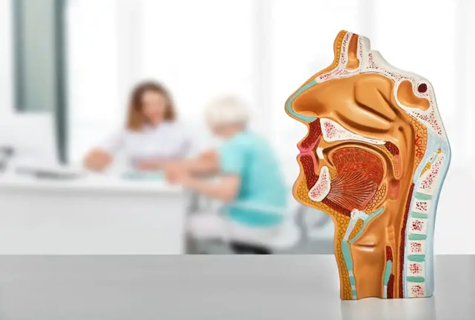
Anatomy and Physiology
The larynx connects the inferior region of the pharynx to the trachea and is located in the anterior neck. There are six cartilages, three of which are singular (thyroid, cricoid, and epiglottic) and three of which are pairs (arytenoid, cuneiform, corniculate). Connective and muscle tissues surround each cartilage. Although the hyoid bone is not a component of the laryngeal structure and is connected to the thyroid cartilage via the thyrohyoid membrane, it plays a key role in the upper aerodigestive tracts' swallowing action. The larynx is divided into three sections for classification purposes, each of which comprises multiple subsites, depending on embryologic development:
- Supraglottis refers to the part of the larynx that is located above the apex of the ventricle. The ventricle, vestibular folding, arytenoids, aryepiglottic folds, and epiglottis are all included.
- The anterior and posterior commissures, as well as the vocal cords, make up the glottis.
- Subglottis runs from the glottis' lower border to the cricoid cartilages' inferior border.
The laryngeal mucosal membrane is made up of squamous epithelium with mucous glands interspersed. Stratified squamous epithelium lines the true vocal cords.
The internal branch of the superior laryngeal nerve supplies sensation to the supraglottic larynx. The superior laryngeal and recurrent laryngeal nerves provide dual sensory nerve input to the mucosa of the true vocal cords. The recurrent laryngeal nerve, on the other hand, provides sensory input to the subglottic larynx. Except for the cricothyroid muscle, which is supplied by the external laryngeal branch of the superior laryngeal nerve, the recurrent laryngeal nerve innervates the intrinsic muscles of the larynx. Branches of the superior and inferior thyroid arteries provide circulation to the larynx.
At levels 2 and 3, the supraglottic larynx possesses a dense lymphatic channel that drains into lymph nodes. The glottic part's lymphatic channel is quite limited, especially near the outer end of the true vocal cord, which is lymphatic-free. The lymph nodes of the paratracheal and deep jugular are drained by the subglottic larynx.
Cricothyrotomy
A tube is inserted through a cut in the cricothyroid membrane during cricothyrotomy. Doctors must have some understanding of the relevant anatomy in order to perform a cricothyrotomy under emergency conditions. The thyroid cartilage, inferiorly the cricoid cartilage, and laterally the bilateral cricothyroideus muscles surround the cricothyroid membrane. Begin by stroking the laryngeal prominence on the thyroid cartilage, sometimes referred to as the Adam's Apple. The vocal cords are located within the thyroid cartilage. Check the cricoid cartilages' spherical signet ring on the lower edge. The cricothyroid membrane is located approximately 2.5 cm below the laryngeal eminence and superior to the cricoid cartilage, and can be felt like a small depression between the two cartilaginous components.
Cricothyrotomy Indications
Emergency cricothyrotomy is the last stage in the emergency airway management protocol, and it is required when you are unable to intubate or oxygenate. Another name for Cannot Intubate, Cannot Oxygenate is Cannot Intubate, Cannot Ventilate. In a Cannot Intubate, Cannot Oxygenate situation, failure to notice and intervene can quickly lead to brain hypoxia and death of the patient. In a Cannot Intubate, Cannot Oxygenate situation, a percutaneous airway must be constructed right away.
In any Cannot Intubate, Cannot Oxygenate circumstance, an immediate cricothyrotomy is necessary. Cannot Intubate, Cannot Oxygenate is a condition that a clinician may meet in the following circumstances:
- Trauma to the mouth or maxillofacial region
- Trauma to the cervical spine
- Oral bleeding in plenty
- Endotracheal intubation is impossible due to anatomical problems
- Copious vomiting
Cricothyrotomy Contraindications
There are no absolute contraindications to emergency cricothyrotomy in the Cannot Intubate, Cannot Ventilate scenario.
Possible or confirmed tracheal surgery, a cracked larynx, laryngotracheal disruption, and children are all relative contraindications. Patients with acute laryngitis are more likely to develop subglottic stenosis, rendering elective cricothyrotomy a risky procedure. Because of the funnel shape of the child’s airway and a potentially higher likelihood of subglottic stenosis, cricothyrotomy is a relative contraindication in children aged 6 to 12.
Cricothyrotomy Preparation
The most significant obstacles to a surgical airway are frequently cognitive. The technique is no more difficult than inserting a chest tube or other common emergency procedure. However, in the physician's view, the expressions "failed airway" and "cannot intubate, cannot oxygenate" may be associated with personal failure, resulting in an unnecessary delay in the plan to reduce the neck. Regular practice on simulators or cadavers is required to become experienced with the technique, which will assist in reducing stress when the procedure is an important prerequisite. A clinician should go through a difficult airway algorithm before any intubation, particularly if it is a prospective difficult airway, to reduce the anxiety and difficulties of the final step in that pathway, emergency cricothyrotomy.
Cricothyrotomy Technique
To gain tracheal entrance, the science describes a variety of procedures that include a variety of instruments, dilators, and special cannulas. The bougie-assisted cricothyrotomy, also known as the Three-Step Technique, integrates the most significant steps in both the Seldinger approach and the rapid four-step surgical approach, making it easy to access technique to increase success in this rarely performed, high-stress situation.
- Locate the cricothyroid membrane with the index finger while supporting the larynx between the thumb and middle finger with your non-dominant hand. In the skin overlying the cricothyroid membrane, produce a 3 cm vertical cut.
- Divide the subcutaneous tissue with your fingers until the cricothyroid membrane is visible.
- Puncture the cricothyroid membrane vertically with the scalpel.
- Make a small cut with your finger inserted inside.
- Insert an elastic bougie into the trachea through the cut, using the finger to direct it inferiorly.
- Pass a cuffed endotracheal tube over the bougie and expand the cuff until the balloon is no longer visible.
- End-tidal capnography is used to validate placement with a Bag-Valve-Mask.
- Use a securement device to keep the endotracheal tube in place.
Standard Cricothyrotomy
- Stabilize the larynx and use the non-preferred hand's index finger to palpate the cricothyroid membrane. The lower margin of the thyroid cartilage and the upper margin of the cricoid cartilage in the center of the neck are used to accomplish this.
- Make a vertical cut in the skin overlying the cricothyroid membrane in the center of the neck, extending the incision roughly 2-4 cm in length while keeping the larynx stable.
- Palpate the cricothyroid membrane and make a horizontal cut into the membrane after you've made the vertical skin cut. Make sure the scalpel is pointed inferiorly to avoid the vocal cords and make the cut gently to avoid the tracheas' posterior wall.
- While inserting a tracheal hook into the opening under the thyroid cartilage, place the tip of the index finger in the cut through the cricothyroid membrane. Grasp Adam’s apple and pull it upwards.
- To widen the horizontal cut vertically, use a dilator.
- Push the tracheostomy tube inferiorly into the trachea through the dilator.
- Release the tracheal hook and dilator.
- Withdraw the tracheostomy tubes' obturator.
- Place the tracheostomy tube's cannula.
- Fill the balloon with air.
- Connect the tube to a ventilator or a Bag-Valve-Mask.
Needle Cricothyrotomy
- Extend the skin in a vertical manner with the thumb and middle finger of the non-dominant hand, palpating the cricothyroid membrane with the index finger to immobilize the larynx and produce tension overlaying the cricothyroid membrane.
- With your dominant hand, puncture the skin covering the cricothyroid membrane at the lower border with a syringe filled with saline connected to a catheter. At a 30–45-degree angle, direct the needle inferiorly. Apply continual negative pressure to the syringe as you advance. You've confirmed placement within the trachea when you see air bubbles in the syringe. Stop moving the needle forward.
- Advance the catheter until the hub comes into contact with the skin. Withdraw the needle.
- Connect the saline syringe to the catheter and aspirate air to check intratracheal insertion.
- In all circumstances, keep the catheter in position. Do not rely on a suture to keep the catheter in the right position.
- Attach the catheter to a Bag-Valve-Mask with 100 percent oxygen or a high-pressure tube. Ventilation should be done at a rate of 9-11 breaths per minute.
Cricothyrotomy Complications
The emergent cricothyrotomy is the final stage in the Cannot intubate, Cannot oxygenate strategy, and it is performed to save a patient's life. As a result, it is a surgery where the benefit far exceeds the danger. The treatment, however, is not without risks. Complication rates vary from study to study, depending on the clinical circumstance, quality of training, and procedure location and can range from 1% to 53%. Hemorrhage is the most prevalent problem. Hemorrhage is to be anticipated during the surgery and should be overlooked. If excessive bleeding occurs, apply pressure or apply packing to the wound. Bruises of the tracheal cartilage, such as the thyroid, cricoid, or tracheal rings, penetration of the trachea, development of a false tract (placement of the endotracheal tube into a possible region other than the trachea), and infection are other urgent problems. Subglottic stenosis and vocal alterations are long-term problems.
Thyrotomy
Thyrotomy is an effective surgical treatment for laryngeal abnormalities; nevertheless, web formation at the anterior section of the vocal cords is a common and annoying side effect. Hayashi's hemi-thyrotomy does not lead to the formation of a web because the anterior commissure is not damaged. However, intralaryngeal access is insufficient for this kind of procedure. Because the anterior inferior section of the thyroid cartilage is excised in Huet's and Thomson's extended thyrotomy, an excellent exposure of the intralaryngeal cavity is gained, more so than in regular thyrotomy. Although the removal of the cartilage has no effect on function, a web is always created at the anterior commissure, just as it is with a standard thyrotomy.
To avoid web development, partial thyrotomy was developed on the basis of two principles: first, not to harm the anterior commissure; and second, to excise the anterior inferior section of the thyroid cartilage to provide a larger operation area.
Thyrotomy Indications
The following are the indications for partial thyrotomy:
- Any tumor in the subglottic space
- Any tumor on the lower surface of the vocal cord, particularly if peroral removal is thought to be tough.
- Glottis aging stenosis or adherence of both vocal cords after laryngeal injury
Thyrotomy Technique
The thyrotomy procedure is done in the following order:
- At the level of the lower border of the thyroid cartilage, a transverse small cut is made over the front part of the neck. If there is a crease at this point, it is quite easy to make a cut along with it. Superior and inferior skin flaps, including the platysma muscles, are produced and stitched to the skin near the cut. The operation area is kept wide during the surgery using this approach, which eliminates the need for a retractor.
- The thyroid and the cricoid cartilages are accessible after the fascia surrounding the strap muscles is dissected in the center. The cricothyroid membranes' pre-laryngeal lymph node must be excised and histopathologically analyzed.
- The perichondral cut is made at the thyroid's lower margin of the thyroid cartilage. This cut does not continue beyond the interior boundary of the point where both cricothyroid muscles meet. A small scoop is used to scrape the outer perichondrium upward to the midpoint between the superior incisura and the inferior margin of the thyroid cartilage. The anterior commissure is located a little higher than this position. The perichondrium's inner layer is likewise pulled away. Then, with little bone forceps, the anterior inferior section of the thyroid ala is excised, being careful not to harm the cricothyroid muscles.
- During intralaryngeal surgery, infiltration anesthesia of the cricothyroid membrane is useful in reducing bleeding. A vertical cut is made on the cricothyroid membrane from a few millimeters above the upper border of the cricoid cartilage to below the anterior commissure of the vocal cords after 4 percent Xylocaine solution is injected into the trachea. A horizontal cut is created from the lower end of this vertical cut toward the uninvolved end. It is sometimes produced on both ends of the cricothyroid muscles' inner edge. With the thread sewn on it, the cricothyroid membrane flap and the mucosal membrane of the subglottal cavity are mirrored laterally upward. This operation provides a clear view of the larynx's lower portion, allowing for intra-laryngeal operations.
- Before beginning the excision, the position and size of the lesion should be thoroughly checked once the larynx has been opened. The intralaryngeal surgery can be performed under a microscope.
- The laryngeal aperture is cautiously closed with interrupted chromic catgut buried stitches when the surgery is completed. Three stitches for the vertical cut and one for the horizontal cut are usually sufficient. The sternohyoid muscles are sutured, and the wound is sealed with continuous subcutaneous stitching to prevent skin scarring.
- A tracheostomy is not required in this procedure. There are no symptoms of breathlessness or hemoptysis. After the surgery, the patient remains silent for four or five days. There are no more issues with the voice.
Cricothyrotomy VS Tracheostomy
A tracheostomy is a procedure that involves making a hole (stoma) in the front of the neck and connecting it to the windpipe (trachea). To assist ventilation, a special tube (tracheostomy) is inserted into the orifice. When the normal breathing pathway is obstructed or limited, a tracheostomy is performed. When health issues necessitate the long-term use of a machine (ventilator) to support breathing, a tracheostomy is frequently required. When the airway is abruptly closed, such as after facial or head trauma, an emergency tracheotomy may be required. Unlike a cricothyroidotomy, a tracheostomy requires anesthesia.
When a tracheostomy is no longer required and the patient can breathe on his or her own, the tube is withdrawn, and the opening is either left to seal naturally or surgically repaired. A tracheostomy might be lifelong in some situations. The majority of tracheotomies are done in a hospital. When there are no other means to protect the airway in an emergency or accident, it may be done.
Cricothyroidotomy is used to construct an airway in an emergency because tracheostomy takes more time and is more challenging to do. It is a less invasive surgical treatment that results in less hemorrhage and consumes less time. An emergency tracheostomy is another term for it. A tracheostomy tube or endotracheal tube with various internal diameters is placed through an incision in the skin around Adam's apple (cricothyroid membrane).
Conclusion
Airway management in critically ill patients remains to be difficult for health care workers, even those with extensive experience and necessitates the use of particular standards and practices to avoid negative outcomes. Professional education, such as anesthesia clinical rotations, airway management classes, seminars, and simulation training, can provide the necessary airway management tools and abilities.
Debriefing and explicit task allocations must precede teamwork as a prerequisite for performance. Failure planning in a step-by-step strategy for success and airway management should be considered as soon as possible to avoid additional challenges and unfavorable outcomes. The delivery of oxygen during the airway intervention must be optimized at all times and should not be compromised for any reason. The front of the neck airway should be considered during initial preparation and completed as soon as possible. All steps of airway care must be documented and reports must contain specifics of challenges.