Introduction
LASIK (Laser-Assisted in Situ Keratomileusis) eye surgery is one of the most popular procedures globally for correcting refractive vision issues such as nearsightedness (myopia), farsightedness (hyperopia), and astigmatism. Over the years, it has become a go-to solution for those looking to reduce or eliminate their dependence on glasses and contact lenses. The procedure uses advanced laser technology to reshape the cornea, improving the way light enters the eye and enhancing vision clarity.
As LASIK continues to gain popularity, millions of people worldwide have benefited from this vision correction method, making it one of the most commonly performed surgeries.
What is LASIK Eye Surgery?
Defining LASIK
LASIK is a minimally invasive eye surgery designed to correct common vision problems. It works by reshaping the cornea—the clear, front part of the eye—so that light can focus properly onto the retina at the back of the eye. This laser treatment improves vision in those with myopia, hyperopia, and astigmatism, offering a permanent solution for many patients.
How LASIK Works
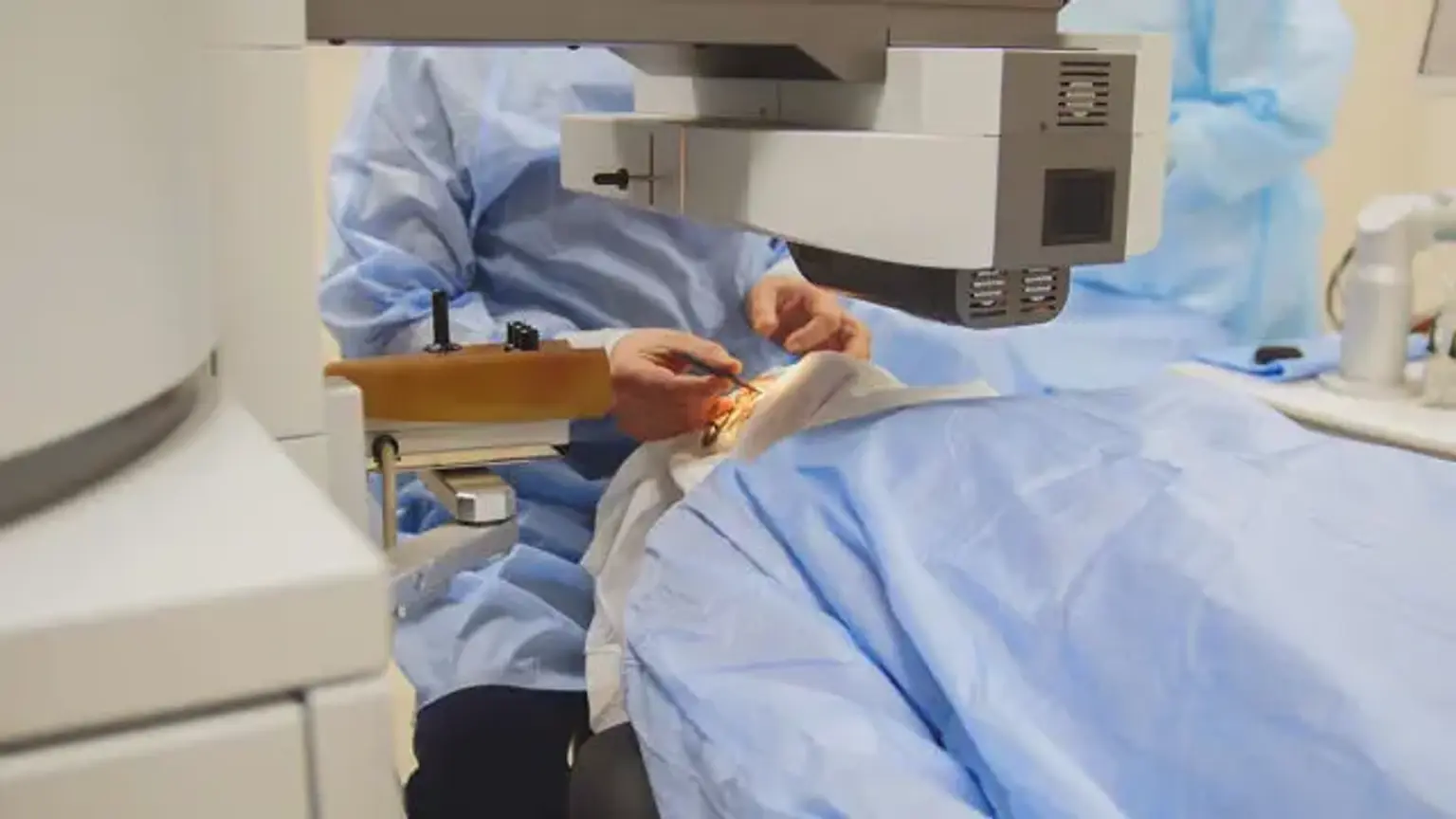
During LASIK surgery, a laser is used to precisely remove tissue from the cornea. The procedure begins with the creation of a thin flap on the cornea, which is lifted to allow the laser to reshape the underlying tissue. Once this is done, the flap is repositioned. Because the cornea heals quickly, stitches are typically not required.
Types of LASIK Procedures
There are several variations of LASIK, including:
Traditional LASIK: This involves using a microkeratome blade to create the corneal flap.
Bladeless LASIK (Femtosecond LASIK): A more advanced technique that uses a laser to create the flap, offering higher precision and a potentially safer procedure.
Custom LASIK: Tailored to individual vision needs, this method uses wavefront technology to map the eye’s unique characteristics and correct irregularities.
LASIK Procedure Details
Step-by-Step Explanation
Consultation: The process begins with a thorough eye exam, where your surgeon will assess your corneal thickness, vision prescription, and eye health to determine if you are a good candidate for LASIK.
The Surgery: On the day of the procedure, you will lie down, and numbing eye drops will be applied. The surgeon then creates a flap in the cornea, either using a blade or a laser, and reshapes the underlying tissue using an excimer laser. The flap is then carefully repositioned to begin the healing process.
Timeframe: The procedure typically lasts between 15 to 30 minutes for both eyes, and most patients experience a significant improvement in vision within hours.
Pain Management and Anesthesia
LASIK surgery is relatively painless. Patients receive numbing eye drops, so the procedure itself is comfortable, and there’s little to no discomfort. You might feel some pressure or a mild sensation in the eye during the surgery, but it’s usually not painful. Afterward, patients may experience mild irritation or dryness, which can be managed with prescribed eye drops.
Post-Surgery Care
After LASIK, you’ll be asked to rest for a few hours before going home. It's essential to avoid rubbing your eyes and to wear protective goggles while sleeping for the first few nights. You may experience blurred vision, glare, or dry eyes during the recovery period, but these symptoms typically subside within a few days to weeks.
Benefits and Risks of LASIK
Key Benefits of LASIK
Quick Results: Most patients notice improved vision within hours of the surgery, with full results appearing in a few days.
Freedom from Glasses and Contacts: LASIK allows many people to live without corrective lenses, making daily life more convenient and cost-effective.
Improved Self-Esteem: With clearer vision, many individuals experience a boost in confidence and an enhanced quality of life.
Common Risks and Complications
While LASIK is generally safe, there are some risks:
Dry Eyes: Temporary dry eyes are common after LASIK, but they usually resolve within a few months.
Visual Disturbances: Some people experience glare, halos, or double vision, particularly at night.
Over or Undercorrection: In some cases, LASIK may not fully correct vision, requiring a touch-up surgery.
It’s important to discuss these risks with your surgeon during the consultation to determine whether LASIK is right for you.
LASIK Success Rate
The procedure has an excellent success rate, with studies showing that over 90% of patients achieve 20/25 vision or better after LASIK. Most patients are highly satisfied with the results, and complications are rare when the surgery is performed by an experienced, qualified surgeon.
LASIK Candidacy and Suitability
Who Is a Good Candidate for LASIK?
Ideal candidates for LASIK are those who:
Are 18 years or older with a stable prescription.
Have healthy eyes without conditions like cataracts or glaucoma.
Have enough corneal thickness for the procedure.
LASIK for Astigmatism and Other Vision Problems
LASIK is effective for treating astigmatism, which causes blurry vision due to an irregularly shaped cornea. However, people with extremely high prescriptions or thin corneas may not be suitable candidates for LASIK, but alternatives like PRK (Photorefractive Keratectomy) may be offered.
Pre-Surgical Assessment
Before surgery, your surgeon will perform tests to evaluate the shape of your cornea, eye health, and the overall suitability for LASIK. This ensures that you’re ready for the procedure and will have the best possible outcome.
Who is a Good Candidate for LASIK?
LASIK Candidacy Requirements
Good candidates for LASIK typically meet the following criteria:
Over 18 years old with a stable vision prescription.
Good overall eye health, with no major conditions like cataracts or glaucoma.
Adequate corneal thickness to allow for reshaping.
LASIK for Astigmatism and Other Vision Issues
LASIK can also treat astigmatism, where the cornea is irregularly shaped, causing blurry vision. It’s important to note that LASIK may not be suitable for people with extremely high prescriptions or thin corneas, but alternative treatments like PRK (Photorefractive Keratectomy) may be recommended.
Pre-Surgical Assessment
Before undergoing LASIK, patients will undergo several tests to evaluate the health of their eyes and determine the best approach. These tests help assess the shape of the cornea, tear production, and overall eye health to ensure that LASIK is the right choice.
Costs and Insurance for LASIK
How Much Does LASIK Cost?
The cost of LASIK surgery varies depending on location and surgeon. Typically, LASIK ranges between $2,000 and $3,000 per eye, including pre-surgery consultations and post-op care. However, this cost can fluctuate based on the type of LASIK (e.g., Bladeless LASIK) and the clinic.
Insurance Coverage for LASIK
Most health insurance plans do not cover LASIK because it's considered an elective procedure. However, many clinics offer financing options, and some employers may provide discounts or flexible spending accounts that can help cover part of the cost.
LASIK Recovery and Long-Term Results
What to Expect Immediately After LASIK Surgery
After LASIK, most patients experience improved vision within hours, though it may be blurry at first. The recovery is quick, with many returning to normal activities within 1-2 days. You might experience mild discomfort, such as dryness or sensitivity to light, but this usually subsides within a few days.
Long-Term Results and LASIK Maintenance
LASIK results are typically long-lasting, with most patients achieving stable vision for years. However, some may experience gradual changes in vision over time due to aging or other factors. If needed, a "touch-up" procedure can be done to refine results, though this is rare.
Global Popularity of LASIK
LASIK’s Popularity Worldwide
LASIK has grown in popularity across the globe, with millions of procedures performed annually. It's particularly common in the US, Europe, and increasingly in Asia. The rise in demand reflects LASIK’s proven effectiveness and its ability to free people from glasses and contact lenses.
Why LASIK Is So Attractive
Its fast recovery time, minimal discomfort, and permanent results make LASIK appealing. The increasing affordability of the procedure and advancements in technology have also contributed to its popularity worldwide.
LASIK Technology and Advancements in the Last Decade
Advancements in LASIK Technology
In recent years, LASIK technology has advanced significantly, making the procedure safer and more precise. The introduction of bladeless LASIK (Femtosecond laser) and wavefront-guided LASIK (which customizes the surgery to the individual’s eye) has improved outcomes and reduced risks.
How Advancements Have Improved LASIK
Modern LASIK technology allows for more accurate flap creation, reducing the risk of complications. Enhanced mapping techniques also ensure that each patient’s eye is treated based on its unique characteristics, resulting in better visual outcomes.
Common Questions and FAQs About LASIK
Is LASIK Painful?
No, LASIK is generally not painful. Numbing eye drops ensure that you won’t feel discomfort during the procedure. Post-surgery, mild irritation may occur, but it is manageable and temporary.
How Do I Prepare for LASIK Surgery?
Preparation for LASIK involves a comprehensive eye exam and discussion with your surgeon about your health and vision needs. You’ll be asked to avoid wearing contact lenses before the procedure for a few weeks to ensure accurate measurements.
Can LASIK Fix All Types of Vision Problems?
LASIK is effective for correcting myopia, hyperopia, and astigmatism. However, it may not be suitable for people with severe vision issues or other eye conditions. Alternatives like PRK may be recommended in these cases.
How Can I Find the Right LASIK Surgeon?
It’s crucial to choose a qualified, experienced surgeon who specializes in LASIK. Look for reviews, ask for referrals, and ensure they use advanced technology. A detailed consultation will help you assess the surgeon’s expertise and approach to your specific needs.
LASIK and Its Impact on Quality of Life
How LASIK Improves Everyday Activities
LASIK can significantly enhance the quality of life for people who struggle with glasses or contacts. Imagine the freedom of waking up in the morning and seeing clearly without reaching for glasses or inserting contact lenses. It allows individuals to participate in activities like swimming, hiking, or playing sports without the limitations of corrective eyewear.
Boost to Self-Esteem and Confidence
For many, LASIK goes beyond just vision correction. It can provide a confidence boost, as individuals no longer have to worry about how they look with glasses or the hassle of contacts. Many patients report feeling more confident in their social and professional lives after undergoing the procedure.
Alternatives to LASIK Surgery
PRK (Photorefractive Keratectomy)
PRK is a similar procedure to LASIK but without creating a corneal flap. Instead, the outer layer of the cornea is removed to reshape the underlying tissue. While the recovery is longer than LASIK, PRK is a good option for individuals with thinner corneas or those who are not candidates for LASIK.
Other Vision Correction Options
In addition to LASIK and PRK, there are several other treatments for vision correction:
Implantable Contact Lenses (ICL): A lens is surgically placed inside the eye to correct vision without altering the cornea.
Cataract Surgery: In older adults, cataract surgery can also provide vision correction, often eliminating the need for glasses.
LASIK for Different Age Groups
Is LASIK Suitable for Younger Patients?
LASIK is usually recommended for people over the age of 18, as vision can still change during teenage years. Younger individuals who have stable prescriptions for at least a year are good candidates. It’s important to wait until the eyes are fully developed to avoid complications later.
LASIK for Older Adults
While LASIK is commonly performed on adults under 40, older adults may also benefit from the procedure, though they may have presbyopia (age-related farsightedness). LASIK can improve vision clarity, but additional treatments like reading glasses may still be necessary.
The Future of LASIK Surgery
Evolving Techniques and Future Possibilities
As technology continues to advance, LASIK is becoming even more precise and accessible. In the future, procedures may involve even less recovery time and improved outcomes, with better customization for patients’ specific needs. Ongoing research into eye health and laser technology could lead to even greater advancements in vision correction.
Expanding Global Access
In the coming years, LASIK surgery is expected to become more available worldwide, especially in developing countries. As technology becomes more affordable and surgeons gain more expertise, LASIK may continue to transform the way people experience and manage their vision.
Cost Considerations for LASIK Surgery
Understanding LASIK Pricing
The cost of LASIK can vary depending on factors like geographic location, the surgeon's experience, and the type of LASIK procedure performed. Typically, LASIK costs between $2,000 and $3,000 per eye. This price often includes the consultation, surgery, and follow-up care. However, more advanced LASIK options like bladeless or custom LASIK may cost more.
Financing and Payment Plans
Since most insurance plans don't cover LASIK as it's considered elective, many clinics offer financing options. Some patients may be able to use health savings accounts (HSAs) or flexible spending accounts (FSAs) to cover part of the expense. It’s important to discuss payment options with your clinic ahead of time to ensure you're prepared.
The Importance of Choosing a Qualified Surgeon
How to Choose a LASIK Surgeon
Choosing a skilled, experienced LASIK surgeon is one of the most crucial steps in ensuring a successful outcome. It’s essential to look for a surgeon who specializes in LASIK and has a strong track record with high patient satisfaction. Consider reading reviews, asking for referrals, and checking for board certifications.
Questions to Ask During Your Consultation
How many LASIK procedures have you performed?
What kind of technology do you use?
What is your complication rate?
Can you provide testimonials or before-and-after photos from previous patients?
A thorough consultation is key to building trust and understanding what you can expect from the procedure.
Safety Protocols and Considerations
Ensuring Safety Before, During, and After Surgery
LASIK is generally a safe procedure when performed by a qualified surgeon. However, safety precautions should be followed:
Pre-Surgery: Ensure your eyes are healthy, and follow the surgeon’s instructions regarding medications and contact lens removal before the procedure.
During Surgery: The procedure is minimally invasive, but ensure the clinic follows strict sterilization and patient safety protocols.
Post-Surgery: Follow all aftercare instructions, avoid rubbing your eyes, and attend follow-up appointments to monitor healing.
By following all safety guidelines, you can reduce the risk of complications and improve your chances for a successful outcome.
Conclusion
Weighing the Pros and Cons
LASIK is a highly effective and safe procedure for correcting common vision problems. With minimal recovery time and lasting results, it can significantly improve your quality of life by freeing you from glasses and contact lenses. However, it's important to carefully consider the costs, risks, and potential for side effects before deciding.
Making an Informed Decision
Consult with an experienced surgeon to determine if LASIK is right for you. A thorough pre-surgical evaluation will help you understand the procedure's suitability based on your eye health and vision needs. LASIK has helped millions worldwide, but it’s essential to make sure that it’s the best option for your specific situation.