Leiomyosarcoma

Last updated date: 09-Jun-2023

Originally Written in English

Leiomyosarcoma

Overview

Leiomyosarcoma is a kind of smooth muscle tumor that is malignant (cancerous). Smooth muscles are found in hollow organs such as the intestines, stomach, bladder, and blood arteries. The uterus contains smooth muscle in females as well. These smooth muscular fibres help carry blood, food, and other materials through the body without you ever realizing it.

Leiomyoma is a benign tumor that arises from the same tissue. While leiomyosarcomas are not expected to develop from leiomyomas, categorization of some leiomyoma types is changing.

Symptoms vary depending on the location and size of the tumor. Some persons with LMS have no symptoms when the malignancy first appears. Later, when the tumor grows in size, symptoms such as pain, unintentional weight loss, nausea and vomiting, and a lump beneath the skin may occur.
Leiomyosarcomas may be quite unexpected; they can be dormant for lengthy periods of time and then resurface years later. It is a resistant cancer, which means it is often unresponsive to chemotherapy or radiation. The best results occur when it can be removed surgically with broad margins when it is still tiny and in situ. 


What is Leiomyosarcoma?

Leiomyosarcoma Definition

Leiomyosarcoma is a cancerous tumor that develops from smooth muscle cells. There are two kinds of muscles in the body: voluntary and involuntary. Smooth muscles are involuntary muscles, meaning they are not controlled by the brain.

Smooth muscles respond automatically to a variety of stimuli. Smooth muscle, which lines the walls of the digestive system, for example, creates wave-like contractions (peristalsis) that help in digestion and food transport. Smooth muscles in the salivary glands cause the glands to pump saliva into the mouth when a bite of food is taken. In response to cold, smooth muscle in the skin causes goose bumps to develop.

Cancer is a category of disorders defined by abnormal, uncontrolled cellular development that invades surrounding tissues and can spread (metastasize) to distant body locations via the bloodstream, lymphatic system, or other ways.

Because smooth muscle is prevalent throughout the body, a leiomyosarcoma can develop practically everywhere there are blood vessels, including the heart, liver, pancreas, genitourinary and gastrointestinal tracts, the region behind the abdominal cavity (retroperitoneum), the uterus, and the skin. A Leiomyosarcoma is most commonly seen in the uterus. The majority of gastrointestinal leiomyosarcomas have been categorized as gastrointestinal stromal tumors. 


How Common is Leiomyosarcoma?

Common of Leiomyosarcoma

Leiomyosarcoma (LMS) is a kind of soft tissue sarcoma that accounts for 10% to 20% of all soft tissue sarcoma cases. Adults are more likely than children to have LMS. It is believed that only approximately 20 to 30 children in the United States are diagnosed with LMS each year. LMS of the uterus affects around 6 per 1 million persons in the United States each year.

According to the American Cancer Society, at least 15,000 new instances of soft tissue sarcoma are diagnosed in the United States each year. Soft tissue sarcomas afflict both men and women equally, and they strike adults more frequently than children or adolescents. Soft tissue sarcomas account for 1% of all adult malignancies in the United States. 


What Causes Leiomyosarcoma?

Causes of Leiomyosarcoma

It is uncertain what causes leiomyosarcoma. Researchers believe that hereditary factors may play a role in the development of LMS.

Malignancies can occur in cancer patients, including leiomyosarcoma patients, owing to aberrant changes in the structure and orientation of specific cells known as oncogenes or tumor suppressor genes. Oncogenes regulate cell proliferation, whereas tumor suppressor genes regulate cell division and ensure that cells die at the appropriate moment. The precise reason of these gene alterations is unclear.

 Current research, however, reveals that DNA (deoxyribonucleic acid), the bearer of the body's genetic code, defects are the fundamental basis of cellular malignant transformation. These aberrant genetic mutations might develop spontaneously for unknown reasons, or they can be inherited in rare cases. 


Risk Factors for Leiomyosarcoma

Risk Factors for Leiomyosarcoma

A risk factor is something that increases your chances of contracting an illness such as cancer. The risk factors for various malignancies varies. Unprotected exposure to bright sunlight, for example, is a risk factor for skin cancer & abnormalities. Smoking is a risk factor for lung cancer and many other cancers. However, risk variables do not tell us everything. Having a risk factor, or even several, does not guarantee that you will get cancer. Furthermore, many people get cancer without having a risk factor.

Still, scientists have found a few risk factors that make a person more likely to develop soft tissue sarcomas:

  • Radiation given to treat other cancers:

Radiation exposure is responsible for fewer than 5% of sarcomas. However, people may acquire sarcomas as a result of radiation used to treat other diseases, such as breast cancer or lymphoma. Sarcomas frequently develop in areas of the body that have been treated with radiation. The typical interval between radiation treatments and a sarcoma diagnosis is roughly ten years.

Radiation therapy procedures have gradually advanced during the last few decades. Treatments are now more accurate in their targeting of malignancies, and more is understood about choosing radiation dosages. These advancements are expected to lower the number of malignancies induced by radiation treatment. 

However, because these tumors take so long to grow, the effects of these mutations may not be recognized for a long time. Still, radiation treatment is utilized only when the benefits (increased survival rate and symptom alleviation) outweigh the hazards. More information may be found at Second Cancers in Adults.


  • Family cancer syndromes

Family cancer syndromes are conditions caused by gene flaws (mutations) that people are born with (typically inherited from a parent) and are associated with an increased risk of developing specific malignancies. Some cancer syndromes in families enhance a person's chance of acquiring soft tissue sarcomas.

1. Neurofibromatosis:

Von Recklinghausen disease is another name for neurofibromatosis. It often runs in families and causes a large number of benign (non-cancerous) tumors to grow in nerves beneath the skin and in other regions of the body (These are called neurofibromas.) It is caused by a flaw (mutation) in the genes NF1 and NF2. A sarcoma in a neurofibroma affects around 5% of persons with neurofibromatosis.

2. Gardner syndrome:

Gardner syndrome is a genetic disorder caused by APC gene mutations. This condition is a kind of familial adenomatous polyposis (FAP), and persons with it have a high risk of colon cancer and numerous polyps in the colon (and intestines). It also produces complications outside of the colon, such as desmoid tumors.

3. Li-Fraumeni syndrome:

Li-Fraumeni syndrome is caused by inherited TP53 gene defects. People who have this condition are at a higher risk of developing cancers such as breast cancer, brain tumors, leukemia, and sarcomas. Despite this, only 10 to 20 persons out of every 100 with Li-Fraumeni syndrome will develop a soft tissue sarcoma.

 People with this disease are more vulnerable to the cancer-causing effects of radiation. So, if they have a cancer that has been treated with radiation, they have a very high likelihood of acquiring a new cancer in the treated area of the body.

4. Retinoblastoma:

Retinoblastoma is an eye cancer in children that can be caused by defects in the RB1 gene. Children with this gene defect also have a higher risk of developing bone cancer, especially if the retinoblastoma was treated with radiation.

5. Werner syndrome:

Defects in the RECQL2 gene cause Werner syndrome. Children with this condition experience issues similar to those experienced in the elderly. Cataracts, skin leions, and blocked heart arteries (arteriosclerosis), which can lead to heart attacks, are examples of these. They are also more likely to develop cancer, especially soft tissue sarcomas.

6. Gorlin syndrome:

Gorlin syndrome is also known as necessitated basal cell carcinoma syndrome (NBCCS). It is caused by mutations in the PTCH1 gene. People who have this condition are at a significant risk of acquiring a variety of basal cell skin malignancies. They are also more likely to develop fibrosarcoma and rhabdomyosarcoma.


  • Damaged lymph system:

Lymph is a transparent fluid that contains immune system cells and is transported throughout the body via a network of lymph veins. These veins link lymph nodes (small bean-shaped collections of immune system cells). Lymph fluid can accumulate and produce edema when lymph nodes are removed or damaged by radiation therapy. This is known as lymphedema.

Lymphangiosarcoma is a relatively unusual consequence of chronic lymphedema. It is a malignant (cancer) tumor that originates in lymph vessels.


  • Chemicals:

Exposure to vinyl chloride (a chemical used in the manufacture of plastics) is a risk factor for developing liver cancer, however it has not been demonstrated to induce soft tissue sarcomas. Arsenic has also been associated to liver sarcoma, but not soft tissue sarcoma. 

Exposure to dioxin and herbicides containing phenoxyacetic acid at large concentrations (as can occur in farm workers) may possibly be risk factors, but this is not proven. There is no evidence that herbicides (weed killers) or insecticides induce sarcomas at levels experienced by the general public. 


Can Soft Tissue Sarcomas Be Prevented?

Preventing Soft Tissue Sarcomas

The only strategy to avoid these soft tissue sarcomas is to minimize risk factors as much as possible. However, because most sarcomas occur in persons with no identified risk factors, there is currently no known strategy to avoid most instances. And, in most cases, patients undergoing radiation therapy have few options.


Disorders Mimics Leiomyosarcoma

Disorders Mimics Leiomyosarcoma

The symptoms of the following illnesses may resemble those of leiomyosarcoma. Comparisons can help in differential diagnosis.

1. Leiomyomas: 

They are smooth muscle tumors that are benign. This tumor is most commonly found in the uterus and the gastrointestinal tract. Multiple leiomyomas can occur in some situations. Leiomyomas frequently do not produce symptoms (asymptomatic). 

They may be linked with discomfort or edema in the afflicted region in certain circumstances. 

Leiomyomas can develop large enough to pressure surrounding tissues, resulting in a range of symptoms and the need for surgical excision. Leiomyomas can develop cancerous in exceedingly rare circumstances (malignant transformation). It is uncertain what causes leiomyomas.


2. Gastrointestinal stromal tumors (GIST)

They are soft tissue sarcomas, which are a kind of cancer. Tumors normally develop in the gastrointestinal system, with the stomach being the most frequent location, followed by the small intestine and the colon/rectum, with rare occurrences developing in the esophagus. Tumors can also form in the membrane tissue lining the stomach wall (peritoneum) or in a fold of such membranous tissue (the omentum). 


Signs & Symptoms of Leiomyosarcoma

Signs & Symptoms of Leiomyosarcoma

Half of all soft tissue sarcomas begin in the arm or leg. The majority of people notice a bulge that has increased over time (weeks to months). The lump may or may not be painful.

When sarcomas develop at the rear of the abdomen (the retroperitoneum), the symptoms are frequently caused by additional issues caused by the tumor. They may, for example, induce stomach or intestinal obstruction or bleeding. They can put pressure on nerves, blood vessels, or organs nearby. They can develop large enough that the tumor can be felt in the stomach. Tumors can cause pain at times. Four out of every ten sarcomas originate in the abdomen (belly). Sarcomas can develop in the chest, the head, or the neck in rare situations.

If you have any of these problems, see a doctor right away:

  • A new lump or a lump that's growing (anywhere on your body).
  • Abdominal pain that's getting worse.
  • Blood in your stool or vomit.
  • Black, tarry stools (when bleeding happens in the stomach or bowels, the blood can turn black as it's digested, and it might make the stool very black and sticky).

These symptoms are more often caused by things other than sarcoma, but they still need to be checked out by a doctor.


How is it Diagnosed?

Leiomyosarcoma diagnosis

A full patient history, a thorough clinical evaluation, and a number of tests, including blood tests, surgical removal and microscopic inspection of tissue (biopsies), and several imaging methods, may be used to make a leiomyosarcoma diagnosis. Individuals may experience a painful lump or mass in the afflicted region in some circumstances. Differentiating malignant leiomyosarcoma from its benign counterpart, leiomyoma, is an important diagnostic component.

1. Biopsy: 

A fine-needle aspiration may be conducted to confirm a diagnosis of leiomyosarcoma. Fine-need aspiration (FNA) is a diagnostic method that involves inserting a tiny, hollow needle through the skin and into a nodule or mass to extract small samples of tissue. The tissue that has been taken is subsequently examined under a microscope. 

FNA may be inconclusive in some circumstances, and clinicians may perform a core (Trucut) biopsy or an incisional biopsy. A tiny sample of tissue is surgically removed and transported to a pathology laboratory where it is processed and examined to establish its microscopic structure and makeup during Trucut or incisional biopsy (histopathology).


2. Imaging studies:

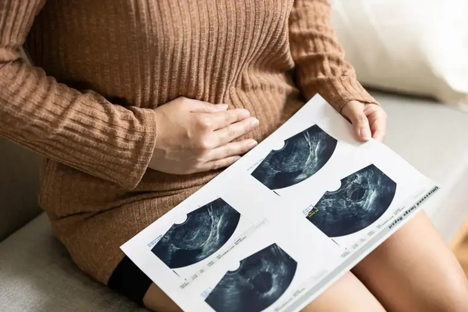
 Individuals with leiomyosarcomas may benefit from specialized imaging techniques to assist evaluate the size, location, and extension of the tumor, as well as to aid in future surgical treatments. Computerized tomography (CT) scanning, magnetic resonance imaging (MRI), and ultrasound are examples of imaging methods. A computer and x-rays are used to make a film that shows cross-sectional pictures of certain tissue features during CT scanning. 

An MRI creates cross-sectional pictures of certain organs and body tissues by using a magnetic field and radio waves. During an ultrasound, reflected sound waves within the body form a picture of interior organs and other structures.

Laboratory testing and specialist imaging tests may also be performed to assess the existence of distant metastases and probable involvement of regional lymph nodes. 


Prognosis for Someone with LMS

LMS Prognosis

Histologic grade, tumor size, and tumor depth are the three most important prognostic variables in patients with LMS. Tumor size, bone or neurovascular involvement, as well as tumor grade, are all related with poor outcomes, particularly in LMS.

The tumor's position is an independent prognostic risk factor. When compared to retroperitoneal LMS, the LMS of the extremities has a superior result. One retrospective investigation found that uterine LMS had a worse survival rate than extra-uterine LMS; however, another small study found that uterine LMS had a better result (most patients with uterine LMS had a near-complete resection in this study).

The histologic grade is a predictive factor that can be used independently. It can evaluate the aggressiveness of cancer, the likelihood of distant metastasis, and disease-specific survival separately. LMS is a very aggressive cancer, with 90% of patients diagnosed with grade 2 to 3 carcinoma. Even after adjusting for tumor grade, histologic subtype is an independent prognostic factor. The histologic subtypes, however, have never been included in the AJCC staging methods, including the most recent AJCC 8th edition. When compared to other histologies, patients with LMS have a higher chance of distant recurrence and a lower disease-specific survival. 


Can Leiomyosarcoma be cured?

Leiomyosarcoma Treatment

Because surgery provides the best chance of curing a soft tissue sarcoma, it is used as part of the treatment for all soft tissue sarcomas whenever possible. It is critical that your surgeon and other clinicians have experience treating sarcomas. These tumors are difficult to treat and need both knowledge and competence. Patients with sarcomas had better results when they are treated in specialist cancer centers with sarcoma experience, according to studies.

Stage I soft tissue sarcoma:

Soft tissue sarcomas in stage I are low-grade tumors of any size. Small (less than 5 cm or around 2 inches wide) arm or leg tumors may be treated surgically alone. The objective of surgery is to remove the tumor along with part of the surrounding normal tissue. If cancer cells are discovered in or around the borders of the excised tissue (referred to as positive or close margins), it may indicate that some cancer was left behind. More surgery is frequently the best option for positive or close margins. Another alternative is to follow up surgery with radiation treatment. This reduces the likelihood of the cancer returning.

If the tumor is not in a limb (for example, in the head, neck, or belly), it may be more difficult to remove the entire tumor while leaving enough normal tissue surrounding it. Before surgery, radiation with or without chemo may be used to treat these tumors. This may be able to reduce the tumor sufficiently for surgery to remove it completely. If radiation is not used before to surgery, it may be utilized after surgery to reduce the likelihood that the tumor will recur.


Stages II and III soft tissue sarcoma:

The majority of sarcomas in stages II and III are high-grade tumors. They have a rapid growth and dissemination. Some cancers in stage III have already migrated to neighboring lymph nodes. Even if these sarcomas have not yet migrated to the lymph nodes, the chance of metastasis (to the lymph nodes or distant places) is quite high. These tumors also tend to regrow in the same location after they've been removed. (This is known as local recurrence.)

The primary therapy for all stage II and III sarcomas is surgical removal of the tumor. If there is cancer in the lymph nodes, these will be removed as well. Radiation may be administered following surgery.

If the tumor is big or in a difficult-to-reach location, but not in lymph nodes, the patient may be treated with chemo, radiation, or both before surgery. (Isolated limb perfusion is another option for treating big tumors in the arms or legs.) The objective of therapy is to reduce the tumor so that it may be removed more easily. Following surgery, chemo, radiotherapy, or both may be administered. These therapies reduce the likelihood of the tumor returning in or around the same location where it first appeared.

Smaller tumors may be treated with surgery initially, followed by radiation to reduce the likelihood of the tumor returning.

In rare cases, amputation is needed to remove the entire limb with the tumor.

Radiation therapy with or without chemo can be used alone when the tumor's location or size or the patient's health in general makes surgery impossible.


Stage IV soft tissue sarcoma:

When a sarcoma has spread to other regions of the body, it is labeled stage IV. Sarcomas at stage IV are seldom curable. However, some people may be cured if the initial tumor and all sites of cancer spread (metastases) can be removed surgically. When it has just gone to the lungs, the chances of success are the highest. The primary tumors in these individuals should be treated as in stages II or III, and metastases should be removed entirely if feasible. Doctors continue to dispute regarding the optimal treatment and which patients are most likely to benefit in this area.


Follow-up Surveillance

Sarcoma specialists

Sarcoma specialists recommend CT or MRI scans of the chest, pelvis and abdomen:

  • every 3-4 months until one is NED (no evidence of disease) for 2 years.
  • every 6 months after you have been NED for 2-3 years.
  • once a year after you have been NED for 5 years and more.


Conclusion

Leiomyosarcoma

Leiomyosarcoma is a rare cancer that starts in smooth muscle tissue. Smooth muscle tissue may be found throughout the body, including the digestive system, urinary system, blood arteries, and uterus.

Leiomyosarcoma is most commonly seen in the abdomen or uterus. It begins as a proliferation of aberrant cells and swiftly spreads to infect and destroy normal bodily tissue.

The signs and symptoms of leiomyosarcoma vary depending from where the disease begins. Pain, weight loss, and a developing lump or swelling that may be felt through the skin are all possible symptoms.

The tests and techniques used to diagnose leiomyosarcoma vary depending on the location of the malignancy, but may include:

  • Physical exam: Your doctor may examine you to learn more about what's causing any signs and symptoms you're having.
  • Imaging examinations: Imaging studies such as MRI, CT, and positron emission tomography may be used (PET).
  • Removing a tissue sample for testing (biopsy): A biopsy involves the removal of a sample of suspicious tissue for testing. The sample is submitted to a lab to be examined to determine whether it is leiomyosarcoma.

The therapies that are most likely to be beneficial for you are determined by the location and size of your leiomyosarcoma, as well as if it has spread to other parts of your body. When developing a treatment plan, your doctor takes into account your general health as well as your preferences.