Limb Deformity

Last updated date: 28-Oct-2023

Originally Written in English

Limb Deformity

Overview

Limb deformities are musculoskeletal anomalies that can be congenital (born with them), developmental, or acquired as a result of a fracture, infection, arthritis, or malignancy. Limb deformity symptoms can range from a little variation in the look of a limb to a significant loss of function of an extremity. Treatment is determined by the anatomic location of the malformation, functional impairment, or aesthetic issue.
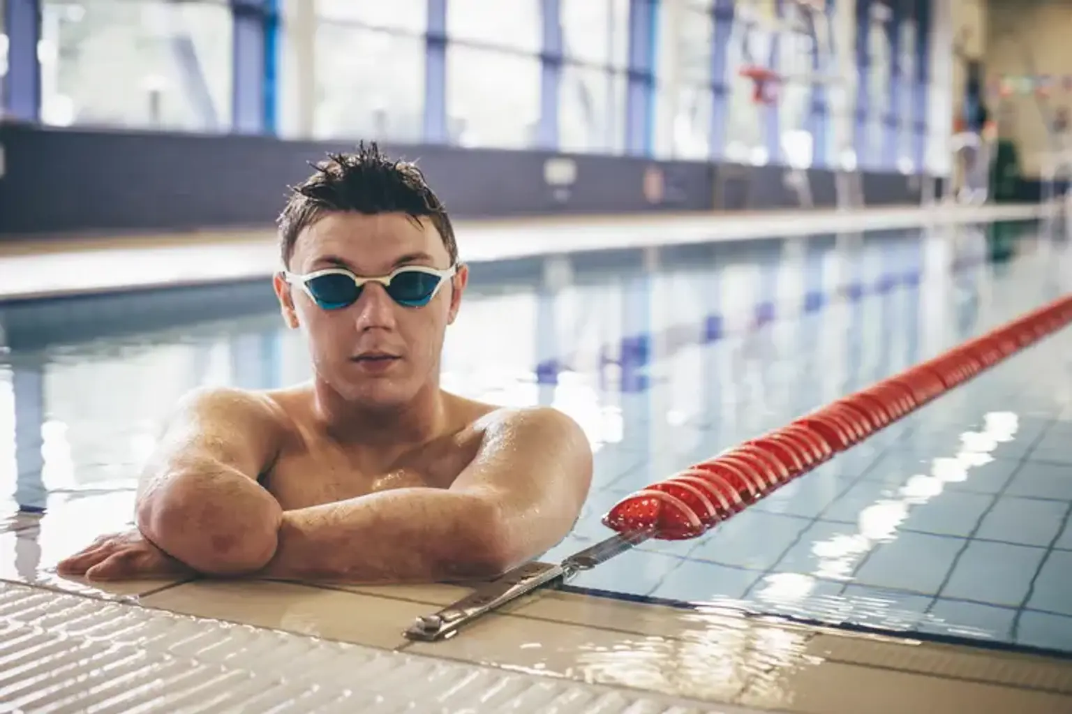
 

What is a Limb Deformity?

Limb Deformity Definition

Limb deformities are any anomalies that occur during the growth and development of the arms or legs. A limb length disparity is more precise and relates to arms or legs that are not the same length.

There are several reasons of limb abnormalities and limb length disparities. These can be the consequence of a congenital difference, be related with an underlying condition, or develop over time as a result of trauma, infection, or unknown reasons.

Symptoms can range from little impairment to significant issues with limb function, such as difficulty using the hands or walking effectively, depending on the origin and degree of the deformity or disparity. 


Structure and Function

Structure and Function of limb

To recognize abnormal, one must first recognize normal. Although it would seem reasonable to expect a person's legs to be straight and of equal length, more than half of the US population has a modest leg length disparity or angulation.

Normal limb alignment varies with aging. Alignment abnormalities can be angular or rotational in nature. Prior to the age of two, children have a relatively varus (bow leg) frontal plane alignment, which subsequently becomes valgus (knock knee) as they develop. By the age of seven, children typically have a final femoral-tibial alignment of 5-6 degrees valgus. The majority of rotational abnormalities are symmetric and are considered variants of normal that improve with age.

The angle of the femoral neck in the axial plane relative to the femoral shaft is termed as femoral rotation. Consider a denuded femur sitting on a level surface to understand femoral rotation. When the posterior femoral condyles are flat, the femoral neck forms an angle with the surface, with the head rising above it.


Limb Length Discrepancies

Limb Length Discrepancies

Limb length discrepancy (LLD) is a type of malformation that occurs when the upper and lower arms or legs are different lengths. A difference in arm length normally creates little or no trouble for patients. However, leg length discrepancies of 3.5 to 4% of total leg length might create apparent walking problems.

Types of limb-length discrepancies

  • Structural discrepancy – a bone in one leg or arm is shorter than the same bone in the other limb.
  • Functional discrepancy – bone lengths are equal in both legs or arms, but a joint contracture (low joint mobility) in a knee, hip, shoulder, elbow, or other joint throws off the symmetry in the limbs.


  • What are the symptoms of a limb-length discrepancy?

The symptoms of a limb-length disparity vary greatly from kid to child, depending on whether a leg or arm is afflicted, the degree of length difference, and the etiology of the problem.

Some common symptoms include:

One of your legs or arms is shorter than the other (this is not always obvious)

  • Signs of leg-length discrepancy
    • Problems with posture, such as a tilting shoulder
    • Walking problems, such as limping or toe-walking
    • A knee that's chronically hyperextended on the short side and flexed on the long side
    • Pain in the back, hip, knee, or ankle


  • How is a limb-length discrepancy diagnosed?

A detailed medical history and physical examination by an orthopedic surgeon are the initial steps in detecting a limb-length difference. During the checkup, the doctor will measure your child's limbs to determine how long they are.

If your kid has a leg length difference, the doctor may observe them walking to search for indicators that they are compensating for their varied leg lengths. Your child's doctor may also ask them to stand with a block under their shorter leg to see how much adjustment is required to bring their hips and the rest of their body into alignment.

For a more exact assessment of the disparity, your child's doctor may also request an x-ray or other imaging tests.

  • How are limb-length discrepancies treated?

Limb length differences can be handled in a variety of ways. Some therapies need surgery, while others do not. Your child's orthopedic surgeon will consult with you about your child's treatment choices based on:

  1. Your child's age, overall health, and medical history
  2. The limbs affected (leg or arm)
  3. The amount of discrepancy
  4. Your child's tolerance for specific medications, procedures, or therapies
  5. Your and your child’s expectations and preferences


Bowed Legs

Bowed Legs

Toddlers with bowed legs are extremely frequent and are a typical aspect of growth. Because every child is unique, some legs will be minimally curled while others will be significantly bowed. Children's legs develop straighter as they grow, allowing them to gently knock knee. Most children's legs are virtually straight by the age of two, and by the age of three, almost all children have outgrown the bending of their legs.

It is crucial to note that each child is unique and grows at a distinct rate, thus some children will not fit into the above-mentioned pattern. Some conditions cause children's legs to bow, such as growth plate disorders like Blount's disease, growth arrest, and metabolic difficulties like rickets.

The majority of bending situations do not need treatment. Your doctor's suggestions will be based on a thorough examination of your kid, which may include x-rays. Treatment may include observation, bracing, or surgery. To address the deformity, surgery may include growth regulation or osteotomies (cutting the bone).


Hammer, Claw, and Mallet Toes

What are Hammer, Claw, and Mallet toes?

Toes twisted into an unusual posture are known as hammer, claw, and mallet toes. They may appear weird, wounded, or both. These toe disorders nearly typically affect the four lesser toes rather than the big toe.

  • At the middle toe joint, a hammer toe bends down toward the floor. As a result, the middle toe joint rises. The second toe is commonly affected. Bunions are frequently associated with hammer toes.
  • Claw toe frequently affects the four lesser toes simultaneously. At the point where the toes and the foot connect, the toes bend up. They bend down at the middle joints and towards the tips of the toes. As a result, the toes curl down toward the floor.
  • A mallet toe bends at the joint closest to the toe's tip. It usually affects the second toe, although it can also affect the other toes.

Consult your doctor if your toe appears abnormal or aches. You might be able to treat your toe at home. If you don't treat the condition immediately away, you may need surgery.


  • What causes them?

The most prevalent cause of these toe issues is wearing shoes that are too small. Wearing tight shoes might throw the toe muscles off balance. When a shoe causes a toe to stay bent for an extended period of time, the muscles tense and the tendons shorten, or contract. This makes straightening the toe more difficult. Even while not wearing shoes, the toe muscles gradually lose their ability to straighten the toe.

These toe issues develop over time and are typical in adults. Women are more likely than males to be impacted because they are more likely to wear shoes with narrow toes or high heels.

Less frequently, these toe difficulties are associated with other illnesses such as diabetes, rheumatoid arthritis, stroke, or a foot or ankle injury.

  • What are the symptoms?

Besides looking odd, hammer, claw, and mallet toes may:

  1. Hurt.
  2. Make it hard to find shoes that fit.
  3. Make contact with your footwear. Calluses or corns can form where a bent toe rubs against another toe or your shoe, or where the tip of the toe contacts the ground.
  4. In more severe cases, these toe problems may affect your balance and make it hard to walk.

  • How are they diagnosed?

Your doctor will ask you questions about your symptoms and medical history, as well as do a physical examination. Your doctor will want to know the following:

  1. When the problems started, what activities or shoes make them worse, and if other parts of the foot are painful.
  2. What kind of shoes you wear and how much time you spend standing or walking every day?
  3. Any previous foot problems you have had.
  4. Any medical conditions you have that could be related, such as arthritis, diabetes, or poor circulation.

During the physical exam, your doctor will examine your foot to determine if the toe joint is fixed or flexible. A movable joint can occasionally be straightened without surgery. A fixed joint frequently need surgery.


If you are considering surgery to fix your ailment, you may require:

  1. An X-ray to help the doctor decide what type of surgery would be most helpful.
  2. If your foot seems to have inadequate blood flow, blood flow testing, which may include Doppler ultrasonography, should be performed.
  3. If your doctor suspects you have nerve abnormalities in your foot, you will undergo nerve testing. If this is the case, you should consult a neurologist, a physician who specializes in brain, spine, and nerve disorders.

  • How can you care for hammer, claw, or mallet toes?

You can probably treat your toe joint problem at home. If you start right away, you may be able to avoid surgery.

Change your shoes. Shoes with large toe boxes, low heels, and strong arch supports are ideal. Sandals or athletic shoes that do not rub on your toes might be an alternative. You might also try custom-made shoes or shoes designed specifically for those with foot difficulties.

Moleskin, toe tubes, arch supports, or other shoe inserts can help cushion the toe or keep the foot in a more comfortable posture (orthotics). These are more effective for treating a flexible toe, although they can also help with a fixed toe.

Take care of any corns or calluses on your feet. Moleskin and other over-the-counter pain relievers may be effective. Never cut corns or calluses since this might result in infection.

Take an acetaminophen, ibuprofen, or naproxen over-the-counter pain reliever. Before using these medications, see your doctor. Take precautions when handling medications. Read and obey all label directions.

Rickets

Rickets

Rickets is an uncommon condition that causes soft, brittle bones. It is mostly caused by a deficiency of vitamin D.

Rickets is most frequent in youngsters, who may have low vitamin D levels owing to a poor diet or a condition (such as celiac disease) that makes the body's absorption of vitamin D and calcium problematic. Rickets can cause bone malformations including bowlegs, knock-knees, an enlarged cranium, and an irregular spine curvature. It can also induce weakness.

The treatment will seek to increase the individual's calcium, phosphate, and vitamin D consumption. A doctor will generally prescribe vitamin D supplements based on the underlying problem.

They may also recommend:

  • Increasing exposure to sunlight
  • Making dietary changes
  • Taking fish oil
  • Getting more exposure to UVB light
  • Consuming calcium and phosphorus

Dietary measures

If rickets results from a poor diet, a doctor may prescribe:

  • Daily calcium and vitamin D supplements
  • An annual vitamin D injection (if a person cannot take supplements orally)
  • A diet plan that focuses on foods rich in vitamin D

To add vitamin D to the diet, a person can consume:

  • Eggs
  • Cod liver oil
  • Oily fish, such as salmon, tuna, sardines, and swordfish
  • Vitamin D-fortified foods, such as milk, some juices, many cereals, some brands of margarine, and some soy milk products
  • Beef liver

Most youngsters may avoid rickets by making dietary modifications and spending time outside each day.


Polydactyly

Polydactyly

Polydactyly, or extra digits, is the most common congenital limb malformation. This deformity is divided into three types: preaxial, central, and postaxial.

  • Preaxial polydactyly is the presence of an additional thumb or great toe. The symptoms range from a wide or duplicated distal phalange to total digit duplication. It can occur alone, possibly with autosomal dominant inheritance, or it can be part of a number of genetic syndromes, such as acrocallosal syndrome (with developmental delay and corpus callosum defects), Carpenter and Pfeiffer syndromes (with craniosynostosis), Fanconi and Diamond-Blackfan anemias, and Holt-Oram syndrome.
  • Central polydactyly is an uncommon condition characterized by the duplication of the ring, middle, and index fingers. It is linked to syndactyly and cleft hand. The vast majority of instances are syndromic in nature.
  • The most prevalent type of polydactyly is postaxial polydactyly, which involves an additional finger on the ulnar/fibular side of the limb. The additional finger is usually rudimentary, although it can be fully developed. This kind of polydactyly is frequently an isolated condition in persons of African origin. It is more commonly linked with a syndrome of several congenital malformations or chromosomal disorders in other groups.


Syndactyly

Syndactyly

Webbing or fusing of fingers or toes is referred to as syndactyly. There are several kinds, with the majority following an autosomal dominant inheritance pattern. Simple syndactyly includes just soft tissue fusion, whereas complicated syndactyly contains bone fusion as well. Apert syndrome is characterized by complex syndactyly (with craniosynostosis). In oculo-dento-digital dysplasia, syndactyly of the ring and tiny fingers is prevalent. Smith-Lemli-Opitz syndrome is characterized by syndactyly of the second and third toes, as well as a number of other congenital malformations.


Bone deformity as a result of trauma

malunited fractures

Fractures that heal at an unusual angle are one example. Malunions are also known as malunited fractures. They do not always create difficulties in the short term, but they can cause problems in the adjacent joints because the 'wear and tear' and stress on the joint is not distributed evenly across the joint.

A malunion can sometimes cause the limb to seem shorter. A limp caused by a malunion in the leg can also cause back pain owing to the tilt when walking or standing.


Treatment of Congenital Limb Abnormalities

Treatment of Congenital Limb Abnormalities

Surgical methods are used to treat polydactyly and syndactyly. Prosthetic devices are particularly beneficial for lower-limb inadequacies and entirely or nearly completely absent upper limbs for treating missing or hypoplastic limbs. If there is any activity in an arm or hand, regardless of the severity of the abnormality, working ability must be extensively evaluated before a prosthesis or surgical operation is proposed. Amputation of any limb or portion of a limb for therapeutic purposes should be undertaken only after assessing the functional and psychological ramifications of the loss and when amputation is required to fit a prosthesis.

An upper-limb prosthesis should be developed to meet as many demands as feasible in order to reduce the number of components. Children are more likely to utilize a prosthesis effectively if it is installed early in their development and forms an intrinsic part of their body and body image. Devices employed in infancy should be as basic and long-lasting as feasible; for example, a hook rather than a bioelectric arm. Most children with congenital amputations enjoy normal lives with good orthopedic and auxiliary assistance.


Conclusion 

Bow legs and knock knees are the most frequent types of limb malformation in children. Limb deformity can also arise in children or adults as a result of an infection or an improperly healed fracture. While certain disorders usually cure on their own as a child develops, some limb issues might interfere with walking if not addressed.