Introduction
What is Fat Grafting (Lipofilling)? Fat grafting, also known as lipofilling, is a cosmetic procedure that uses your own body fat to enhance or reshape different areas of your body. The process involves harvesting fat from one part of the body, like the abdomen or thighs, purifying it, and then injecting it into areas that need volume or contouring. This procedure is commonly used for breast augmentation, facial rejuvenation, and body sculpting.
Popularity of Fat Grafting Globally Fat grafting has become a popular choice in aesthetic surgery around the world. Unlike synthetic implants or fillers, fat grafting offers a natural solution that blends seamlessly with your body. Many people are drawn to the idea of using their own fat for enhancements, leading to its increasing demand, especially in countries like the United States, Brazil, and South Korea.
How Fat Grafting Works: The Procedure Explained
Fat Harvesting and Liposuction The procedure begins with liposuction, where excess fat is gently removed from areas like the abdomen, thighs, or flanks. A small tube (cannula) is inserted into the skin to suction out fat cells. This is usually done under local anesthesia, making the process relatively comfortable for the patient.
Fat Processing and Injection Once the fat is harvested, it's processed to remove impurities and excess fluids. The purified fat is then injected into areas of the body where volume or contouring is needed, such as the face, buttocks, or breasts. These injections are carefully placed to ensure smooth, natural results.
Minimal Invasive Nature of Lipofilling One of the key advantages of fat grafting is that it’s minimally invasive. Unlike traditional surgeries that require large incisions, fat grafting uses tiny incisions, resulting in less downtime and a quicker recovery. This makes it an appealing option for many patients seeking body enhancements without major surgery.
Benefits of Fat Grafting (Lipofilling)
Natural Results and Long-Term Effects Since fat grafting uses your own fat, the results are entirely natural, avoiding issues like rejection or allergic reactions that may occur with synthetic implants. The fat that "takes" after the procedure can provide long-lasting results. Though some of the fat may be absorbed by the body, the remaining fat will typically stay in place for years, making it a reliable option for body enhancement.
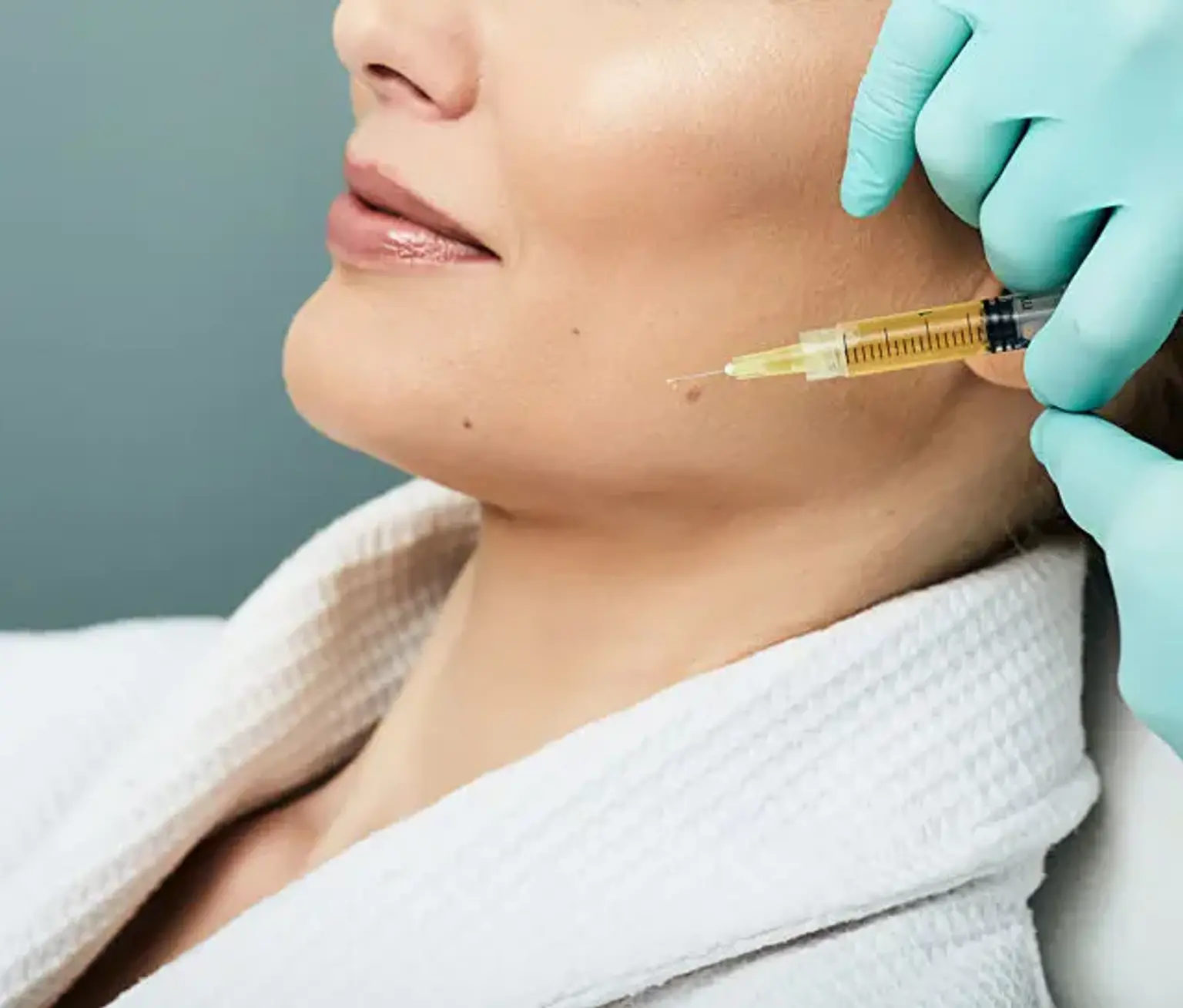
Facial Rejuvenation with Lipofilling Fat grafting can effectively restore volume to the face, a process that is especially beneficial as we age. It can be used to fill in hollow areas under the eyes, add fullness to the cheeks, and soften deep lines and wrinkles. The result is a more youthful, refreshed appearance without the need for synthetic fillers.
Body Contouring and Sculpting In addition to facial applications, fat grafting is popular for body contouring. Patients who have undergone significant weight loss often turn to fat transfer to restore lost volume in areas like the buttocks and thighs. Lipofilling provides a natural solution for improving body proportions and achieving a more balanced silhouette.
Breast Augmentation Without Implants Fat grafting offers a more natural alternative to breast implants. Whether used for breast reconstruction after surgery or for elective augmentation, fat transfer provides a subtle, soft result that mimics the natural feel and appearance of breast tissue. This approach is ideal for women who prefer a more natural look over artificial implants.
Fat Grafting Applications: Areas of Treatment
Fat Grafting for the Face One of the most common uses of fat grafting is for facial rejuvenation. The procedure can restore youthful fullness to the face by adding volume to the cheeks, under-eye areas, and jawline. It also works well to soften deep lines, such as nasolabial folds (smile lines), and the marionette lines around the mouth. The fat used in these areas can help create a smoother, more youthful appearance.
Fat Grafting for the Body Fat grafting is also used for body contouring. By transferring fat to areas like the buttocks, hips, and thighs, patients can achieve enhanced curves and more defined contours. For those who have lost a significant amount of weight or undergone liposuction, fat grafting can smooth out any remaining irregularities or areas of hollowing, providing a more balanced and natural look.
Fat Grafting for Breast Reconstruction Fat grafting has become a key tool in breast reconstruction following mastectomies. It can help reshape the breast or fill in areas that may have been left uneven after surgery. For some patients, fat grafting can also be used as a complement to breast implants, enhancing the shape and feel of the breasts for a more natural outcome.
What to Expect During the Fat Grafting Procedure
Pre-Surgical Consultations Before undergoing fat grafting, you'll have a thorough consultation with your surgeon. This is a crucial step to ensure you're a good candidate for the procedure. Your surgeon will assess your overall health, medical history, and the areas you'd like to enhance. They’ll also explain the process, answer your questions, and set expectations for recovery and results.
Step-by-Step Overview of Lipofilling Surgery Fat grafting is typically performed under local anesthesia, though general anesthesia may be used depending on the areas treated. First, your surgeon will mark the areas from which fat will be harvested, often from areas like the abdomen, thighs, or flanks. Using a thin cannula, they will gently remove the fat. After the fat is purified, it will be injected into the treatment area in layers to create a smooth and natural result. The entire procedure usually takes between 1 to 3 hours, depending on the extent of the treatment.
Recovery and Aftercare: What You Need to Know
Fat Grafting Recovery Timeline The recovery process for fat grafting is generally quicker than for more invasive surgeries. You may experience some swelling and bruising in both the area where fat was harvested and where it was injected. Most patients can return to light activities within a few days, although full recovery may take a few weeks. You’ll need to avoid strenuous activities and exercise for a few weeks to ensure proper healing.
Long-Term Results and Fat Retention After the procedure, some of the fat that was transferred will naturally be absorbed by the body. However, the fat that "takes" will remain long-term. While results vary, many patients see lasting improvements in volume and contour, especially in facial and body areas. If desired, follow-up treatments may be necessary to enhance or maintain results.
Post-Procedure Care Tips To maximize the success of your fat grafting procedure, it’s important to follow your surgeon's aftercare instructions. This may include wearing compression garments to reduce swelling, avoiding direct pressure on the treated areas, and attending follow-up appointments. Your surgeon may also recommend gentle massage to help the fat integrate into the surrounding tissue.
Risks and Considerations with Fat Grafting (Lipofilling)
Potential Risks of Fat Transfer Surgery Like any surgical procedure, fat grafting carries some risks. These may include infection, bleeding, and the possibility of fat resorption (where the body absorbs the transferred fat). There is also a small risk of asymmetry, where one side of the treated area may appear slightly different from the other. However, these risks are relatively low when performed by an experienced, board-certified surgeon.
Long-Term Safety Concerns While fat grafting is generally considered safe, some patients may experience complications over time. Fat may shift or become lumpy in certain areas, and there’s also the possibility that the body may absorb the fat, resulting in less noticeable results. These issues can often be corrected with additional touch-up procedures.
Ensuring a Safe Procedure Choosing a qualified, experienced surgeon is crucial to minimizing the risks associated with fat grafting. Board-certified plastic surgeons who specialize in fat transfer procedures are most likely to achieve the best results with minimal complications. Be sure to check your surgeon's credentials and review patient testimonials before proceeding.
Fat Grafting for Post-Surgical Reconstruction
Breast Reconstruction After Mastectomy Fat grafting has become a valuable tool in breast reconstruction after mastectomy. It helps to rebuild the breast’s shape and volume following the removal of breast tissue due to cancer. Fat transfer can be used to smooth out any irregularities or provide additional volume after breast implants are placed, improving the overall aesthetic result.
Reconstructing Defects After Trauma Fat grafting is also useful for patients recovering from traumatic injuries that result in facial or body deformities. It allows surgeons to restore lost tissue and contour the body naturally. Fat transfer can provide a smoother, more consistent appearance, reducing the need for synthetic implants or other artificial fillers.
Enhancing Skin Grafting Results Fat grafting can enhance the results of skin grafting, particularly in areas where the skin has been damaged or thinned. The added volume can improve skin elasticity and texture, providing a more youthful, natural-looking finish to the area.
Cost of Fat Grafting: What You Should Know
Average Costs for Lipofilling Procedures The cost of fat grafting varies depending on factors such as the areas being treated, the amount of fat needed, and the surgeon's experience. On average, fat grafting can cost anywhere from $3,000 to $10,000 or more. For breast augmentation or body contouring, the price may be higher, depending on how many areas are treated.
Insurance and Financing Options Since fat grafting is typically considered a cosmetic procedure, it is not usually covered by insurance. However, many plastic surgery clinics offer financing options to help make the procedure more affordable. You can discuss payment plans with your surgeon during your consultation to find a solution that fits your budget.
The Effectiveness of Fat Grafting: How Well Does It Work?
Fat Grafting for Aesthetic Improvement Fat grafting is highly effective in enhancing areas that need volume or contour, such as the face, breasts, and body. It can restore a youthful appearance by filling in wrinkles, hollow areas, and volume loss. Results are typically long-lasting, with many patients seeing improvement for several years.
How Permanent Are Fat Grafting Results? The results of fat grafting can be long-term, but not all the transferred fat will survive. On average, 50-70% of the fat will remain, with the rest being absorbed by the body. Follow-up treatments may be needed to maintain or enhance results over time, but most patients are satisfied with the final outcome.
Fat Grafting vs. Other Options (Fillers, Implants) Compared to synthetic fillers or implants, fat grafting offers more natural-looking and longer-lasting results. Unlike fillers that need to be reapplied, fat transfer uses your own tissue, which integrates seamlessly with your body, reducing the risk of rejection or allergic reactions.
Global Popularity: Why People Choose Fat Grafting
Fat Grafting in Different Regions Fat grafting has gained global popularity, especially in cosmetic surgery hubs like the United States, South Korea, Brazil, and Europe. In countries like South Korea, where beauty standards emphasize natural, subtle enhancements, fat grafting is particularly favored over implants or fillers.
Cultural Preferences and Attitudes Towards Fat Grafting In many cultures, using your own body fat for aesthetic enhancement is seen as a more natural and safer option than synthetic alternatives. This has contributed to the rising demand for fat grafting, as patients increasingly prefer non-invasive, autologous treatments that give a more subtle and individualized result.
Celebrity Influence on Fat Grafting Celebrities and influencers have significantly influenced the popularity of fat grafting. Public figures who openly share their experiences with fat transfer procedures have helped normalize the practice and inspire more people to consider it for aesthetic and reconstructive purposes.
FAQs About Fat Grafting (Lipofilling)
Is Fat Grafting Safe?
Fat grafting is generally safe, especially when performed by a qualified, board-certified surgeon. Like any procedure, it carries some risks, such as infection or uneven results, but these are rare when proper precautions are taken.
How Much Fat Can Be Transferred?
The amount of fat that can be transferred depends on the areas treated and the fat available for harvesting. In general, surgeons can safely transfer several hundred milliliters of fat, but the amount may vary depending on the patient’s body type and the desired outcome.
What Are the Risks of Fat Grafting for Breast Augmentation?
Fat grafting for breast augmentation is generally safe, but there are risks, including the potential for uneven results, fat absorption, and cyst formation. It’s important to discuss these risks with your surgeon to ensure you have realistic expectations.
Can Fat Grafting Be Combined with Other Procedures?
Yes, fat grafting can be combined with other cosmetic procedures, like liposuction, facelifts, or tummy tucks. Many patients choose to combine treatments for a more comprehensive, transformative result.
Innovations and Advancements in Fat Grafting
Recent Developments in Fat Grafting Techniques Advancements in fat grafting techniques have improved both the safety and effectiveness of the procedure. New technologies, such as advanced fat processing systems and enhanced cannula designs, allow for more precise fat transfer and better fat survival rates. These innovations contribute to smoother, more natural results with minimal risk of complications.
Stem Cell Enrichment in Fat Grafting Some surgeons are now incorporating stem cell technology into fat grafting to improve the outcomes. By isolating stem cells from the harvested fat and enriching the fat tissue with these cells, surgeons may be able to enhance fat retention and promote better healing. While this is still an emerging technique, early results suggest it could offer even more long-lasting and natural outcomes.
The Role of 3D Imaging in Planning Fat Grafting Procedures Advanced 3D imaging technology is also playing a key role in fat grafting. It allows surgeons to plan the procedure with greater precision, helping them visualize the anatomy and create more symmetrical, personalized results. This technology also assists in predicting the amount of fat that will survive after grafting, improving patient expectations.
Patient Stories: Real-Life Experiences with Fat Grafting
Success Stories: Enhancing Appearance and Confidence Many patients who undergo fat grafting report significant boosts in their confidence and self-esteem. For instance, individuals who have had facial rejuvenation often see a dramatic improvement in their appearance, with smoother skin and restored volume. Similarly, those who use fat grafting for breast or body augmentation often express satisfaction with the natural, subtle outcomes.
Challenges and Considerations: Learning from Patient Experiences While most patients are happy with their results, some face challenges during the recovery phase. Issues like swelling, bruising, or slight asymmetry may occur but can usually be addressed with follow-up treatments or minor touch-ups. Listening to patient experiences can help new patients understand the full scope of the procedure and set realistic expectations.
Emotional Impact of Fat Grafting The emotional impact of fat grafting can be profound, particularly in reconstructive cases. Patients who have undergone breast reconstruction or facial trauma often report feeling a sense of wholeness and improved quality of life after the procedure. For many, the results are not just about physical appearance but also about regaining confidence and comfort in their own bodies.
Conclusion
Assessing Your Goals and Expectations Before deciding on fat grafting, it’s important to assess your goals and understand what the procedure can realistically achieve. If you're looking for natural enhancement or a subtle, long-lasting improvement, fat grafting may be the right choice. However, if you’re seeking dramatic changes or have unrealistic expectations, it’s essential to have an honest conversation with your surgeon about what can be achieved.
Consult with a Qualified Surgeon Choosing the right surgeon is key to a successful fat grafting experience. Look for a board-certified plastic surgeon with extensive experience in lipofilling. They should provide a detailed consultation, clearly outline the risks and benefits, and work with you to ensure the procedure aligns with your aesthetic goals.
The Bottom Line: Natural Enhancement with Fat Grafting Fat grafting offers a safe, natural solution for a variety of cosmetic and reconstructive needs. It enhances body contour, restores facial volume, and provides a more organic, long-term alternative to other procedures. With advancements in technology and growing popularity, fat grafting continues to be an excellent option for patients seeking subtle, natural-looking results.