Low Dose Chest CT
The National Lung Cancer Screening Study recently revealed that using low-dose computed tomography to check high-risk populations lowers lung cancer deaths. The National Comprehensive Cancer Program recommendations advised low-dose computed tomography for chosen patients who are at risk of lung cancer depending on this encouraging result. As a result, it's likely that more CT screenings will be conducted in the future. Although the low-dose computed tomography approach is straightforward, key CT settings are critical and must be precisely adjusted in order to obtain high diagnostic quality while reducing the delivered radiation. Furthermore, low-dose computed tomography scans are not as simple to read as they may appear; multiple strategies and instruments for nodule diagnosis and assessment are known. Furthermore, dealing with positive results can be a complicated function that differs greatly from standard clinical practice. As a result, the study focuses on the low-dose computed tomography methodology, reading approaches, and analysis in lung cancer screening, especially for radiologists who are unfamiliar with the technique.
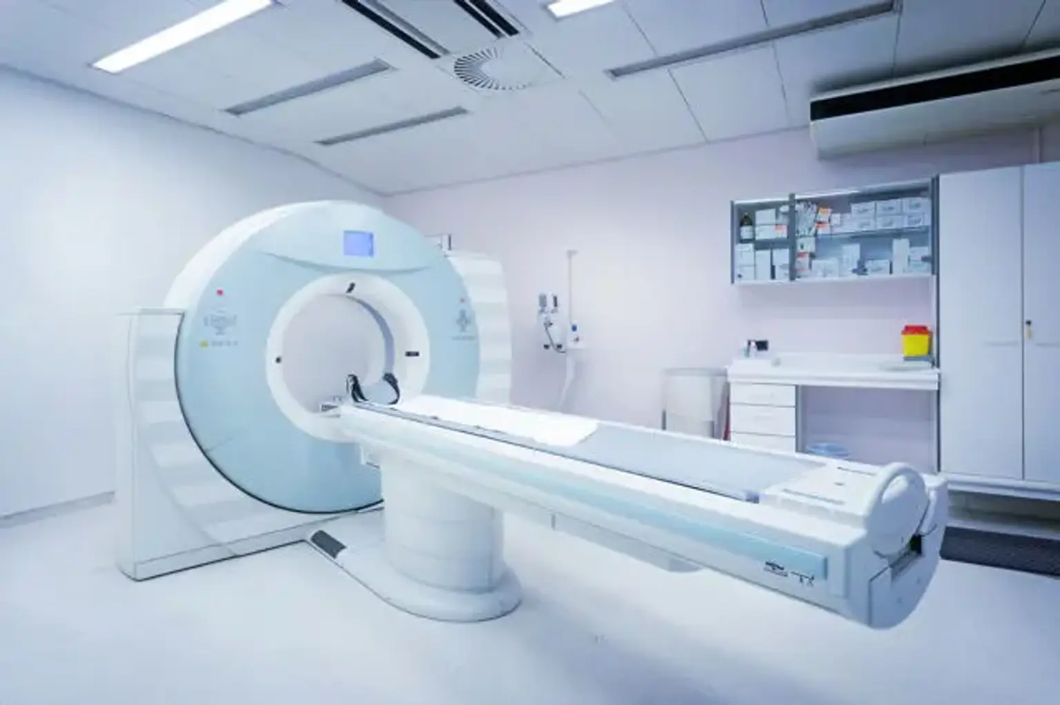
What is Low Dose CT?
The conventional CT scan exposes you to a lot of radiation, but it's necessary to investigate and treat some medical conditions. Patients who get this procedure on a regular basis are reasonably concerned about their frequent radiation exposure.
A low-dose CT scan, also known as an LDCT scan, produces considerably less radiation, minimizing these concerns. Newer scanners, such as those used at Victorian Radiology Clinic, provide pictures with extremely low levels of radiation without compromising their quality. It's a quick and painless procedure that just takes a few minutes.
Low Dose CT Scan Technical Notes
Even while single-slice spiral computed tomography can be used to provide low-dose computed tomography, the usage of multidetector CT scanners is generally suggested. The justification for employing multidetector CT scanners is that most of the nodules discovered on screen are tiny and require good spatial resolution for computer-aided detection and nodule size assessment.
As per the National Lung Cancer Screening Trial and the International Early Lung Cancer Action Program, low-dose computed tomography scanners with at least four detectors should be used to guarantee that the chest may be imaged in a single breath-hold and that better spatial resolution is achieved.
Low-dose computed tomography allows for a minimal radiation dosage while maintaining good diagnostic performance due to a high contrast resolution between air and lung lesions. There is no consensus on what constitutes a low dosage, and the parameters that determine the dose in CT vary, including tube voltage, tube current, and tube rotation speed.
Multidetector CT scanners lung cancer screening programs have used a variety of kilovoltage peak and milliampere-second values, with varying estimated effective doses. A new study aiming at establishing the optimal dose distribution associated with a single low-dose computed tomography examination found that satisfactory CT screening can be achieved with an average total effective dose of roughly 2 mSv.
Instead of filtered back projection, CT producers have actually updated iterative reconstruction. Iterative methods have the advantage of reducing noise while maintaining spatial resolution, CT number precision, and uniformity. Unfortunately, there is a lack of comprehensive evidence on the use of iterative reconstruction-based techniques with low-dose computed tomography, and more research is needed to determine the technique's utility.
The low-dose computed tomography collimation should be tuned so that thin-section reconstruction pictures may be obtained, allowing for the application of computer-aided detection and an ideal volumetric analysis. As a result, reconstructed segment thicknesses of at least 1.5 mm or less should be used. In order to minimize partial volume artifacts, the section interval should be set lower than the slice thickness. This is crucial for determining the dimensions of nodules accurately.
Low Dose CT Pulmonary Nodules
The pulmonary nodules are the focus of low-dose computed tomography for screening objectives. Solid nodules and subsolid nodules are two types of lung nodules. Non-solid nodules and part-solid nodules are two types of subsolid nodules. This distinction is important because various nodules necessitate distinct detection, assessment, and management strategies.
Solid and subsolid nodules develop at different rates, and subsolid nodules are more likely to become cancerous. In lung cancer screening investigation, Li et al. examined malignant and benign nodules and discovered that malignancy was found in 60 percent of non-solid nodules, 49 percent of part-solid nodules, and 10 percent of solid nodules. In the Early Lung Cancer Action Project, 35% of subsolid nodules diagnosed at baseline were cancerous, compared to only 8% for solid nodules. Malignant growth rates for part-solid and non-solid nodules were 64 percent and 17 percent, respectively.
Even while non-solid nodules are more prone to be cancerous, their growth rate is much slower than solid lesions, which has a significant impact on the follow-up interval and care of these lesions. When cancerous, solid, and part-solid nodules, on the other hand, are more prone to be invasive and grow rapidly.
Atypical adenomatous hyperplasia, adenocarcinoma in situ, and minimally invasive cancer are the most common subsolid nodules. The names adenocarcinoma in situ, and minimally invasive cancer were adopted by the new categorization of lung adenocarcinoma, and if surgically removed, 5-year disease-free survival can reach 100%.
Low Dose CT Pulmonary Nodules Detection
The perceptual mistake is a common cause of false-negative diagnoses in lung cancer diagnosis. Several variables, such as CT characteristics, reader knowledge, and nodule position, can influence radiologists' nodule detection performance. If the use of thin-slice multi-detector Computed tomography improves the reader's sensitivity for lung nodule detection, the rate of identification is also influenced by the presence of the nodule. Perihilar lung nodules were discovered with a sensitivity of 37 percent compared to 74 percent for peripherally situated nodules. They also discovered that vessel-attached nodules had a low sensitivity when detected.
For low-dose computed tomography, a dual reading approach and computer-aided detection systems have been suggested to enhance the radiologist's detection accuracy. The identification rate of pulmonary nodules was greatly enhanced by double reading. Wormanns et al. found that employing dual reading, the average sensitivity for detecting 390 nodules improved from 63 to 79 percent for single readers. However, a new study found that dual reading in lung cancer detection did not improve cancer detection rates, but did result in the discovery of 20 percent more nodules.
Small pulmonary nodules can be detected with the help of a computer-aided detection system. Rubin found that single reading, double reading, and reader utilizing computer-aided detection had a sensitivity of 51 percent, 62 percent, and 77%, respectively. Computer-aided detection recognized 79 percent of missing lung nodules and revealed 94 percent of lesions undiagnosed owing to interpretation mistakes. Thinner reconstruction photos and overlapped reconstructed sections improve the efficiency of computer-aided detection for nodule detection, even if this results in a higher percentage of false-positive results. Dose level appears to have little effect on the use of computer-aided detection systems. Lee et al. tested 25 volunteers with four different tube’s current flow and reported no significant variations in nodule detection between the 8 mA and 32 mA scanners. It's also important to consider the reader's expertise with computer-assisted detection techniques. In a study comparing the actual quality of experienced (6–8 years of experience) and inexperienced (6 months of experience) radiologists using computer-aided detection as a second reader, the actual quality of an experienced radiologist with computer-aided detection assistance was considerably better than that of an inexperienced radiologist using computer-aided detection as a second reader.
Maximum intensity projection can also increase the detection rate of lung nodules. In a lung cancer screening investigation, Bastarrika et al. found that non-overlapping 10-mm-thick axial maximum intensity projection reconstructions allowed for more accurate detection of pulmonary nodules than 1.25-mm conventional axial images. When compared to 1-mm low dose computed tomography pictures, Jankowski et al. assessed the diagnostic advantages of maximum intensity projection and a computer-aided detection method for lung nodule detection. The researchers discovered that, as compared to low-dose computed tomography, maximum intensity projection and computer-aided detection decreased the number of missed nodules, and that maximum intensity projection was more sensitive than computer-aided detection. M multiplanar reconstruction images have been found to be a sensitive technique for detecting lung nodules.
However, there is no agreement on which methodology should be used to interpret low-dose CT images, and more research is needed to determine the best way for increasing the rate of lung nodule diagnosis.
Low Dose CT Beyond Pulmonary Nodules
Low-dose computed tomography of the chest can reveal extra details about cardiovascular events and chronic obstructive pulmonary disease (COPD). Some studies suggest that using coronary artery calcium scoring as a component of ungated low-dose computed tomography to estimate the risk of cardiovascular disease is a smart option. In a study comparing coronary artery calcium and thoracic aorta calcium as potential predictors of mortality and cardiovascular problems in a lung cancer screening trial, researchers found that coronary artery calcium is a better predictor of mortality, cardiovascular, and coronary events than thoracic aorta calcium. Low-dose computed tomography has also been found to be a reliable method for determining the severity and progression of emphysema. The sensitivity and specificity of COPD diagnosis with low dose computed tomography have lately been investigated; inspiratory and expiratory low dose computed tomography images may identify participants with COPD with a sensitivity and specificity of 64 percent and 89 percent, respectively. The lung parenchyma should not be the only part of a low-dose computed tomography examination. Extrapulmonary malignancies are found at a rate of 1 case per 201 people evaluated in a lung cancer screening trial.
Low Dose CT Pulmonary Nodule Measurement
The most essential components in the therapy of pulmonary nodules discovered during screening are their size and development. As a result, it appears that using a consistent and accurate approach for measuring pulmonary nodules is critical. Manual and volumetric approaches are being utilized to determine nodule size, but no agreement on which method should be employed in lung cancer screening has yet been established. The two largest randomized screening studies utilize distinct methods: the NLST study uses manual diameter, whereas the NELSON trial combines volume and diameter.
Solid Pulmonary Nodules
In solid nodules, both manual and volumetric measurements of lung nodules can be used. Some writers claim that volumetric assessment is a more accurate method because there are valid uncertainties about the reliability of manual measurements due to high intra- and interobserver variability. Semi-automated measurements were not reproducible in 12% of solid nodules in the NELSON study (in which solid nodules were quantified by volumetric technology apart from pleural-attached nodules) and hence may lead to mistakes in the assessment of nodule expansion.
The volumetric assessment of lung nodules might be influenced by a variety of CT characteristics. The thickness of the slices is the most critical of these. The volumetric measures of thin and thick portions differed by 20 percent on average. Nietert et al. found that slice thickness of less than 2.5 mm is insufficient for detecting 1-mm variations in nodule diameter in lung nodules.
The level of precision of volumetric assessments can also be influenced by size and shape. The mistake in estimating the volume of a lung nodule increases as the nodule size decreases. Yankelevich et al. looked at the nodule shape and discovered that volume measurements for elongated shapes had higher measurement error than spherical shapes. Marten et al. came to similar conclusions. Furthermore, juxta-pleural and juxta-vascular nodules are more difficult to segment and may necessitate manual adjustment, resulting in greater variability in nodule measurements.
Subsolid Pulmonary Nodules
Subsolid nodule assessment is more challenging than solid nodule assessment, and the sluggish growth of these nodules makes accurate growth assessment more challenging during follow-up. Even though some commercially available technology has volumetric techniques for non-solid nodules, segmentation is frequently poor, necessitating human rectification. There is less evidence to support the use of the volumetric method for subsolid nodules at this time. A subsolid nodule with a constant diameter but an increasing solid portion at follow-up is also indicative of malignancy. Neither a manual nor a volumetric evaluation is applicable in this circumstance.
To solve this problem, de Hoop et al. developed a method for quantifying change in non-solid nodules called estimation of nodule mass. They showed that mass measurements, rather than volume or diameter measurements, can help detect expansion earlier and are less variable. However, determining the mass of a nodule necessitates a manual evaluation of the lesion's volume, which lengthens reporting time and increases observer variability.
Pulmonary Nodule Growth Follow-up
In low-dose CT follow-up tests, the same CT scanning and reconstruction settings should be employed. To reduce measurement errors, it is advised that when nodule size is measured volumetrically, follow-up exams use the same software version and segmentation method.
The lung cancer screening definition of nodule growth is controversial. In the I-ELCAP trial, nodule growth was defined as a 50 percent growth in mean nodule diameter for nodules less than 5 mm, a 30 percent growth for nodules 5–9 mm, and a 20 percent increase for nodules bigger than 10 mm. In the NLST study, growth was defined as an increase in nodule diameter of 10 percent or more. Nodule growth was defined as a 25 percent change in volume after at least a three-month gap in the NELSON research.
The doubling time is dependent on an exponential growth pattern, regardless of diameter or volume. Lindell et al., on the other hand, found that lung malignancies are not confined to exponential growth in a series of 18 screen-detected lung malignant tumors. This is significant because research and calculations based on exponential growth could mislead an uncertain nodule or lung cancer aggressiveness.
Malignant nodules are typically fast-growing tumors. According to Revel et al., benign solid nodules with a doubling time of more than 500 days account for 98 percent of all cases. Even if some lesions grow more gradually, especially the spectrum of adenocarcinomas, the time of 500 days is typically considered as the top range of the doubling time for malignant pulmonary lesions. This means that the generally held belief that two years of constancy is required to distinguish malignant from benign nodules can only be applicable to solid nodules, not subsolid nodules, which are primarily adenocarcinomas.
Benign nodules can double in size in as little as 20 days or as long as 450 days. However, the NELSON study found that benign nodules can grow significantly: 85% of nodules with a size doubling time of 400 days after 3 months were benign.
Low Dose CT Pulmonary Nodule Management
The analysis of low-dose CT studies is complicated by the presence of a lung nodule. Different lung cancer screening trials have recommended various nodule treatment regimens; however, it is presently impossible to determine the best accurate lung cancer screening procedure due to varying patient groups and research settings. To determine which work-up is best for every nodule, the size and growth rate of the nodule must be considered. Most notably, radiologists, pulmonologists, surgeons, and nuclear medicine specialists all contribute to the management of lung nodules. As a result, we would recommend evaluating the primary variables that can affect the diagnostic strategy in lung cancer screening rather than recommending a specific regimen for nodule care.
Pulmonary Nodule Size
The size of the nodule is one of the most crucial criteria, as there is a direct link between size and the risk of cancer. The prevalence of malignancy varied depending on nodule size, ranging from 0 to 1 percent for nodules under 5 mm, from 7 to 28 percent for those between 5 and 10 mm, and from 65 to 82 percent for nodules above 20 mm, according to a meta-analysis of 8 large low-dose CT screening trials.
All screening regimens propose a one-year follow-up if a nodule was detected at the baseline scan and no growth was seen with the yearly repeat low dose CT scan. The concept of 2-year constancy on follow-up CT as a sign of a nodules' benign character should be confined to solid nodules.
To minimize the number of false-positive results in lung cancer screening studies, it is widely agreed that a low-dose CT scan is negative when the diameter of the lung nodule is less than 4 or 5 mm, or when the volume is less than 50 mm3. Presently, if a nodule size at baseline or a new nodule at a follow-up scan is larger than these values, it is deemed a positive finding, and more testing is advised based on nodule size and attenuation.
The size of a new nodule discovered on a repeated low-dose CT scan is the determining factor in its care. If it's less than 5 mm, a one-year follow-up is recommended. A low-dose CT follow-up at 3 or 6 months is usually recommended if it is between 5 and 8 mm. Low-dose CT follow-up at 1 or 3 months or positron emission tomography (PET scan) and/or biopsy should be performed if the nodule size is larger than 10 mm, depending on nodule attenuation.
Pulmonary Nodule Growth Rate
The growth in the volume of a nodule over time can be utilized to distinguish between benign and malignant nodules. If a nodule has expanded, its size and rate of growth should be evaluated when determining how it should be managed. A quick growth rate (doubling time shorter than one month) is more indicative of a benign lesion, and in this situation, antibiotics can be given followed by a CT scan one month later. If the doubling time of the nodule is less than 400 days, a low-dose CT follow-up after 3 months, PET scan, or biopsy can be done; if the doubling time is 400–600 days, a follow-up scan can be conducted at 6 months or 1 year after. If the doubling time exceeds 600 days, a low-dose CT scan should be repeated after a year.
Subsolid Pulmonary Nodules
Despite proper management recommendations for subsolid nodules having yet to be published, Godoy and Naidich have recently offered interim management standards. Non-solid nodules, as previously noted, grow more slowly yet have a high percentage of malignancy. This means that for non-solid nodules, a minimum of three years of follow-up is needed to distinguish malignant from benign nodules and that any change in the size or the emergence of a solid component is suggestive of malignancy. When a non-solid nodule is less than 10 mm in diameter, close monitoring is recommended. A PET and/or CT-guided biopsy or surgical biopsy should be done if a non-solid lesion grows in size or if a solid component emerges. Non-solid nodules can be caused by a variety of benign disorders, such as inflammatory disease or fibrosis.
Invasive Procedures and Complications
CT-guided biopsy, bronchoscopic biopsy, or wedge resection can all be used to obtain histologic specimens of pulmonary nodules. As per different study designs, invasive interventions in lung cancer screening varied from 10 to 43 percent. A recent study looked at how many invasive treatments were indicated for suspicious nodules in 4783 high-risk smokers. The researchers discovered that 84 percent of the patients were appropriately diagnosed with cancer. The NLST is the only study that reports on low-dose CT screening consequences. The incidence of serious problems during the diagnostic evaluation was 34 per 10,000 people evaluated with low-dose CT in this trial.
Conclusion
The National Lung Screening Trial has shown that screening high-risk populations with low-dose CT decreases lung cancer deaths. Although recording low-dose CT scans may seem straightforward, radiologists working in lung cancer screening should be aware that interpreting low-dose CT scans is not as simple as it may appear. To avoid false-negative diagnosis and unwanted follow-up tests, various considerations such as unique nodule detection, techniques, and measurement evaluations should be taken into account. Furthermore, understanding the relevance of various nodule subtypes is critical, as distinct nodules require different detection, assessment, and management approaches.
The therapy of pulmonary nodules in asymptomatic high-risk patients is difficult, and clinical practice might differ greatly. As a result, radiologists' experience is essential. Quality control in low-dose CT image interpretation, as well as an interdisciplinary approach to managing good outcomes, are critical. Radiologists who have received specialized training should read CT scans.