Lumbar Disc Fusion
Overview
Spinal fusion, also known as spondylodesis or spondylosyndesis, is a neurosurgical or orthopedic surgical procedure that connects two or more vertebrae. This technique, which may be done at any level of the spine (cervical, thoracic, or lumbar), restricts movement between the fused vertebrae.
There are several types of spinal fusion techniques, and each one requires the use of bone grafting—either from the patient (autograft), a donor (allograft), or artificial bone substitutes—to assist the bones knit together. Additional hardware (screws, plates, or cages) is frequently utilized to keep the bones together while the graft fuses the two vertebrae together. Fluoroscopy, navigation systems, and robots can all be used to guide the placement of devices.
Lumbar disc fusion is most usually used to treat discomfort and pressure from mechanical pain in the vertebrae or on the spinal cord caused by the wear and tear of a disc (cartilage between two vertebrae) (degenerative disc disease). Spinal stenosis, spondylolisthesis, spondylosis, spinal fractures, scoliosis, and kyphosis are some of the other prevalent pathological diseases treated by spinal fusion.
There are many approaches to lumbar spine fusion surgery, and all involve the following process: adding bone graft to a segment of the spine, set up a biological response that causes the bone graft to grow between the two vertebral elements, and the bony fusion technique- which results in one fixed bone replacing a mobile joint - stops the motion at that joint segment
Complications, like with any operation, may include infection, blood loss, and nerve injury. Fusion also alters the natural mobility of the spine, putting additional strain on the vertebrae above and below the fused segments. As a result, long-term issues such as degeneration at these contiguous spine segments might occur.
When Lumbar Disc Fusion is indicated?
Spinal fusion can be used to treat a variety of conditions affecting any level of the spine (lumbar, cervical and thoracic). In general, spinal fusion is performed to decompress and stabilize the spine. The greatest benefit appears to be in spondylolisthesis, while evidence is less good for spinal stenosis.
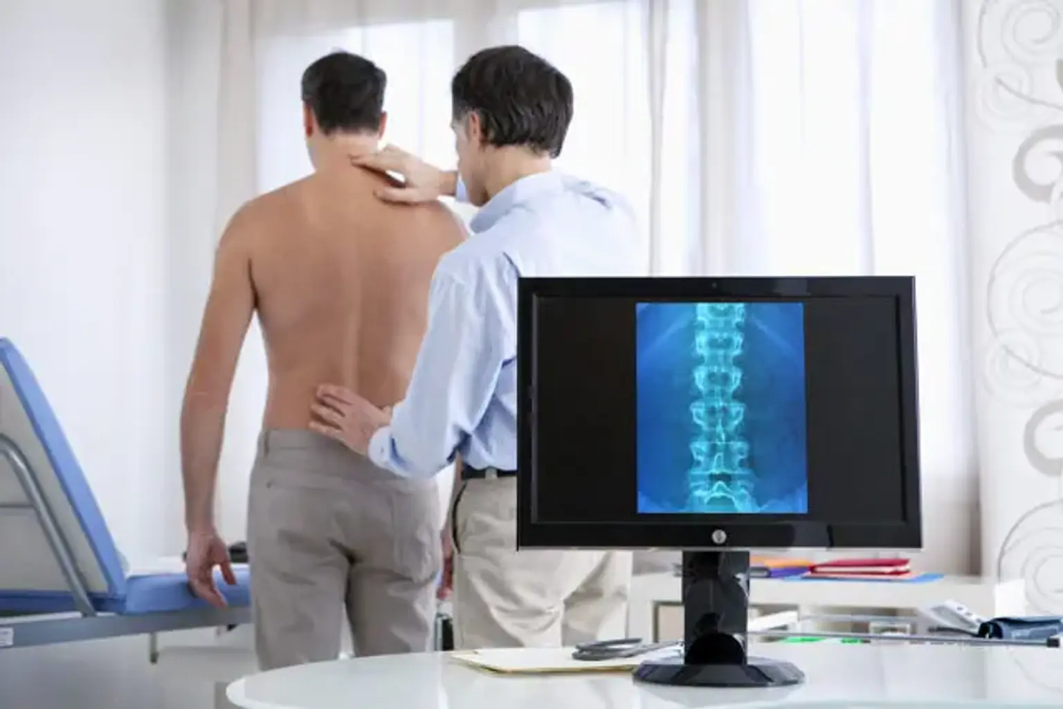
Degenerative disc disease is the most prevalent cause of pressure on the spinal cord/nerves. Disc herniation, spinal stenosis, trauma, and spinal malignancies are other prevalent causes. Spinal stenosis is caused by bone growths (osteophytes) or thicker ligaments that cause the spinal canal to narrow over time. This results in leg discomfort with increasing activity, which is known as neurogenic claudication. Radiculopathy, or pressure on the nerves as they depart the spinal cord, produces discomfort in the location where the nerves originated (leg for lumbar pathology). This pressure can produce neurologic impairments such as numbness, tingling, bowel/bladder malfunction, and paralysis in extreme cases.
Lumbar spinal fusions are more commonly performed than thoracic fusions. Degeneration happens more frequently at this level due to increased motion and stress. Conditions where lumber disc fusion may be considered include the following:
- Degenerative disc disease.
- Spinal disc herniation.
- Discogenic pain.
- Spinal tumor.
- Vertebral fracture.
- Scoliosis.
- Kyphosis (e. g., Scheuermann's disease).
- Lordosis.
- Spondylolisthesis.
- Spondylosis.
- Posterior rami syndrome.
- Other degenerative spinal conditions.
- Any condition that causes instability of the spine.
Epidemiology of Lumbar Disc Fusion surgery
According to the Agency for Healthcare Research and Quality, about 488,000 spinal fusions were done during hospital stays in the United States in 2011 (a rate of 15.7 stays per 10,000 people), accounting for 3.1 percent of all operating room operations. This is a 70% increase in operations since 2001. Lumbar fusions are the most common type of fusion, with 210,000 done each year.
According to a 2008 study of spinal fusions in the United States, the following features were found:
- Average age for someone undergoing a spinal fusion was 56.3 years for primary lumbar fusions.
- 45.5% of all spinal fusions were on men.
- 83.8% were white, 7.5% black, 5.1% Hispanic, 1.6% Asian or Pacific Islander, 0.4% Native American.
- Average length of hospital stay was 3.7 days –8.5 days for primary thoracic fusion, and 3.9 days for primary lumbar fusion.
- In-hospital mortality was 0.25%.
Is Lumbar Disc Fusion effective?
Although spinal fusion surgery is commonly used, there is insufficient evidence that it is useful for a variety of common medical disorders. In a randomized controlled trial of patients with spinal stenosis, for example, there were no significant therapeutic advantages of lumbar fusion in conjunction with decompression surgery compared to decompression surgery alone after 2 and 5 years. This Swedish study, which included 247 patients enrolled from 2006 to 2012, discovered that those who got fusion surgery had higher medical costs as a result of increased operation time, hospital stay duration, and implant cost.
Furthermore, a 2009 systematic review on surgery for lower back pain discovered that there was no benefit in health outcomes (improvement in pain or function) of performing fusion surgery versus intensive rehabilitation including cognitive-behavioral treatment for nonradicular low back pain with degenerative disk disease. Similarly, Washington State researchers evaluated lumbar fusion surgery as having doubtful medical value, increased expenses, and increased hazards when compared to intense pain treatments for persistent low back pain with degenerative disk disease.
Different techniques of Lumbar Disc Fusion
There are several techniques for lumbar spine fusion. Depending on the level of the spine and the location of the compressed spinal cord/nerves, each technique differs. Following decompression of the spine, bone graft or artificial bone replacement is inserted between the vertebrae to aid in their healing. Fusions are often performed on the anterior (stomach), posterior (back), or both sides of the spine.
Most fusions are now accompanied with hardware (screws, plates, and rods) since it has been demonstrated that they have greater union rates than non-instrumented fusions. Minimally invasive procedures are also gaining popularity. These procedures employ modern imaging guidance systems to put rods/screws into the spine through smaller incisions, resulting in reduced muscle injury, blood loss, infections, discomfort, and hospital stay.
There are several types of spinal fusion surgery options. The most commonly employed surgical techniques include:
- Posterolateral fusion is a bone graft between the transverse processes in the back of the spine. These vertebrae are then fixed in place with screws or wire through the pedicles of each vertebra, attaching to a metal rod on each side of the vertebrae.
- Interbody Fusion is a graft where the entire intervertebral disc between vertebrae is removed and a bone graft is placed in the space between the vertebra. A plastic or titanium device may be placed between the vertebra to maintain spine alignment and disc height. The types of interbody fusion are:
1. Anterior Lumbar Interbody Fusion (ALIF):
ALIF has proven to be an effective and widely used surgical procedure, particularly in the treatment of discogenic low back pain. The anterior retroperitoneal method allows for appropriate access to the whole ventral surface of the exposed disc, enabling for full discectomy and direct implant placement. The patient is prepared and positioned supine for this procedure. Midline, paramedian (all levels), or Mini-Pfannenstiel (L5/S1) incisions with a retroperitoneal corridor and vascular mobilization and dissection are all options for incision and approach.
Because of the vascular structure, the ALIF technique is most suited for levels L4/L5 and L5/S1 (Figure 4). Although uncommon, the ALIF method is restricted for L2/3 and L3/4 due to substantial peritoneal and kidney (L2/3) retraction and the possibility of superior mesenteric artery thrombosis.
An ALIF operation may be appropriate for degenerative disc disease, discogenic disease, and unsuccessful posterior fusion revision. Significant past abdominal surgery with adhesions or unfavourable vascular architecture, severe peripheral vascular disease, solitary kidney on the side of exposure, spinal infection, and high-grade degenerative spondylolisthesis in the absence of posterior fusion are all contraindications to ALIF. Isthmic spondylolisthesis at L5/S1 is a relative contraindication, and posterior fixation should be used in conjunction with the ALIF approach.
The ALIF technique has numerous significant advantages. At beginning, this approach provides a direct midline view of the disc space as well as significant lateral exposure of the vertebral bodies, allowing for effective disc space clearing and quick endplate preparation. Furthermore, anterior access enables for increased implant size and surface area, allowing for aggressive lordosis correction and foraminal height restoration.
2. Posterior Lumbar Interbody Fusion (PLIF):
PLIF is an innovative method to LIF. The PLIF approach provides surgical access to the intervertebral disc from the back. The patient is initially placed on an Andrews or Jackson table in a prone posture. To reach the posterior column of the vertebral body, either an open midline approach with bilateral muscle strip dissection or a MIS paramedian Wiltse muscle splitting method can be employed. Once the spinous process and laminae at the proper levels (L1-S1) have been identified, a laminotomy medial to the facet and the dura retracted to expose a corridor to the disc space can be done. After that, the endplates and disc space can be prepared for implant/spacer insertion.
The posterior approach may be appropriate for degenerative reasons that necessitate a fusion operation. A PLIF procedure may also assist patients with segmental instability, recurrent disc herniation, symptomatic spinal stenosis, and pseudoarthrosis. Extensive epidural scarring, arachnoiditis, and current infection are all contraindications to posterior fusion surgery.
There are various advantages to having PLIF surgery. To begin, the PLIF method is a typical lumbar approach in which the majority of spinal surgeons are highly educated and at ease. A posterior exposure enables for great visibility of the nerve roots while not interfering with blood flow to the transplant. PLIF restores proper interbody height and enables for neural decompression while preserving posterior support systems.
3. Transforaminal Lumbar Interbody Fusion (TLIF):
TLIF is another posterior surgical method for fusion that is utilized for stabilization and treatment of degenerative lumbar disease when conservative therapy has failed. The level of neural retraction necessary for the posterior fusion technique was one of the key issues, with special worries about probable nerve root damage, dural tears, and epidural fibrosis. To overcome this issue, the TLIF method was devised, which involves direct, unilateral access to the intervertebral foraminal area while minimizing direct dissection, surgical damage, and structural integrity.
By opening the neural foramen on one side only, damage to important anatomical structures such as nerve roots, dura and ligamentum flavum may be reduced. Like other fusion procedures, TLIF can be performed via an open procedure or MIS “mini-open” technique with smaller incision sizes and use of microscopy.
After putting the patient under general anesthesia, the TLIF approach involves placing the patient prone. A midline or bilateral paramedian mini-open incision is performed to provide access to the L1-S1 disc area. The spinal canal is accessed by a unilateral laminectomy and inferior facetectomy, which allows for the implantation of bone grafts.
All degenerative diseases, including broad-based disc prolapses, degenerate disc disease, recurrent disc herniation, pseudoarthrosis, and symptomatic spondylosis, are indications for a TLIF approach. Similar to PLIF, contraindications include significant epidural scarring, arachnoiditis, active infection, conjoined nerve roots (which may prevent access to the disc space), and osteoporotic patients.
Advantages of the TLIF approach include relatively easier access to the posterior structures including the lamina, ligamentum flavum and facet joints. Compared to a traditional PLIF technique, the TLIF approach preserves ligamentous structures which are instrumental to restoring biomechanical stability of the segment and adjacent structures
4. Lateral Lumbar Interbody Fusion (LLIF):
Ozgur et al. described the LLIF or extreme lateral interbody fusion (XLIF) procedure in 2006, which entails accessing the disc space via a lateral retroperitoneal, transpsoas corridor. LLIF is appropriate for problems that necessitate access to the interbody disc space between T12/L1 and L4/5.
Due to the location of the iliac crest, which obstructs lateral access, this procedure is not appropriate for the L5/S1 level. Furthermore, at higher caudal levels of the lumbar spine, the lumbar plexus runs anteriorly and the iliac vessels run laterally, increasing the risk of damage from a lateral approach. Depending on the surgeon's preference and ease of access, the patient is positioned laterally, either left or right side up.
A small lateral incision is performed based on position and angulation of the disc on image intensification when the patient is positioned. Neuromonitoring is essential for the transpsoas access to the disc space.
The LLIF method is appropriate for all degenerative indications. It is an excellent option for lumbar degenerative scoliosis with laterolisthesis sagittal and coronal deformity correction. The LLIF method, however, may not be appropriate for severe central canal stenosis, bony lateral recess stenosis, or high-grade spondylolisthesis.
Facet arthropathy, instability, deformity, next to a prior fusion, and numerous levels should not be employed by operators employing a freestanding LLIF approach without posterior instrumentation. The lateral technique is also not appropriate in patients who have had previous retroperitoneal surgery or who have a retroperitoneal abscess, as well as in individuals who have aberrant vascular architecture. The advantages of LLIF include a MIS muscle-splitting technique that allows for quick postoperative mobility.
Is Lumbar Disc Fusion too risky?
Spinal fusion is a high-risk procedure with catastrophic consequences, including death. In general, elderly adults with an elevated BMI, other medical problems, poor nutrition, and nerve symptoms (numbness, weakness, bowel/bladder disorders) prior to surgery have a higher risk of complications. Complications are also affected by the kind and extent of spinal fusion surgery. There are three major time periods when problems are most likely to arise:
During surgery:
- Patient positioning on operating table.
- Blood loss.
- Damage to nerves and surrounding structures during procedure.
- Insertion of spinal hardware.
- Harvesting of bone graft (if autograft is used).
Within a few days:
- Moderate to severe postoperative pain.
- Wound infections - risk factors include old age, obesity, diabetes, smoking, prior surgery.
- Deep vein thrombosis (DVT).
- Pulmonary embolism (PE).
- Urinary retention.
- Malnutrition.
- Neurologic deficit.
Weeks to years following surgery:
- Infection: sources of bacterial bioburden that infiltrate the wound site are several, but the latest research highlights repeated reprocessing of implants before surgery and exposure of implants (such as pedicle screws) to bacterial contaminants in the "sterile-field" during surgery as major risk factors.
- Deformity: loss of height, alignment, and failure of fusion
- Pseudarthrosis: nonunion between fused bone segments. Risk factors include tobacco use, nonsteroidal anti-inflammatory drug use, osteoporosis, revision procedures, decreased immune system.
- Adjacent segment disease: degeneration of vertebrae above/below the fused segments due to increased stress and motion.
- Epidural fibrosis: scarring of the tissue that surrounds the spinal cord.
- Arachnoiditis: inflammation of the thin membrane surrounding the spinal cord, usually caused by infection or contrast dye.
Recovery time following Lumbar Disc Fusion Surgery
Recovery time after spinal fusion varies greatly based on the surgeon's preference and the type of procedure performed. For spinal fusions, the typical hospital stay is 3.7 days. Minimally invasive operations are also drastically lowering hospitalization time. Recovery usually entails limiting certain activities as well as rehabilitation exercises. Restrictions after surgery are mostly determined by the surgeon's preference.
A typical timeline for common restrictions after a lumbar fusion surgery are listed below:
- Walking – most people are out of bed and walking the day after surgery.
- Sitting – can begin at 1–6 weeks following surgery.
- Lifting – it is generally recommended to avoid lifting until 12 weeks.
- Driving – usually can begin at 3–6 weeks.
- Return to sedentary work – usually between 3–6 weeks.
- Return to manual work – between 7–12 weeks.
Rehabilitation after spinal fusion is not mandatory. There is some evidence that it improves functional status and low back pain so some surgeons may recommend it.
Conclusion
Degenerative disc and facet joint degeneration of the lumbar spine is widespread among the elderly and is one of the leading causes of disability. Mechanical back pain, radicular and claudicant sensations, decreased mobility, and a poor quality of life can all be indications of lumbar spondylosis.
Surgical lumbar disc fusion at degenerative levels is a viable therapy option for stabilizing the uncomfortable motion segment, as well as providing indirect decompression of the neural components, restoring lordosis, and correcting deformity.
The following lumbar spine fusion procedures are available: posterior lumbar interbody fusion (PLIF), transforaminal lumbar interbody fusion (TLIF), minimally invasive transforaminal lumbar interbody fusion (MI-TLIF), oblique lumbar interbody fusion/anterior to psoas (OLIF/ATP), lateral lumbar interbody fusion (LLIF), and anterior lumbar interbody fusion (ALIF).
Discogenic/facetogenic low back pain, neurogenic claudication, radiculopathy owing to foraminal stenosis, lumbar degenerative spinal deformity including symptomatic spondylolisthesis, and degenerative scoliosis are all possible indicators.
Traditional posterior methods are often employed with good fusion rates and minimal complication rates, although they are restricted by thecal sac and nerve root retraction, as well as iatrogenic paraspinal muscle damage and disruption of the posterior tension band.
In an effort to lessen approach-related problems, minimally invasive (MIS) posterior techniques have emerged. Although anterior methods bypass the spinal canal, cauda equina, and nerve roots, they are associated with stomach and vascular problems. Furthermore, lateral and OLIF procedures may be harmful to the lumbar plexus and psoas muscle.