Mesenchymal Stem Cells

Last updated date: 13-Jun-2023

Originally Written in English

Mesenchymal Stem Cells

Overview

Mesenchymal stem cells (MSCs) are a common cell type employed in regenerative therapy. They might be suitable for future experimental or clinical uses. MSC differentiation, mobilization, and homing pathways are complicated. Because of their multipotency, mesenchymal stem cells are an appealing candidate for therapeutic applications.
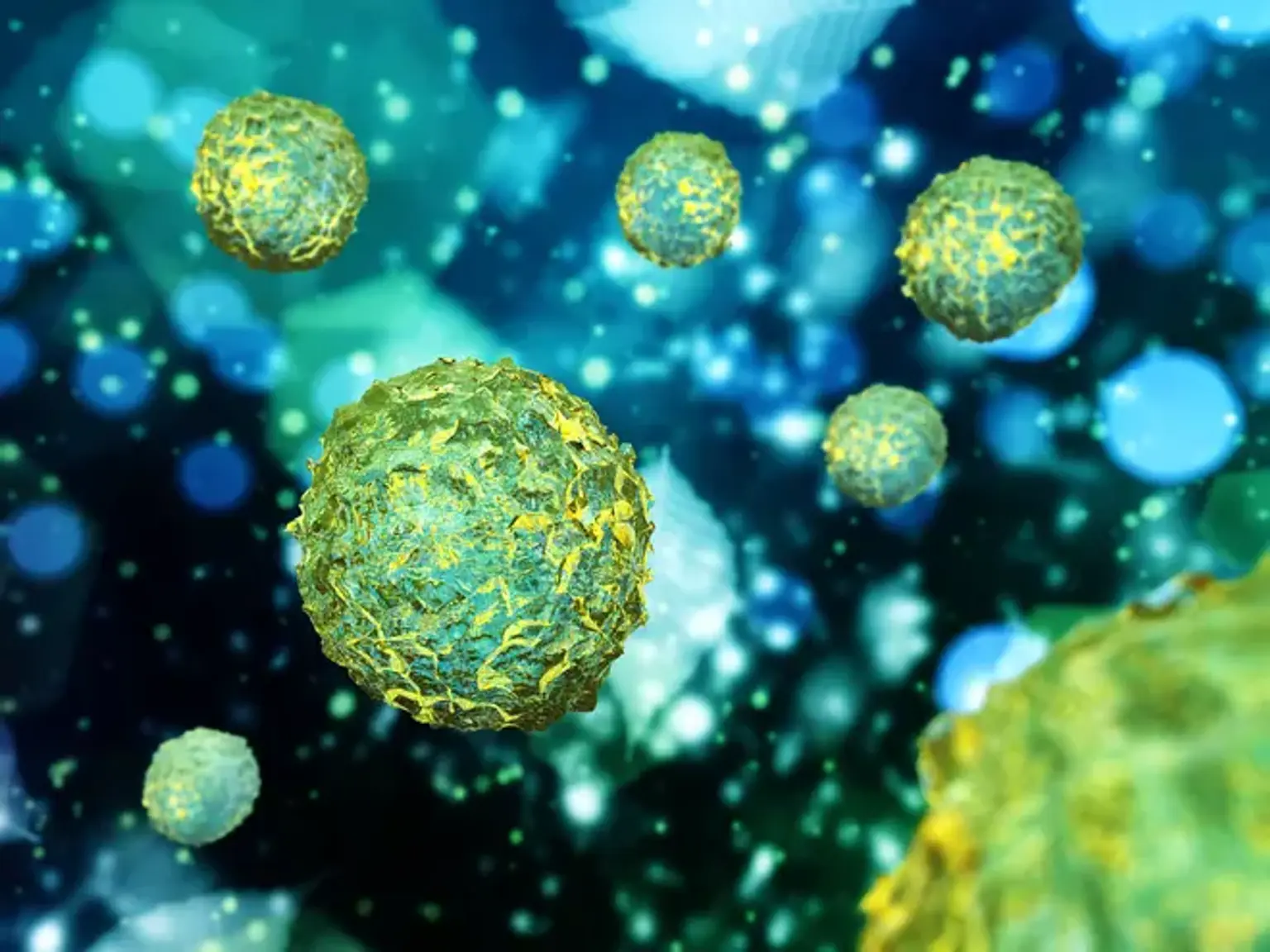
 

What are Mesenchymal Stem Cells (MSCs)?

Mesenchymal Stem Cells (MSCs)

Mesenchymal stem cells (MSCs) are multipotent stem cells that may develop into bone cells (osteoblasts), cartilage cells (chondrocytes), muscle cells (myocytes), and fat cells that give birth to marrow adipose tissue (adipocytes). The MSC reservoir in bone marrow is limited and depletes with age and illness. Surprisingly, a decrease in functional MSCs frequently coincides with an increase in adiposity. This is one of the primary reasons bones fail to repair in osteoporosis.

A great number of studies have indicated that MSC-based treatments are effective in treating a variety of pathologies, including neurological illnesses, heart ischemia, diabetes, and bone and cartilage diseases. However, the therapeutic potential of MSCs in cancer remains debatable. While some research suggest that MSCs may play a role in cancer etiology, new evidence suggests that MSCs have anti-cancer properties. Because of this, a continuous effort to understand whether MSCs promote or restrict tumor formation is required before developing an MSC-based cancer treatment. 

Mesenchymal stem cells (MSCs), also known as stromal stem cells, have the ability to develop into a variety of different types of cells inside the body, including:

  • Bone cells
  •  Cartilage
  •  Muscle cells
  •  Neural cells
  • Skin cells
  • Corneal cells

Historical Overview

bone marrow (BM)

Friedenstein and colleagues established in 1970 that bone marrow (BM) includes a population of hematopoietic stem cells (HSCs) as well as a rare population of plastic-adherent stromal cells (1 in 10 000 nucleated cells in BM). These malleable adherent cells, formerly known as stromal cells but now more popularly known as MSCs, were capable of generating single-cell colonies. When the plastic-adherent BM cells were cultured, circular colonies resembling fibroblastoid cells developed, earning the moniker colony forming unit-fibroblasts.

Friedenstein was the first to show that MSCs may develop into mesoderm-derived tissue and to recognize their relevance in directing the hematopoietic niche. Control of stem cell niches is emerging as a major role for MSCs in a wide range of organs, including hair follicles and the gut, and MSC ablation has recently been demonstrated to affect hematopoiesis.

MSCs were discovered to develop into osteoblasts, chondrocytes, and adipocytes in the 1980s. Caplan established that MSCs influenced bone and cartilage turnover, and that the surrounding environment was crucial in triggering MSC differentiation. MSCs were proved to differentiate into a myogenic phenotype in the 1990s, and Pittenger and colleagues revealed that individual adult human MSCs may be grown to colonies while keeping their multilineage potential.

In the early twenty-first century, in vivo investigations revealed that human MSCs transdifferentiate into endoderm-derived cells and cardiomyocytes, while in vitro coculturing of ventricular myocytes with MSCs caused cardiomyocyte transdifferentiation. During this period, MSCs were also shown to decrease T-lymphocyte proliferation, paving the door for the use of MSC therapy in allogeneic transplantation and as a possible immunomodulatory therapy.

Large-animal preclinical trials of MSC treatment in post-MI hearts indicated MSC engraftment, differentiation, and significant functional recovery. MSC treatment has recently been incorporated into clinical studies for ischemic heart disease.


Where are Mesenchymal Stem Cells sourced?

Mesenchymal Stem Cells

For many years, scientists assumed that mesenchymal stem cells could only be found in bone marrow. However, research has discovered that MSCs may be obtained from a variety of sources, including umbilical cord tissue, body fat, molar teeth, and amniotic fluid.

The cells generated from cord tissue, notably Wharton's Jelly, are the most basic and juvenile MSCs accessible. This supply is both non-harmful and widely available, as the majority of umbilical cords are just discarded after childbirth.

Because these cells are so immature, they have the ability to convert into any sort of cell that the body requires. Youthful cells also reproduce more quickly, and MSCs can not only develop into other cell types but also proliferate to boost their therapeutic effect on the body.

Researchers have also shown that the potency of a stem cell is related to its age, making cord tissue MSCs among of the most competent cells available.

Treatments that use MSCs from a patient's fat (adipose) sample, on the other hand, have demonstrated weak or unpredictable results. A stem cell is only as good as its source, and if the cells come from an elderly person, no amount of growth will boost their potency.

Stem cell numbers and effectiveness begin to decline significantly as we age. Stem cells from a person in their twenties, for example, are not nearly as good quality as brand new cells derived from cord tissue. Although MSCs can be obtained from any source, patients risk receiving little to no benefit from employing poorer grade stem cells.


What are mesenchymal stem cells used for?

Mesenchymal Stem Cells

The medical world has established the capacity to augment a person's stem cell count through transplantation with younger, more capable cells by collecting mesenchymal stem cells (MSCs) from donated cord tissue and increasing them to larger quantities.


Mesenchymal stem cells are immunopriviledged

MSCs have demonstrated the capacity to escape a negative immunological reaction, allowing the cells to be transplanted into a wide spectrum of patients without worry of rejection. These transplants significantly boost the body's natural healing capacities and have powerful anti-inflammatory and immunosuppressive effects.


Mesenchymal stem cells can treat a variety of conditions

MSCs have been utilized to treat a variety of autoimmune disorders, including Crohn's disease, Multiple Sclerosis, Lupus, COPD, Parkinson's, and others.

While MSCs do not give a cure for these ailments, the concept is that they let the body to recover itself sufficiently to alleviate the symptoms of the disorders for extended periods of time. In many situations, this alone provides for a significant improvement in patients' quality of life.


Mesenchymal stem cells have little adverse side effects

There have been very few side effects discovered, with the primary disadvantage being the necessity for repeated treatments to maintain high stem cell counts in the body. Without more treatments, the cells will gradually get accustomed to the point where a patient's healing abilities will revert to normal after a few years. However, patient reports indicate that the results of proper therapy persist 5-10 years.‍


How do native MSCs support tissue regeneration?

MSCs support tissue regeneration

Although the multipotent, immunomodulatory, and trophic functions of MSCs exhibited in culture offer tremendous promise for cell treatment, it is not yet clear if these MSCs also play a natural role in tissue regeneration in vivo. The finding that pericytes are innate MSCs has considerably aided our knowledge of MSC native functions, since these cells can now be investigated in vivo and their destiny probed using recognized pericyte markers.

Recent studies have shown that perivascular progenitor cells have a direct role in the genesis and/or regeneration of white adipocytes, skeletal muscle, follicular dendritic cells, and dental pulp. Overall, these findings point to the presence of regeneration cells linked with all blood arteries in adults, which are therefore widely distributed throughout the organism.

Surprisingly, the contribution of pericytes to musculoskeletal tissue formation and regeneration is not absolute and varies depending on anatomical location. The contribution of pericytes to myofibers, for example, varies amongst muscles, ranging from 1% (tibialis anterior muscle) to 7% (diaphragm), and is augmented (but only slightly) by acute or chronic muscle regeneration.

The number of chemicals known to mediate MSC paracrine activity is increasing. In vitro MSC paracrine actions can be classified as trophic, immunomodulatory, or chemoattractant. MSCs' trophic activities include apoptosis suppression, regeneration support, and stimulation, maintenance, proliferation, and differentiation of tissue-specific progenitors.

In a variety of circumstances, including ischemic brain damage, perivascular cells have been demonstrated to stimulate the development and differentiation of local stem cells. Hypoxia occurs during the early phases of tissue damage, and MSC release of anti-apoptotic proteins reduces the degree of cell death in the tissues surrounding the affected regions. The restoration of blood flow is critical for the healing of injured tissues, and the pro-angiogenic action of MSCs has been shown in mouse models of hind-limb ischemia.

Furthermore, the return to a pericyte phenotype helps to maintain the developing vasculature in vitro and in vivo. The immunomodulatory features of bone marrow-derived MSCs have been widely characterized, including their immunosuppressive effects upon allogeneic stem cell transplantation. Direct cell-cell interaction and secreted bioactive chemicals involving dendritic cells and B and T cells, including T regulatory cells, T helper cells, and killer cells, mediate the cells' immunoactivity. MSCs release a range of chemoattractant chemicals as well.

Monocytes, eosinophils, neutrophils, basophils, memory and naive T cells, B cells, natural killer cells, dendritic cells, and endothelial cell progenitors are all potential targets. Exposure to other cell types, particularly immune cells, is likely to alter the pattern of chemokine production by MSCs. As seen above, natural MSCs stimulate tissue regeneration through a number of processes and chemicals.


Do native MSCs exert exclusively beneficial effects?

native MSCs

No. There is mounting evidence that pericytes play a critical role in tissue fibrosis. Fibrosis is defined as the abnormal buildup of collagenous extracellular matrix, which can compromise tissue function in a wide range of important organs and tissues. Despite the wide variety of tissues that can be affected by fibrosis, all fibrotic responses have basic cellular and molecular pathways.

Fibrosis is defined by the persistent activity of matrix modifying myofibroblasts and often begins as a good physiological repair response to organ damage with hemostatic, inflammatory, and remodeling phases. Several recent investigations have revealed pericytes as important myofibroblast progenitors in several organs utilizing cutting-edge mouse genetic cell labeling methods.

Similarly, studies have arisen that link MSC pathology to the development of heterotopic ossification. As a result, the negative consequences are unlikely to constitute a 'normal' function, but rather a disease that develops when normal function gets dysregulated.


Clinical significance 

Clinical significance 

A tubular cartilaginous construct produced from amniotic mesenchymal stem cells has a typical physical appearance. Mesenchymal stem cells can be activated and deployed if necessary, although their effectiveness, for example, in muscle regeneration, is still rather low. Further research into the mechanisms of MSC function may lead to new ways of enhancing their potential for tissue healing.


Autoimmune disease

Clinical trials to investigate the efficacy of mesenchymal stem cells in the treatment of disorders are now underway, with a focus on autoimmune diseases, graft versus host disease, Crohn's disease, multiple sclerosis, systemic lupus erythematosus, and systemic sclerosis. As of 2014, no high-quality clinical research offers proof of effectiveness, and the study techniques are riddled with inconsistencies and flaws.


Other diseases

Many early therapeutic successes with intravenous transplantation were in systemic disorders including graft versus host disease and sepsis. Because vascular delivery suffers from a "pulmonary first pass effect," when intravenous infused cells are sequestered in the lungs, direct injection or implantation of cells into a region in need of repair may be the preferable form of treatment.


Intravenous MSC Therapy

Intravenous MSC Therapy

Intravenous infusion of MSCs is the simplest and most practical form of administration since it only needs peripheral venous access; nonetheless, the cells must travel via the pulmonary circulation to reach the heart, where cell entrapment is a problem. Intravenous infusion of MSCs was performed 15 minutes after blockage of the left anterior descending (LAD) coronary artery in pigs randomized to vehicle or different dosages of allogeneic BM-MSCs.

At 12 weeks after MI, LV ventriculography revealed no difference in EF between MSC-treated and placebo-treated rats. When MSC-treated animals were compared to placebo, pressure-volume loop analysis revealed a substantial improvement in the end-systolic pressure-volume relationship and preload recruitable stroke work. In MSC-treated rats, histological examination of postmortem hearts revealed considerably higher density of von Willebrand factor-positive blood arteries and vascular endothelial growth factor expression. Although steady-state coronary blood flow reserve was comparable across groups, adenosine-recruited coronary blood flow reserve was enhanced in MSC-treated rats.

The hemodynamic and electrophysiological effects of intravenous infusion of allogeneic MSCs were examined in a pig model of acute MI. EF improved and less eccentric hypertrophy was seen in MSC-treated mice at 3-month follow-up than in placebo-treated animals using transthoracic echocardiography

Electrophysiological investigations 3 months after MI revealed that MSC-treated rats had shorter epicardial effective refractory periods compared to placebo. Shorter effective refractory periods can cause ventricular tachycardia, raising the notion that MSCs might cause proarrhythmic remodeling.


The future: activating stem cells in their native environment?

Yes, perhaps. The presence of MSCs in vascularized organs indicates the availability of a reservoir of potentially therapeutic regenerating units throughout the body. This suggests that treatment techniques might be used to 'recruit' or enhance the regeneration ability of local MSCs following damage. Adult stem cell 'activation' is known to be regulated in part by neighboring cells; manipulation of this communication via local infiltration of growth factors or molecules integral to this interaction may represent such an approach, with the ultimate goal being the manipulation and activation of the regenerative potential of both local stem cells and those recruited from distant sites.

Because MSCs are common in vascular tissues, regulated activation of their potential would provide an alternative to purification and transplanting in circumstances when the original structural context is sufficiently intact to allow healing. MSC stimulation in situ might be used to speed healing and allow for an early return to function following a variety of musculoskeletal ailments.

Other possible uses of activating resident MSCs include the therapy of osteoporosis and muscular dystrophies in cases where local administration of MSCs is impractical. To summarize, regulated stimulation of MSCs in their natural environment will be a significant future therapeutic technique.


Conclusion 

Mesenchymal stem cells are adult stem cells obtained from various sources that have the ability to develop into numerous types of cells. These sources include bone marrow, fat (adipose tissue), umbilical cord tissue, and amniotic fluid. Future research should look at the role of MSCs in disease differentiation, transplantation, and immune response.