Micro Tese (1 testicle)

Last updated date: 16-Aug-2023

Originally Written in English

Micro TESE (1 Testicle)

Micro TESE ( 1 Testicle)

Overview

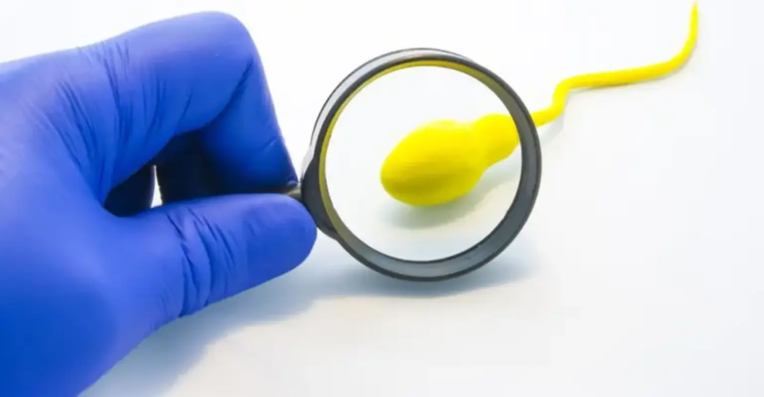
MicroTESE (microscopic testicular sperm extraction) is a process that extracts sperm straight from a man's reproductive system's testicular tissue. If a man is unable to produce or release enough quality sperm on his own, this medical treatment may be prescribed for reproductive concerns. The testicular tissue is present in the two testes, which produce sperm. The testes are located inside the scrotum, which is a tiny sac behind and under the penis.

The goals of the microTESE procedure are to obtain the best quality sperm, get enough sperm to fertilize an egg from a woman & minimize damage to the reproductive organs.


What is Micro TEZE?

Micro TEZE

Microsurgical testicular sperm extraction (microTESE) is a surgical treatment used to remove sperm from a male's testes' seminiferous tubules. It is prescribed for men who have non-obstructive azoospermia, a condition in which a man cannot generate enough sperm to have a measurable quantity in his sperm – a common cause of male infertility. If a man has a sufficient level of testosterone in his blood after a hormone test and additional checks show that his testicles are not producing typical levels of sperm, doctors will usually offer microTESE.

If a man remains azoospermic after therapy and testosterone levels that have been normal for at least four months.

Men have a high success rate with microTESE. During microTESE treatments, doctors are able to detect sperm around 60% of the time.


What is Male Infertility?

Male Infertility

Infertility is a reproductive system disorder that makes it difficult for the body to complete the basic functions of reproduction. Both men and women are affected. Most instances of infertility are addressed with medication or surgery.

Male infertility can be caused by either nonobstructive azoospermia (the male does not make sperm) or obstructive azoospermia (sperm is created but obstructed and cannot be released from the body).

Many times, the causes of infertility are unexplained. Sometimes, the difficulties may be from a genetic disease such as cystic fibrosis, a birth defect, a medical problem, or the result of an earlier treatment that may cause infertility (such as certain cancer treatments). In other cases, we do not know what the cause of the infertility is, but we still have treatments that may help.


What Makes Micro TESE Successful?

Micro TESE Successful

A skilled surgeon and an outstanding andrology technologist searching for sperm are required for microTESE to be effective. An andrology lab technician will be present in the operating room throughout your microTESE procedure to analyze your seminiferous tubules for sperm.

If sperm is discovered during your microTESE, it will be collected and preserved so that it can be utilized in future reproductive therapies such as in-vitro fertilization (IVF) with intracytoplasmic sperm injection (ICSI).

According to current research, frozen sperm may outperform fresh sperm during IVF (in-vitro fertilization).

Please keep in mind that any sperm discovered during this treatment must be used for IVF (in-vitro fertilization). Because the sperm in your testis hasn't learnt to swim yet, it won't be able to fertilize an egg if it's placed inside the uterus.


How is Infertility Diagnosed?

Infertility Diagnosis

If a couple is unable to conceive after several months of trying without using birth control, they should consult a doctor. If the lady is over the age of 35, the pair should contact a doctor as soon as possible. Your healthcare professional will examine both spouses to rule out any physical concerns that may be causing infertility. The physician will also ask several questions and go over each individual's medical history.

To examine male fertility, the following tests may be advised or ordered:

  • Semen analysis: Determines the number and quality of sperm. Semen is a body fluid that is secreted by the male reproductive organs. It carries sperm and other nutrients that help the sperm survive to enable successful fertilization.
  • Blood test: Checks for genetic or hormone problems. (Hormone levels are important in both male and female fertility.)
  • Ultrasound of the scrotum: Looks for abnormalities in the veins that carry blood from the testicles and back to the rest of the body.

In-home testing kits may be available for semen analysis. Ask the doctor for more information.


What Will the Infertility Tests Determine?

 Infertility Tests Determine

Semen analysis will give the doctor necessary information to help assess fertility and create a treatment plan. The tests should show some of the following:

  • Amount of semen: At least 1.5 milliliters is considered normal. A lower number may mean that there is an internal issue with a part of the reproductive system, such as the seminal vesicles or a prostate gland, blocking the release of semen.
  • Sperm count: Fifteen million to 300 million per milliliter is considered a normal range. Below 15 million is abnormal.
  • Morphology: The size and shape of the sperm. Four percent of normal-shaped sperm (using the “strict” criteria) is ideal to be able to fertilize an egg. Some doctors consider even a lower percentage as normal.
  • Motility: The movement of the sperm. Approximately 40% of sperm should be moving. The quality of movement is graded from 0 to 4, with a score over 3 considered good.


How Does Micro-TESE Work?

Micro-TESE

Micro-TESE is normally done in combination with ICSI since the concentration of sperm collected is generally low and inappropriate for IVF.

You and your doctor will agree on a date for the operation once you have decided to have micro-TESE, which will only be after you have had an in-depth consultation with a member of our staff and undertaken a series of tests to see if this is the right choice for you. This is normally done prior to the start of IVF therapy.

During the procedure, the surgeon opens the testicles and examines the tissues under a microscope, removing any areas that he believes are likely to contain sperm - he can tell by looking at the seminiferous tubules (the parts of the testicles that produce and transport sperm) and determining which ones are dilated.

Once all of these areas have been extracted, they are taken to the lab, where they are examined to see if any sperm is present. Any sperm that is found will then be used for treatment or frozen until the point of the IVF treatment cycle when it is needed. At that stage, the sperm will be defrosted and the strongest specimens selected to be injected directly into the eggs that have been harvested.


Optimization Prior to Micro TESE

Optimization of Micro-TESE

Medical management

Most men presenting with a central cause of azoospermia due to hypothalamic-pituitary axis dysfunction will dramatically benefit from medical management with gonadotropin releasing hormone (GnRH) or gonadotropins (i.e., hCG, hMG, recombinant FSH). Common regimens include pulsatile GnRH 25–600 ng/kg subcutaneously (SC), intravenous (IV) or by pump every 120 minutes, hCG 1,000–2,500 IU injected intramuscularly (IM)/SC twice per week, or hMG 75–150 IU injected 3 times per week, recombinant FSH 75 IU every 1–2 days. Most of these men will not require sperm retrieval if testicular function is optimized with hormonal therapy.

Medical optimization strategies have been investigated for patients with azoospermia due to testicular dysfunction, with the goal of increasing both testosterone and FSH levels to stimulate spermatogenesis. A multi-institutional study evaluated the effectiveness of using a combination of clomiphene citrate (CC), hCG, and/or hMG to increase serum testosterone to 600–800 ng/dL and serum FSH to 1.5 times baseline in 612 men with NOA. 

In this study, the intervention group received hormonal optimization according to a defined protocol while the control group received no hormonal optimization, resulting in a significantly higher SRR for the intervention group (SRR 57% vs. 34% for intervention group vs. control group). While this study supports the medical hormonal optimization of FSH and endogenous testicular testosterone levels to improve SRR, it is important to note that the control group had a lower SRR than previous reports in the literature. 

Furthermore, a large retrospective study of 1,054 men, did not find benefit of hormonal therapy in men undergoing micro TESE. Here, no difference in SRR was identified among men with baseline testosterone >300 ng/dL, compared to men with baseline testosterone <300 ng/dL and not receiving medical hormonal treatment, or men receiving aromatase inhibitors, selective estrogen receptor modulators, or any combination with hCG.

Another class of medication that has been used to optimize hormonal parameters in NOA and cryptozoospermia patients is aromatase inhibitors, such as anastrozole and letrozole. Although most studies were performed using anastrozole, which appears to have fewer side effects, one study comparing letrozole to placebo in cryptozoospermia and NOA patients suggested that letrozole may help enhance spermatogenesis with sperm return to the ejaculate in some NOA patients. Candidates for management with aromatase inhibitor include men with low serum testosterone (<300 ng/dL) and low testosterone: estradiol ratios (<10), in whom aromatase therapy has been suggested to enhance intratesticular testosterone levels and improve spermatogenesis.


Varicocele repair (VR):

VR has been demonstrated to result in return of sperm to the ejaculate in 10% of patients, thus obviating the need for surgical sperm retrieval. VR is more likely to result in sperm return to the ejaculate in patients with hypospermatogenesis or late maturation arrest on testicular histology when compared to more severe NOA histologies. Of course, these patients are the same individuals likely to have sperm detected in the ejaculate on a more detailed or repeat semen analysis. 

A meta-analysis demonstrated that VR increases the likelihood of successful sperm retrieval at microTESE by an odds ratio of 2.65. Other large studies, apparently excluded from the meta-analysis show no effect of prior VR on SRRs. In one series, no increase in post varicocele microTESE SRR was identified and sperm returned to the ejaculate among 22% of men postoperatively, but only 9.6% have adequate motile sperm for ICSI. 

Both female partner age and number of children desired are important considerations when discussing VR in the management of the azoospermic male since the potential benefits of VR are not realized until at least 3–6 months after the repair.


What Happens During Micro TEZE?

Micro-TESE during anesthesia

MicroTESE should preferably be conducted under general anesthesia. To get simple access to both testes, an incision is made in the median raphe. By incising through the dartos and tunica vaginalis, the larger of two testicles is delivered first.

The tunica albuginea of the testicle is then incised transversely under the operating microscope with a 15-degree micro-knife, taking care to avoid equatorial testicular vessels. Mosquito clamps are inserted on each side of the tunical incision, including the margin of the seminiferous tubules, to avoid tissue avulsion while the testicle is bivalved using the surgeon's fingers.

Right-handed surgeons should stand to the patient's left, allowing the surgeon's left hand to rest between the patient's legs rather than on the abdomen. To support the testicle and keep it exposed, a three-finger method is used with the left hand. The third digit here supports the posterior side of the testis, while the thumb and index finger offer exposure on the testis' sliced surface. The seminiferous tubules are then checked systematically for dilated, opaque tubules.

Excellent visibility of the seminiferous tubules is crucial, and this is accomplished by keeping the seminiferous tubules within the focus distance of the microscope and providing appropriate hemostasis by bipolar electrocautery. After identifying dilated, opaque tubules, the full length of the centrifugally-oriented tubule is extracted using tissue micro-forceps and deposited in a tiny petri dish containing sperm transport buffer.

After searching the whole cranial or caudal half of the testis, the tissue is readied for transfer to an embryologist in the operating room, who will check the extracted testicular tissue for sperm. If sperm is found, the contralateral testicle is not dissected; however, if sperm is not found, the contralateral side is dissected.

Tissue processing of excised tubules is critical for effective identification of sperm contained inside the tissue. Prior to embryologist observation, excised tubules are minced with scissors until the suspension is thin enough to be aspirated in and out of a 24-gauge angio-catheter. This approach has increased sperm recovery by 300-fold.

Using bipolar electrocautery, hemostasis is carefully maintained during dissection. After a testis has been entirely dissected, a succession of mosquito clamps is used to re-approximate the tunica albuginea margins, which are subsequently closed in a running fashion with a 5-0 non-absorbable monofilament suture. This serves to indicate the site of dissection in the event that a repeat surgery is necessary in the future. The testicle is restored to its anatomical location within the tunica vaginalis, which is subsequently closed in a running fashion using an absorbable monofilament suture. If the contralateral testicle needs to be dissected, it is done at this time as well.

Otherwise, the tunica vaginalis is injected with local anesthesia and the dartos layer is closed in a running fashion with an absorbable suture, taking care to incorporate the whole cut edge in the closure for maximum hemostasis. Before tying the knot, each hemi-scrotum receives 5 mL of local anesthetic. The knots are hidden behind the dartos layer. Finally, an adequate dressing is placed and the skin is closed using interrupted horizontal mattress stitches.


Complications & Considerations

Complications in Micro-TESE

It is important to obtain a semen analysis prior to the planned microTESE, to assess for sperm. Since 5–10% of men with NOA will have sperm in the ejaculate viable for use in ICSI, thus obviating the need for a surgical sperm retrieval. Another important consideration prior to microTESE is the use of fresh or frozen sperm for IVF-ICSI. 

Among men with NOA that have sperm retrieved, frozen and later thawed, only 33% of sperm will be viable for use with ICSI. Thus, we recommend performing simultaneous ICSI with fresh testicular sperm harvested by microTESE in NOA patients. Clinical pregnancy rates with IVF-ICSI using sperm retrieved from microTESE range between 20–50%.

Studies have found serum testosterone levels following microTESE to decrease from 316 to 251 ng/dL, but return to 95% of baseline at 18 months, of these 5–10% of men will have a decrease in testosterone significant enough to warrant subsequent androgen replacement. Ultrasound changes in the testes are seen in 18.3% of microTESE testes at 1 month, 10–44% of testes at 3 months, but in only 3.3–10% of testes at 6 months. 

Early testicular ultrasound findings following microTESE include hypoechoic changes while late findings at 6 months tend to be limited to focal echogenic lesions of fibrosis and calcification. Since time is required for recovery of the limited sperm production that is present in men with NOA, at least 6–12 months should be allowed after microTESE, before considering repeat microTESE procedures if additional attempts are required.

When compared to conventional TESE, microTESE results in lower complication rates, with fewer hematomas, less testicular fibrosis, and less frequent testicular atrophy with higher SRRs. We have not experienced testicular loss following microTESE, although some anecdotal reports exist among very small testes.


Will the Micro TEZE Procedure Need to Be Repeated?

Micro TEZE Procedure

Any extra sperm collected during testicular sperm extraction should be frozen wherever possible to preserve the sperm and avoid further operations.

Although freezing the sperm may have some negative consequences, medical experts believe that the outcome is generally the same as using non-frozen sperm in fertility procedures such as intracytoplasmic sperm injection (ICSI), a type of in vitro fertilization in which one sperm is injected directly into one egg. In vitro fertilization is a laboratory operation in which a sperm and an egg are fertilized outside of the body. The fertilized egg is subsequently implanted in the uterus of the female.

If the microTESE procedure needs to be repeated, it is usually best to wait six to 12 months


Conclusion

Nonobstructive azoospermia (NOA)

The lack of sperm in the ejaculate due to spermatogenesis failure is known as nonobstructive azoospermia (NOA). It is the most severe kind of infertility in men caused by intrinsic testicular failure or insufficient gonadotropin production. However, microsurgical testicular sperm extraction (microTESE) may provide a road to biological parenthood for men with NOA.

Men with NOA should be provided microTESE, according to the most recent American Urological Association and American Society for Reproductive Medicine male infertility recommendations. MicroTESE is a surgical treatment that extracts sperm from the testis's seminiferous tubules.