Minimally invasive thoracic procedures (without opening the chest)
Overview
Thoracic surgery has changed throughout the years. Accessing the intrathoracic organs through the bony ribcage has proven problematic for thoracic surgeons. Large incisions across the chest, such as posterolateral thoracotomies with rib spreading, were the conventional way to accessing the lungs in the past. These procedures involve significant trauma to the patient and have high rates of death and morbidity.
However, as technology and surgical techniques have advanced, thoracic surgery has developed to reduce patient stress while still adhering to oncological and surgical principles. Modern technology, along with a wide range of old and new surgical procedures, provides the thoracic surgeon with a strong weapon.
What is Minimally Invasive Thoracic Surgery?
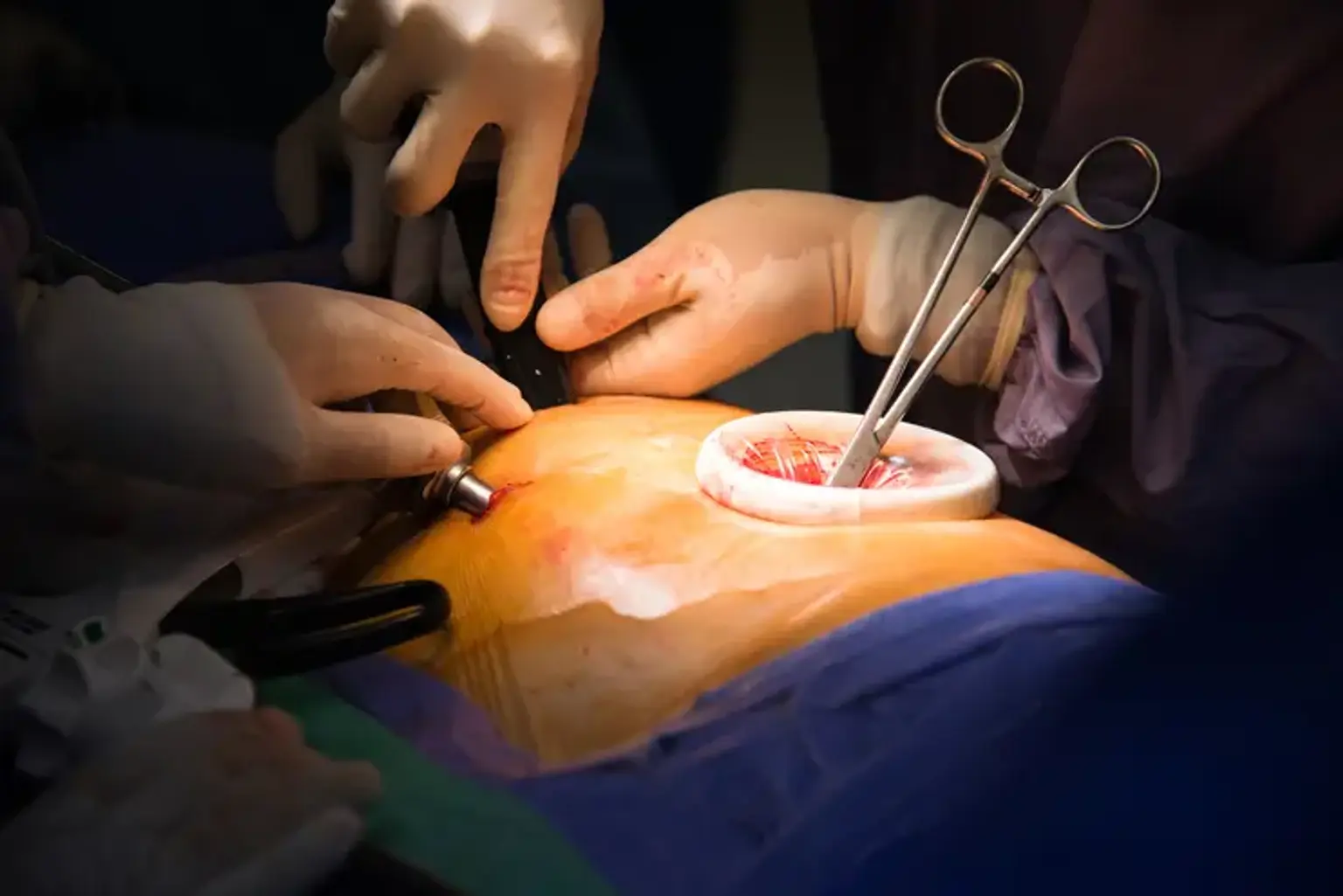
Minimally invasive thoracic surgery is a method of doing surgery in the chest with small incisions rather than major cuts or incisions in the body, and it does not require the ribs to be moved apart. Surgeons enter the lung by tiny incisions between the ribs with a camera and equipment. For minimally invasive thoracic surgery, there are two options: video-assisted thoracoscopic surgery (VATS) and robotic-assisted surgery.
A thin tube called a thoracoscope is placed through a small incision between the ribs during video-assisted thoracoscopic surgery (VATS). A tiny camera is located at the tube's end. This allows the surgeon to see the whole chest cavity without opening the chest or spreading the ribs. The lung tissue is subsequently removed by the surgeon using specially developed equipment inserted through one or two further minor incisions.
In robotic-assisted surgery, a surgeon will sit at a console next to the patient in the operating room and manage the tools on the robotic surgical system, including a camera. To offer an excellent view of the inside of the chest cavity, a tiny 3D high-definition camera is put via one of the small incisions, while wristed robotic devices are entered through the other small incisions created between the ribs. Through one of the small incisions, the surgeon extracts lung tissue. The wristed devices enable the physician to do the operation without the need for bigger incisions to open up the chest or spread the ribs.
What conditions can be treated with minimally invasive thoracic surgeries?
On a case-by-case basis, surgeons determine if a patient is a candidate for minimally invasive thoracic surgery. When determining whether a patient is a candidate for minimally invasive surgery, surgeons assess the patient's individual condition, medical history, and anatomy. VATS and robotic surgery are used to treat a variety of disorders, including:
- Emphysema — One incision allows access to the viewing tool (thoracoscope) during Lung Volume Reduction Surgery (LVRS). Through two further incisions, forceps and a surgical stapling equipment are utilized to remove the afflicted tissue.
- Interstitial Lung Disease — As a result of VATS, the diagnosis of interstitial lung disease has become substantially more accurate. To determine the existence of this condition, many sections of the lung can be biopsied and scanned without the need for a big incision.
- Lung Cancer — VATS is commonly used to help in the diagnosis, staging, and therapy of lung cancer. VATS can greatly minimize the morbidity associated with traditional surgical excision of lung nodules, in addition to assisting in the detection of pulmonary nodules.
- Myasthenia Gravis — VATS is a technique for removing the thymus gland via incisions under the arm. Transcervical thymectomy, a less invasive operation in which the thymus is removed through a tiny incision in the lower region of the neck, can also be performed by experienced surgeons.
- Spontaneous Pneumothorax — In individuals with spontaneous pneumothorax, VATS allows for improved imaging of the whole lung surface (collapsed lungs). Patients who undergo this therapy also have less postoperative discomfort than patients who undergo standard surgical procedures.
- Lobectomy and mediastinal lymph node dissection for lung cancer
- Wedge resection for solitary pulmonary nodules
- Wedge resection for pulmonary metastases
- Esophageal mobilization for esophageal cancer
- Decortication for empyema (infection within the pleural space)
- Insertion of pleurex catheters for recurrent effusions
- Thoracic sympathectomy for hyperhidrosis (excessive sweating)
- First rib resection for thoracic outlet syndrome
- Heller myotomy for achalasia
What are the advantages of minimally invasive thoracic surgeries?
Prior to the development of minimally invasive thoracic procedures, surgeons relied on big open thoracotomies to get access to the chest cavity. To merely access the interior chest cavity, a big incision in the chest is required, as well as the shattering and spreading of several ribs and substantial tissue damage.
VATS and robotic thoracic surgery are conducted through a few tiny incisions in the chest using high-tech camera-equipped tools. These methods result in no rib breakage or spreading. Unfortunately, many surgeons are not fully educated in modern minimally invasive thoracic surgical procedures, particularly the gold standard for early-stage lung cancer therapy, minimally invasive lobectomy.
Because the benefits of minimally invasive thoracic surgery for patients are significant, surgeons use these surgical procedures almost exclusively. Among the advantages are:
- Reduced scarring
- Decreased postoperative pain
- Shorter hospital stays
- Lower risk of complications
- Better overall surgical outcomes
- Reduced immunosuppression following surgery
Video-assisted thoracic surgery
Video-assisted thoracoscopic surgery (VATS) is a minimally invasive surgical method used to diagnose and treat a range of chest cavity disorders. When compared to typical thoracic surgeries, the VATS approach provides significant benefits to patients. Aside from lung cancer surgery, the VATS method can be utilized for different sorts of chest operations involving the lungs, esophagus, thymus, pleural or pericardium.
Video-assisted lobectomy
The most common operation for lung cancer is a lobectomy (removal of a substantial piece of the lung). Lobectomy has historically been performed as part of thoracotomy surgery. An incision is created on the side of the chest between the ribs during thoracotomy surgery. The ribs are then stretched apart to provide the surgeon access to the chest cavity in order to remove the tumor or damaged tissue.
To gain access to the chest cavity without spreading the ribs, three 1-inch incisions and one 3- to 4-inch incision are performed during video-assisted lobectomy. When compared to standard thoracotomy surgery, video-assisted lobectomy results in a faster recovery with less discomfort and a shorter hospital stay (typically 3 days). Video-assisted lobectomy surgical results are equivalent to standard lobectomy outcomes.
Although minimally invasive techniques are evaluated for all patients, patients with big or central tumors may not be candidates for video-assisted lobectomy in some situations.
Wedge resection
The surgical excision of a wedge-shaped segment of tissue from one or both lungs is known as a wedge resection. A wedge resection is often used to diagnose or treat tiny lung lesions.
Lung biopsy
A lung biopsy is a procedure that involves removing a small sample of lung tissue through a small incision between the ribs. Expert pathologists examine the lung tissue under a microscope and may send it to a microbiological laboratory to be cultured. The existence of lung disorders such as infectious or interstitial lung disease is investigated in the lung tissue.
Drainage of pleural effusions
A pleural effusion is an accumulation of extra fluid between the layers of the pleura, which is the thin membrane that borders the exterior of the lungs and the interior of the chest cavity. Normally, there is relatively little fluid in this region. The extra fluid is removed (drained) through a thoracoscopic operation known as thoracentesis and may be collected for study to rule out potential causes of pleural effusion such as infection, malignancy, heart failure, cirrhosis, or renal disease. To avoid the recurrence of fluid build-up, sterile talc or an antibiotic may be introduced during surgery.
Mediastinal, Pericardial, and Thymus thoracoscopic procedures
The mediastinum is the space between the lungs in the center of the chest. The pericardium is the tissue that surrounds the heart. The thymus is a tiny organ that runs from the base of the throat to the front of the heart in the upper/front section of the chest. The thymus cells are part of the body's regular immune system. The thymus plays a vital function in the development of the immune system early in life.
Thoracoscopic procedures can be used to evaluate the mediastinum, pericardium, or thymus, as well as to take tissue samples and surgically remove malignant growths in the affected area.
Preoperative preparation
Ask your healthcare provider about what you need to do to get ready for your VATS. In general:
- You may need to stop taking certain medications, such as blood thinners, before the surgery. Discuss with your healthcare practitioner all of the medications you use, including over-the-counter medications.
- If you smoke, you need to quit before your surgery. Ask your healthcare provider for resources to help you.
- Daily exercise is an important part of getting ready. Ask your healthcare provider what kind is best for you.
- You might need to do breathing exercises with a device called a spirometer.
- Avoid eating or drinking anything after midnight before your surgery.
- Any hair over the surgical area may be removed with clippers before the surgery.
Your doctor may order tests to determine how well your lungs are functioning. Before your operation, he or she may want to check on your overall health. These will vary depending on the cause for your VATS. Some examples may be:
- Chest X-ray to see the heart and lungs
- Chest CT scan to get more detailed pictures of the lungs
- Positron emission tomography to look for cancer tissue
- Electrocardiogram to check the heart rhythm
- Pulmonary function tests to see how well your lungs are working
- Blood tests to check overall health
How VATS is performed?
- Your surgery will begin with you being placed under general anesthesia.
- Once you are asleep, a breathing tube is placed into your airway to allow each lung to be separately inflated during surgery.
- You are then positioned on your side.
- The surgeon will make several small cuts (incisions) in your thoracic (chest) area between your ribs.
- The thoracoscope and other tools are inserted through these incisions.
- The surgeon uses these tools to remove part of the lung and lymph nodes, drain fluid from around the lung, or do a procedure on other chest organs.
- At the end of the surgery, the surgeon will insert a chest tube through one of the small incisions to drain fluid or air leaking into the chest cavity and to help your lungs re-inflate. This tube remains in place for a few days and is typically removed at the bedside before you go home.
- Patients are usually discharged from the hospital a few days after surgery.
- It is important to give yourself time to rest when you go home. Gradually you will get stronger and feel like yourself in a few weeks. Follow any post-operative instructions from your doctor.
Postoperative care
When you first wake up, you may be confused. You may awaken several hours or even longer following the procedure. You will be connected to various equipment so that the medical personnel may monitor your heart rate and other vital signs.
You may receive oxygen by inserting little tubes into your nose. It is typically just transitory. You will experience some discomfort. However, you should not experience excruciating pain. You can request pain medication if necessary. A chest tube will most likely be used to collect fluid from your lungs.
To avoid an illness, you will utilize a gadget that helps you to breathe deeply. Wearing specific stockings can help avoid blood clots. You will be admitted to the hospital for a few days.
The length of your hospital stay will be determined by the operation performed. Patients who get thoracoscopic lung biopsies or wedge resections can usually go home the same day. VATS lobectomy patients are generally able to go home 3 to 4 days following surgery.
Make sure you have someone to drive you home when you leave the hospital. You will also require some assistance at home for a time. Your stitches or staples will most likely be removed during a follow-up appointment. Keep all of your follow-up appointments.
You may feel tired after the procedure. However, you will gradually regain your strength. It might take many weeks for you to fully recover. You must get up and walk numerous times every day. Inquire with your doctor about when it will be safe for you to drive.
For many weeks, avoid lifting anything heavy. Follow all medication, exercise, food, and wound care advice given to you by your healthcare professional. If you see any indications of infection, fever, edema, or worsening discomfort, contact your healthcare professional straight away. It is typical to have some discharge from your incision.
A follow-up visit will be scheduled 7 to 10 days after your operation. At your follow-up appointment, your surgeon will check the wound sites and your recuperation, as well as offer advice for your activities and return to work. Most persons who have minimally invasive thoracic surgery may return to work within 3 to 4 weeks.
What are the possible complications of VATS?
Each person reacts differently to surgery, and your outcomes may vary depending on a variety of factors such as your health history, the type of lung cancer you have, the planned treatment, and/or your surgeon's experience.
There are dangers associated with all forms of surgery. Blood clots, infection, hemorrhage, air leaking from your lungs, and discomfort are all possibilities. Other VATS complications include:
- Partial collapsed lung (atelectasis)
- Abnormal heart rhythms
- Pneumonia
- Collection of thick pus in the chest cavity (empyema)
- Blood clot. This can lead to pulmonary embolism or stroke.
- Shock
- Complications from anesthesia
Robotic-assisted thoracic surgery
Robotic thoracic surgery is a type of minimally invasive surgery used in thoracic procedures for some lung cancer cases. It can be used to remove diseased lung tissue and possibly surrounding lymph nodes. In robotic surgery, a surgeon will sit at a console next to the patient in the operating room and control the instruments on the robotic surgical system.
First, a small 3D high-definition camera is placed through one of the cuts (incisions) to provide a view of the inside of the chest cavity. Then robotic instruments are placed through the other small incisions made in between the ribs. The robotic instruments are completely controlled by the doctor’s hands at the console.
Through one of the incisions, the surgeon extracts lung tissue. The magnified image and wristed devices enable the surgeon to make precise, controlled motions to remove lung tissue without opening up the chest or spreading the ribs. Aside from lung cancer surgery, the robotic approach may be utilized for various types of chest treatments involving the lungs, esophagus, thymus, and some cardiac procedures.
Your care team will decide your eligibility for robotic thoracic surgery on a case-by-case basis. Patients with early-stage lung cancer are often eligible for lung cancer surgery as a first-line therapy (stage 1 or 2). Lung cancer surgery is sometimes performed after another sort of treatment, such as chemotherapy or radiation, has been used to decrease the tumor. When considering lung surgery, look for a surgeon that is educated in minimally invasive surgical procedures.
How Robotic thoracic surgery is performed?
- Your doctor will go through everything you need to do to prepare for surgery with you.
- You will be put under general anesthesia before your procedure.
- After you've fallen unconscious, a breathing tube is inserted into your airway to allow each lung to be inflated individually during operation.
- You are then positioned on your side.
- The surgeon will normally make four tiny cuts (incisions) between your ribs to provide the robot access to your thoracic region.
- The surgical assistant will place the instruments and cameras attached to the “arms” of the robot into the incisions.
- The surgeon sits at a control console nearby where they can see magnified, clear images of the surgical area.
- The robot reacts in real time to the surgeon's hand motions when the diseased section of the lung and perhaps lymph nodes are removed.
- At the end of the procedure, the surgeon will introduce a chest tube through one of the minor incisions to drain fluid or air seeping into the chest cavity and aid in the re-inflating of your lungs. As you heal, your care team will remove this tube.
- Patients are frequently discharged from the hospital sooner after undergoing minimally invasive surgery.
- Follow any post-operative instructions from your doctor.
- It is necessary to allow yourself time to recuperate when you get home. You will gradually gain strength and feel more like yourself.
Risks and benefits of Robotic thoracic surgery
There are risks associated with all forms of surgery. Blood clots, infection, bleeding, irregular heartbeats, air leaks from your lungs, and discomfort are examples. Longer operational hours and time under anesthesia may be related with robotic thoracic surgery. If necessary, your surgeon may have to switch to a more intrusive strategy to complete the procedure.
There are other advantages as compared to open chest surgery through thoracotomy:
- The robotic tools are very accurate and flexible. The camera also helps the surgeon to receive a good view of the chest during surgery and to reach regions of the chest that are otherwise difficult to reach.
- Minimally invasive surgery may result in reduced blood loss, shorter recovery periods, and fewer problems.
Your outcomes may be influenced by a variety of factors such as your medical history, the kind of lung cancer you have, the planned technique, and/or the experience of your surgeon.
Conclusion
Minimally invasive approaches for thoracic and bronchial operations using fiberoptic video technology have been developed to lower the risk, trauma, and cost of intrathoracic surgical therapies. A video-assisted technique conducted through a few tiny incisions can help the surgical treatment of practically every intrathoracic disease.
Many surgical procedures may now be performed successfully, affordably, and with minimum patient stress thanks to video-assisted thoracoscopic surgery. Surgeons have begun to better identify which minimally invasive thoracic surgical procedures are beneficial and which patients are candidates for them. Patient acceptability has been favorable as a result of reduced morbidity and speedier return to exercise.
A surgeon and a highly trained team of surgical professionals and anesthesiologists undertake video-assisted thoracoscopic surgery. Three or four small incisions (1-2 cm) are made in the chest cavity while under general anesthesia, and a tiny camera called a thoracoscope is placed between the ribs and into the chest. This device sends a live video stream from within the chest to monitors in the operating room, which our surgeons may observe during the procedure.
To complete the process, more tools are put via the other incisions. The incisions are kept as tiny as possible, and the ribs are never shattered or spread. When the procedure is finished, the equipment and camera are withdrawn, and the incisions are sutured closed.
There are potential dangers, as with any operation. These include an air leak, which necessitates the use of the chest tube for a longer period of time than anticipated; bleeding; wound infection; pneumonia, harm to the surrounding lung tissue, heart, liver, and kidneys, and possibly death. Conversion from a minimally invasive to a classic, open method may be required.