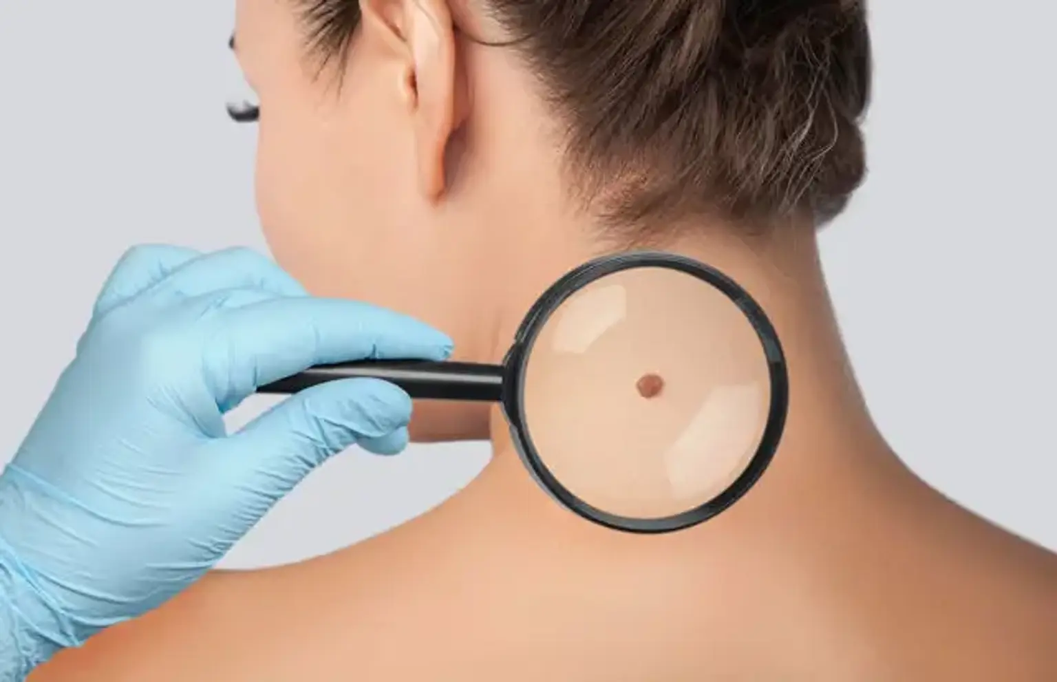
Introduction
Mohs surgery is one of the most effective treatments for skin cancer, particularly basal cell carcinoma (BCC) and squamous cell carcinoma (SCC). It is a specialized microsurgical procedure that removes cancerous tissue layer by layer, ensuring that only the affected area is excised while preserving as much healthy skin as possible.
This technique is widely regarded as the gold standard for skin cancer removal because of its high success rate and minimal scarring. Patients with skin cancer in sensitive areas, such as the face, hands, or neck, often prefer Mohs surgery because it leaves a smaller scar compared to traditional excision.
In this article, we will explore what Mohs surgery is, how it works, and why it is a leading choice for many skin cancer patients.
What is Mohs Surgery?
Mohs micrographic surgery is a precise and highly effective technique for treating skin cancer. Named after Dr. Frederic Mohs, who developed it in the 1930s, the procedure involves removing thin layers of cancerous tissue and examining them under a microscope in real time. This allows surgeons to identify and eliminate all cancer cells while sparing healthy tissue.
Unlike traditional skin cancer removal methods, where a larger area is excised with wide margins, Mohs surgery is performed in stages. After each layer is removed, the surgeon checks for remaining cancer cells before deciding whether further tissue removal is necessary.
The key advantage of Mohs surgery is its precision. It offers up to a 99% cure rate for basal cell carcinoma and 94% for squamous cell carcinoma, making it one of the most reliable skin cancer treatments available.
Why is Mohs Surgery Performed?
Mohs surgery is recommended for skin cancers that:
Are located in high-risk areas (face, scalp, ears, hands, feet, genitals).
Have irregular or undefined borders, making them difficult to remove with traditional methods.
Have returned after previous treatment.
Are aggressive or fast-growing.
This procedure is most commonly used for basal cell carcinoma (BCC) and squamous cell carcinoma (SCC), but in some cases, it can also be effective for early-stage melanoma. Because it minimizes damage to healthy tissue, Mohs surgery is ideal for areas where cosmetic appearance and function are important.
Patients who undergo Mohs surgery benefit from its high success rate, reduced risk of recurrence, and smaller scars compared to other skin cancer treatments.
The Step-by-Step Mohs Surgery Process
Mohs surgery is performed as an outpatient procedure under local anesthesia. The process typically follows these steps:
Preparation
The surgeon marks the cancerous lesion, cleans the area, and injects a local anesthetic to numb the skin. The patient remains awake during the procedure.
Layer-by-Layer Tissue Removal
The surgeon removes a thin layer of skin containing the tumor and immediately sends it to a lab for microscopic analysis.
Tissue Examination
The removed tissue is frozen, sliced into thin sections, and examined under a microscope to detect any remaining cancer cells. If cancer is still present, another layer is removed. This process repeats until all margins are cancer-free.
Wound Closure
Once the surgeon confirms that the cancer has been completely removed, the wound is either left to heal naturally, closed with stitches, or in some cases, repaired with a skin graft. The patient is then given post-operative care instructions to ensure proper healing.
This method allows for maximum precision, ensuring complete cancer removal while preserving as much healthy skin as possible.
Benefits of Mohs Surgery
Mohs surgery is widely recognized as one of the most effective treatments for skin cancer. Its advantages include:
High Success Rate
Mohs surgery offers a 99% cure rate for basal cell carcinoma (BCC) and a 94% cure rate for squamous cell carcinoma (SCC). These rates are significantly higher than those of traditional excision or radiation therapy.
Precise Cancer Removal
Because the surgeon examines each tissue layer under a microscope, only cancerous tissue is removed, sparing as much healthy skin as possible. This makes it ideal for treating skin cancer in cosmetically sensitive areas like the face, nose, or hands.
Minimal Scarring
Unlike standard excision, which removes a larger margin of skin, Mohs surgery preserves healthy tissue, resulting in smaller scars and better cosmetic outcomes.
Same-Day Results
Since tissue is examined immediately, Mohs surgery provides real-time confirmation of complete cancer removal. This eliminates the need for additional surgeries or follow-up procedures.
Mohs Surgery vs. Other Skin Cancer Treatments
Mohs surgery is not the only treatment for skin cancer. Here’s how it compares to other common options:
Mohs Surgery vs. Standard Excision
FactorMohs SurgeryStandard ExcisionPrecisionRemoves cancer layer by layer, sparing healthy skinRemoves cancer with a larger margin of healthy tissueCure Rate99% for BCC, 94% for SCC90-95% for non-Mohs excisionScarringMinimized due to tissue preservationLarger scar due to wider excisionSame-Day ResultsYesNo, pathology results take days
Mohs Surgery vs. Radiation Therapy
Mohs surgery physically removes the cancer, while radiation therapy uses X-rays to destroy it.
Radiation may be used for elderly or high-risk patients who are not good candidates for surgery.
Mohs is preferred for cancers in visible areas, as radiation can cause skin discoloration over time.
Mohs Surgery vs. Topical Treatments
Topical creams (imiquimod, 5-FU) are only effective for very early-stage skin cancers.
Mohs surgery is necessary for deeper or aggressive tumors.
Mohs is often the best choice for skin cancers in cosmetically sensitive areas, where tissue preservation and high cure rates are priorities.
Risks and Side Effects
While Mohs surgery is a safe and highly effective procedure, like any surgery, it carries some risks.
Common Side Effects
Pain and swelling: Mild discomfort, redness, and swelling around the surgical site are common.
Bruising: Some patients experience bruising, especially if the surgery is performed on the face or near the eyes.
Temporary numbness: If nerves are affected during surgery, numbness around the area may persist for weeks or months.
Potential Complications
Scarring: Although Mohs surgery minimizes scarring, some patients may still develop a visible scar. Over time, scars generally fade and improve in appearance.
Infection: The risk of infection is low, but patients should follow post-operative care instructions to prevent complications.
Bleeding: Some bleeding may occur, but it typically resolves with proper wound care.
Despite these risks, serious complications are rare, and most patients recover without any major issues.
How to Prepare for Mohs Surgery
Proper preparation can improve the success of Mohs surgery and ensure a smooth recovery.
Before the Procedure
Medical Evaluation: Your doctor will review your medical history and may request blood tests.
Medication Adjustments: Patients taking blood thinners, aspirin, or anti-inflammatory drugs may need to pause these medications before surgery (with doctor approval).
Avoid Alcohol & Smoking: Alcohol and smoking can slow healing and increase the risk of complications.
Day of Surgery
Eat a Light Meal: Since Mohs surgery is performed under local anesthesia, you don’t need to fast.
Wear Comfortable Clothing: Loose, easy-to-remove clothing is best.
Plan for a Ride Home: Although you’ll be awake, some patients prefer to have a friend or family member drive them home.
Being well-prepared helps reduce anxiety and ensures a smooth surgical experience.
Recovery and Healing After Mohs Surgery
The healing process after Mohs surgery varies depending on the size and location of the wound. Most patients experience mild discomfort, but recovery is generally quick and manageable.
Healing Timeline
First few days: Swelling, redness, and mild pain are normal. Patients may need over-the-counter pain relievers.
1-2 weeks: Stitches (if used) are removed, and the wound starts to heal significantly.
1-3 months: Scar fading begins, and numbness or tightness in the area improves.
6-12 months: The final scar appearance settles, and skin texture normalizes.
Post-Surgical Care Tips
Keep the wound clean and follow your doctor’s dressing change instructions.
Avoid strenuous activities for at least one to two weeks to prevent bleeding or reopening of the wound.
Apply sunscreen to protect the healing skin and minimize scar formation.
Most patients return to normal activities within a few days, and the cosmetic results are often very favorable.
Scarring and Cosmetic Concerns
One of the biggest concerns for patients undergoing Mohs surgery is scarring. While Mohs surgery is designed to minimize tissue removal, some scarring is inevitable. However, there are ways to improve healing and reduce visible scars.
How Mohs Surgery Minimizes Scarring
Preserves more healthy tissue compared to traditional excision.
Careful wound closure techniques help improve cosmetic outcomes.
Performed by specialists trained in skin cancer and reconstruction.
Scar Healing and Treatment Options
Silicone gel sheets or creams can soften and flatten scars over time.
Laser therapy can improve scar color and texture.
Steroid injections may help reduce raised scars (hypertrophic or keloid scars).
Most scars fade significantly within a year, and in many cases, they become barely noticeable.
Success Rate and Long-Term Outcomes
Mohs surgery is one of the most effective skin cancer treatments, with an extremely high cure rate.
Mohs Surgery Success Rates
Basal cell carcinoma (BCC): Up to 99% cure rate.
Squamous cell carcinoma (SCC): About 94% cure rate.
Recurrent skin cancers: Mohs is still highly effective in removing previously treated tumors.
Long-Term Follow-Up
Even after successful Mohs surgery, regular skin checks are essential to monitor for new cancers. Patients who’ve had one skin cancer are at a higher risk of developing others in the future. Dermatologists recommend:
Annual skin exams for early detection.
Self-checks at home for new or changing moles.
Consistent sun protection to prevent further skin damage.
With proper monitoring and prevention, most Mohs surgery patients remain cancer-free.
Cost and Insurance Coverage
Many patients wonder, how much does Mohs surgery cost? The answer depends on several factors, including location, surgeon expertise, and insurance coverage.
Average Cost of Mohs Surgery
In the United States, Mohs surgery typically costs $1,000 to $3,000 per session.
Additional costs may apply if reconstructive surgery is needed.
Pathology and lab fees are usually included in the total price.
Does Insurance Cover Mohs Surgery?
Most insurance plans do cover Mohs surgery, as it is a medically necessary procedure for skin cancer treatment. Coverage may vary depending on:
Your insurance provider and plan details.
Whether your Mohs surgeon is in-network.
If additional reconstructive surgery is needed.
Financial Assistance Options
For uninsured or underinsured patients:
Hospital payment plans may allow for monthly installments.
Non-profit organizations offer grants for skin cancer treatment.
Medicare and Medicaid typically cover Mohs surgery for eligible patients.
It’s always best to check with your insurance provider before the procedure to understand your coverage.
Who Performs Mohs Surgery?
Mohs surgery is a specialized procedure that requires advanced training. It is typically performed by board-certified dermatologic surgeons with expertise in both skin cancer treatment and reconstructive surgery.
Qualifications of a Mohs Surgeon
Completed medical school and dermatology residency.
Received fellowship training in Mohs micrographic surgery (usually 1–2 years).
Specialized in skin cancer pathology and reconstructive techniques.
Choosing a Qualified Mohs Surgeon
When selecting a Mohs surgeon, look for:
Board certification in dermatology or dermatologic surgery.
Membership in the American College of Mohs Surgery (ACMS) or a similar professional body.
Experience in high-risk skin cancer cases.
Positive patient reviews and before-and-after photos of past procedures.
Since Mohs surgery is both a cancer treatment and a cosmetic procedure, it’s crucial to choose a skilled surgeon to achieve the best medical and aesthetic outcomes.
Common Patient Questions About Mohs Surgery
Does Mohs Surgery Hurt?
Patients receive local anesthesia, so they feel no pain during the procedure. Some mild discomfort may occur afterward, but it’s usually managed with over-the-counter pain relievers.
How Long Does Mohs Surgery Take?
The procedure typically lasts 2 to 4 hours, depending on:
The number of layers that need to be removed.
How quickly the lab processes microscopic tissue analysis.
Whether the wound requires stitches or reconstruction.
Will I Need Reconstructive Surgery?
Most Mohs surgery wounds heal naturally or with simple sutures. However, for larger or complex cases, reconstructive options include:
Skin grafts (taking skin from another part of the body).
Flaps (repositioning nearby skin for better healing).
Laser treatment to reduce scar visibility over time.
Can Skin Cancer Return After Mohs Surgery?
Although Mohs surgery has one of the highest cure rates, no procedure is 100% guaranteed to prevent recurrence. Patients should schedule regular follow-ups and practice sun protection to lower their risk.
Mohs Surgery for Melanoma: Is It Effective?
While Mohs surgery is mainly used for basal cell carcinoma (BCC) and squamous cell carcinoma (SCC), it can also be used for certain types of melanoma.
When Is Mohs Surgery Used for Melanoma?
Early-stage melanoma in situ (cancer that has not spread).
Lentigo maligna melanoma, a slow-growing form often found on the face.
Cases where tissue preservation is important, such as the nose, eyelids, or ears.
Why Isn’t Mohs Surgery Commonly Used for Melanoma?
Unlike BCC and SCC, melanoma is more aggressive and can spread beyond the skin. Traditional melanoma surgery typically involves:
Wide excision with larger safety margins.
Sentinel lymph node biopsy to check for cancer spread.
New techniques, such as immunohistochemistry (IHC) staining, are making Mohs surgery more effective for melanoma, but it is still not the first-line treatment for deeper or advanced cases.
Mohs Surgery for Special Cases
Mohs surgery is not just for common skin cancers. It is also used in complex or high-risk cases, including:
1. Large or Aggressive Tumors
Some aggressive subtypes of basal and squamous cell carcinoma grow deep into the skin. Mohs surgery helps ensure that all cancerous cells are removed while avoiding excessive tissue loss.
2. Immunocompromised Patients
People with weakened immune systems, such as organ transplant recipients or those with HIV, have a higher risk of recurring skin cancer. Mohs surgery is often recommended because of its high cure rate and tissue-sparing benefits.
3. Pediatric Cases
Although rare, children can develop skin cancer due to genetic conditions or excessive sun exposure. Mohs surgery is sometimes performed in pediatric cases to minimize scarring and maximize precision.
This specialized use of Mohs surgery helps ensure optimal cancer removal while maintaining function and appearance in sensitive or complicated cases.
Advances and Innovations in Mohs Surgery
Mohs surgery has continuously evolved with new technologies and techniques that improve precision, efficiency, and patient outcomes.
1. Digital Mapping and Artificial Intelligence (AI)
AI-assisted tools help detect cancerous cells more accurately, reducing the chance of missing any residual cancer.
Digital pathology allows real-time mapping of excised tissue, making the procedure even more precise.
2. Faster Tissue Processing
Traditional Mohs surgery requires freezing and slicing tissue for microscopic examination, which can take time.
Newer automated processing systems help speed up lab analysis, shortening procedure time.
3. Non-Invasive Imaging Techniques
Optical coherence tomography (OCT) and confocal microscopy allow dermatologists to visualize skin layers before making incisions, reducing unnecessary tissue removal.
These techniques may eventually help replace some biopsies and improve early detection of skin cancer.
With continued research and advancements, Mohs surgery is becoming even more efficient and effective.
Preventing Skin Cancer After Mohs Surgery
Having undergone Mohs surgery, it’s essential to take preventive measures to reduce the risk of future skin cancer.
1. Sun Protection
Wear sunscreen (SPF 30+) daily, even on cloudy days.
Use protective clothing, hats, and sunglasses.
Avoid prolonged sun exposure, especially between 10 AM and 4 PM.
2. Regular Skin Examinations
Self-examine your skin monthly for new or changing spots.
Visit a dermatologist at least once a year for a professional skin check.
3. Healthy Lifestyle Choices
Quit smoking: Smoking can slow skin healing and increase cancer risk.
Eat a diet rich in antioxidants (fruits, vegetables, omega-3 fatty acids).
Stay hydrated to maintain healthy skin.
Preventive care is key to reducing recurrence and ensuring long-term skin health.
Conclusion
Mohs surgery is a highly effective and precise treatment for skin cancer, offering the highest cure rates while preserving healthy tissue. It is especially beneficial for cancers in cosmetically sensitive areas like the face, scalp, hands, and neck, where minimizing scarring is important.
With a 99% success rate for basal cell carcinoma (BCC) and 94% for squamous cell carcinoma (SCC), Mohs surgery provides peace of mind for patients seeking the best possible outcome. The same-day results, low recurrence rates, and minimized scarring make it the gold standard in skin cancer treatment.
If you or a loved one has been diagnosed with skin cancer, consider consulting a board-certified Mohs surgeon to discuss if this procedure is the right option. Early detection and timely treatment can make all the difference in achieving a cancer-free future with the best cosmetic and medical results.
Protect your skin, schedule regular check-ups, and stay proactive in preventing skin cancer!