MRI (Spine, Knee)
An MRI is an imaging scan that combines magnets and radio waves to obtain images within the body. This is without the creation of a surgical cut or incision. This test may be done in any region of the body. A spine MRI evaluates the spine and the nearby tissues, while an MRI knee examines the knee and the surrounding areas in detail.
An MRI allows your doctor to view the soft tissues, blood vessels, as well as bones. Hence, it makes it easy for them to examine the parts of the spine or the knee that may have been damaged in physical activity or due to wear and tear.
How MRI Works
An MRI scan produces comprehensive 3-dimensional photographs of the body using a strong magnetic field and radio waves.
Because the human body is composed primarily of water, it has millions of hydrogen atoms. These atoms all line up in a similar direction when they come in touch with the magnetic field of the MRI. When radio waves from an MRI are introduced to the magnetic field, they interrupt this alignment.
The atoms revert to their initial location once the radiofrequency is switched off. The amount of time it takes varies depending on the kind of tissue. The length of time it takes for the atoms and the magnetic field to realign together is calculated by the MRI machine sensor. Images are thus created from the results.
Before the MRI procedure, a contrast dye is sometimes administered intravenously (via a vein). It may be easy to view the blood vessels and tumors in more detail as a result of this. A magnetic resonance angiogram (MRA) is an MRI that uses contrast dye.
Reasons for Knee MRI
If your doctor suspects any problems in the knee joint and spine, he or she may conduct an MRI for the knee. Without the need for surgery, the test allows the doctor to view the structure of the knee, including bones, tendons, cartilages, muscles, ligaments, and blood vessels. They can also assess the underlying reason for your inflammation, discomfort, or weakness.
Doctors often use an MRI knee scan to diagnose and treat a variety of disorders, including;
- An accumulation of fluid in the knee
- An injury from sports or a traumatic event
- Arthritis and other joint degenerative diseases
- Cartilage, tendons, ligaments, or meniscus damages
- Fractures of the bones
- Issues with medical implanted devices
- Knee infection
- Reduced knee joint movement
- Tumors
Reasons for spine MRI
Your medical provider can order an MRI spine to help diagnose and address any spine abnormality. A spine condition could result due to an injury-associated discomfort, infection, disease, or other reasons.
If you have any of the following symptoms, your doctor may likely recommend an MRI spine scan:
- A backache that is accompanied by a fever
- Birth abnormalities that affect your spinal cord or vertebrae
- A back injury
- Back pain that is persistent or severe
- Multiple sclerosis
- Difficulties with your bladder
- Signs of cancer of the brain or spine
- Numbness, weakness, or other issues in your legs
- Infection of the discs, spinal cords, vertebrae, or meninges
If you're having spine surgery, your doctor can recommend an MRI. It helps plan for the operation before creating an incision. An MRI of the spine enables the doctor to view the spinal anatomy, including the spinal cord, discs, bones, and gaps between the vertebral bones that nerves run through.
How to Prepare for an MRI
MRI preparations differ depending on the testing facility. However, the doctor or technician will give you detailed guidelines to help you get ready for your particular test.
Before undergoing an MRI test, your provider will discuss the procedure in detail. They will also perform a thorough physical examination and review your medical history. Inform them of any medications you are currently using, including herbal supplements and over-the-counter drugs.
Also, let your doctor know if you are allergic or have had a previous adverse reaction to contrast dye, or have been diagnosed with renal disease.
If you're pregnant, think you might be pregnant, or are breastfeeding, let them know as well. For pregnant women, MRIs with radioactive contrast dye are not recommended. On the other hand, breastfeeding women should wait two days following the test to resume breastfeeding.
The MRI machine comprises a small and enclosed area. Therefore, if you are afraid of tight spaces or are claustrophobic, discuss with your provider about other alternatives. To make you relax, they can administer a sedative. If you have severe claustrophobia, the doctor may recommend an open MRI. It utilizes an MRI machine that isn't enclosed around your body.
Prior to the MRI scan, your provider will ask you to take off all jewelry as well as piercings and put on a hospital gown. The magnets used in MRIs may sometimes attract metals. Also, if you have any metallic implants or any objects in your body, make sure to inform your doctor. Examples of these objects are artificial heart valves, plates, pins, clips, staples, screws, stents, and prosthetic limbs or joints.
What Happens During MRI Procedure?
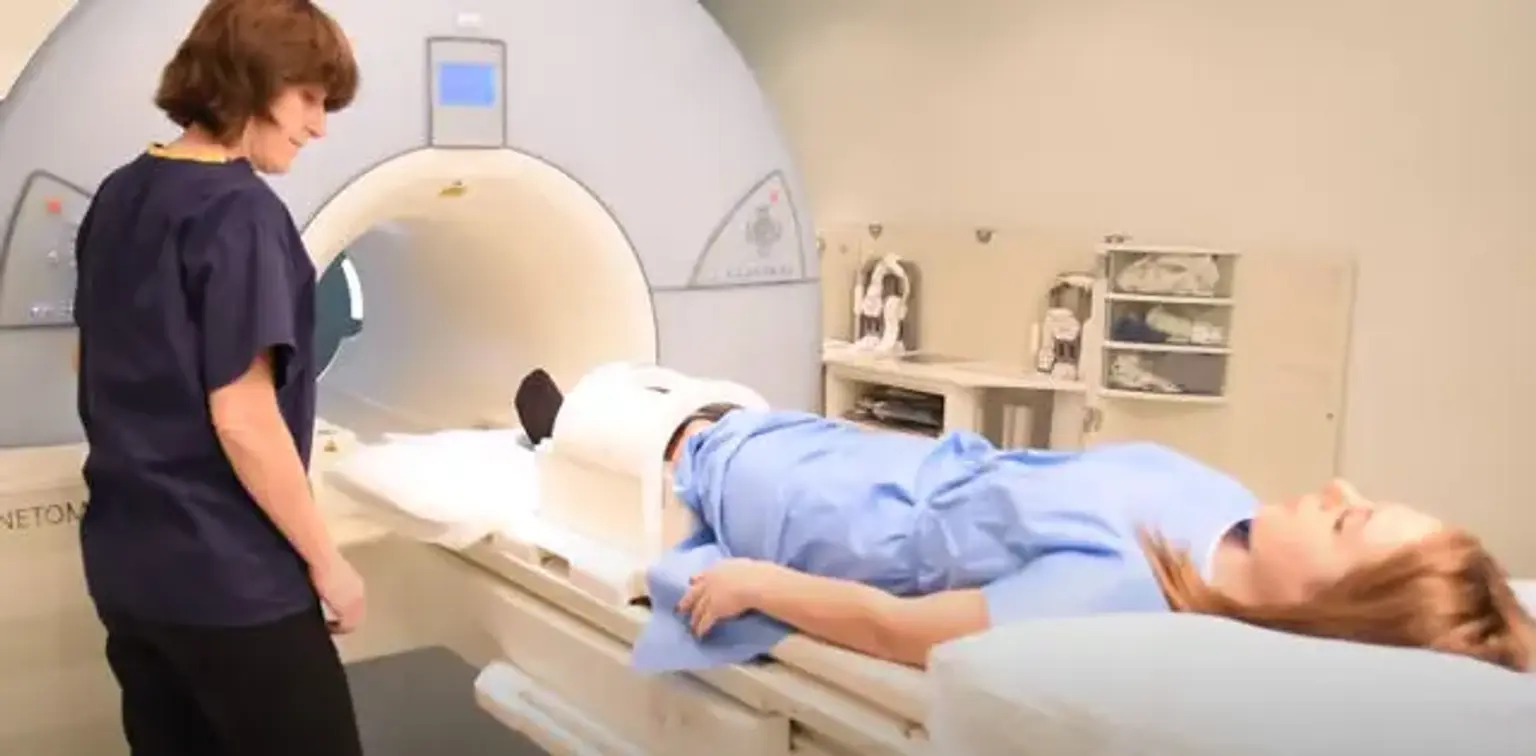
An MRI machine resembles a big metallic and plastic doughnut and a bench that slowly slides you into the middle. By following your provider’s directions and removing all the metals in your body, you are going to be entirely safe inside and about the machine. The whole procedure can take approximately 30 to 90 minutes.
If the procedure involves the use of contrast dye, the doctor will inject it via a tube placed in one of the veins. In some situations, the dye might take up to one hour to find its way through the bloodstream and into the target area (spine or knee).
The technician will ask you to lie flat on your back, sideways, or on your stomach on the device bench. If you have any problems lying on the bench, you may be given a pillow or blanket. From a different room, the technician will regulate the bench's movement. He or she will also converse with you via the machine's speaker.
As it takes pictures, the equipment will produce some loud humming and pounding sounds. Several hospitals provide earplugs to make the procedure much simpler, whereas others have televisions or music headphones.
You can change back to your clothing and carry on with your day once the technician has captured the images required.
What to Expect after MRI
In most cases, you are free to continue with your day following the MRI test. However, you should not drive if you received sedatives before undergoing the test procedure.
The film may take several hours to form if your MRI photos were projected on the film. Your doctor will need some time to analyze the photographs and evaluate the results. Modern devices use a computer to display images; hence the doctor can quickly check and interpret them.
The MRI results might take up to one or even more weeks to obtain. Your doctor will contact you once the results are out to review them and talk about the following steps regarding your treatment.
Overall Benefits of MRI
- MRI is a typically noninvasive imaging method that does not require radiation exposure.
- Spine and knee images obtained using MRI are clear and detailed, unlike images produced using other imaging techniques. MRI can reveal anomalies, injuries, and diseases in the spine or knee that aren't visible through other imaging tests.
- With conventional imaging modalities, anomalies may be concealed by bone. However, MRI can easily detect them.
- When it comes to evaluating knee and spinal injuries, an MRI is beneficial. When a physical examination reveals paralysis or muscle weakness, MRI might assist diagnose or rule out the possible causes.
- MRI can identify minor changes in the spinal column or knee joint that could indicate an infection or malignancy. For examining tumors, abscesses, and other soft tissue masses, MRI is highly sensitive, unlike CT scanning.
Risks and Complications of MRI
An MRI scan, unlike CT scans and x-ray, does not involve the use of radiation. Hence, it is a safer option for every person, including minors and pregnant mothers. CT scans comprise safe levels of radiation for adults. However, they are not suitable for growing fetuses and should be used carefully in minors.
People who have metallic implants are at a high risk of complications. This is because an MRI's magnets can interfere with pacemakers or cause implanted plates, pins, or screws to shift from their original location.
The contrast dye used when performing an MRI might cause adverse reactions for some people. Gadolinium is the most prevalent form of contrast dye. Luckily, these reactions are frequently minor and treatable with medications.
Conclusion
Magnetic resonance imaging (MRI) creates detailed images of the entire body without using an x-ray. Instead, it uses radio waves and powerful magnetic fields to capture the images. This enables the doctor the view the spine and knee structure and determines if there is any abnormality.
If you have medical devices or other metallic objects implanted in your body, be sure to inform your doctor. Special instructions and other schedule adjustments may be necessary with MRI scanning.