Multi-detector computed tomography (MDCT)

Multi-detector computed tomography

Overview

The invention of multidetector computed tomography (MDCT) is regarded as a significant advancement in CT imaging, with direct implications in the imaging of many systems, most notably the cardiovascular system. The benefits of MDCT include a significant improvement in image acquisition speed, increased patient coverage, and excellent spatial resolution.
 

What is Multi-detector computed tomography (MDCT)?

Multi-detector computed tomography

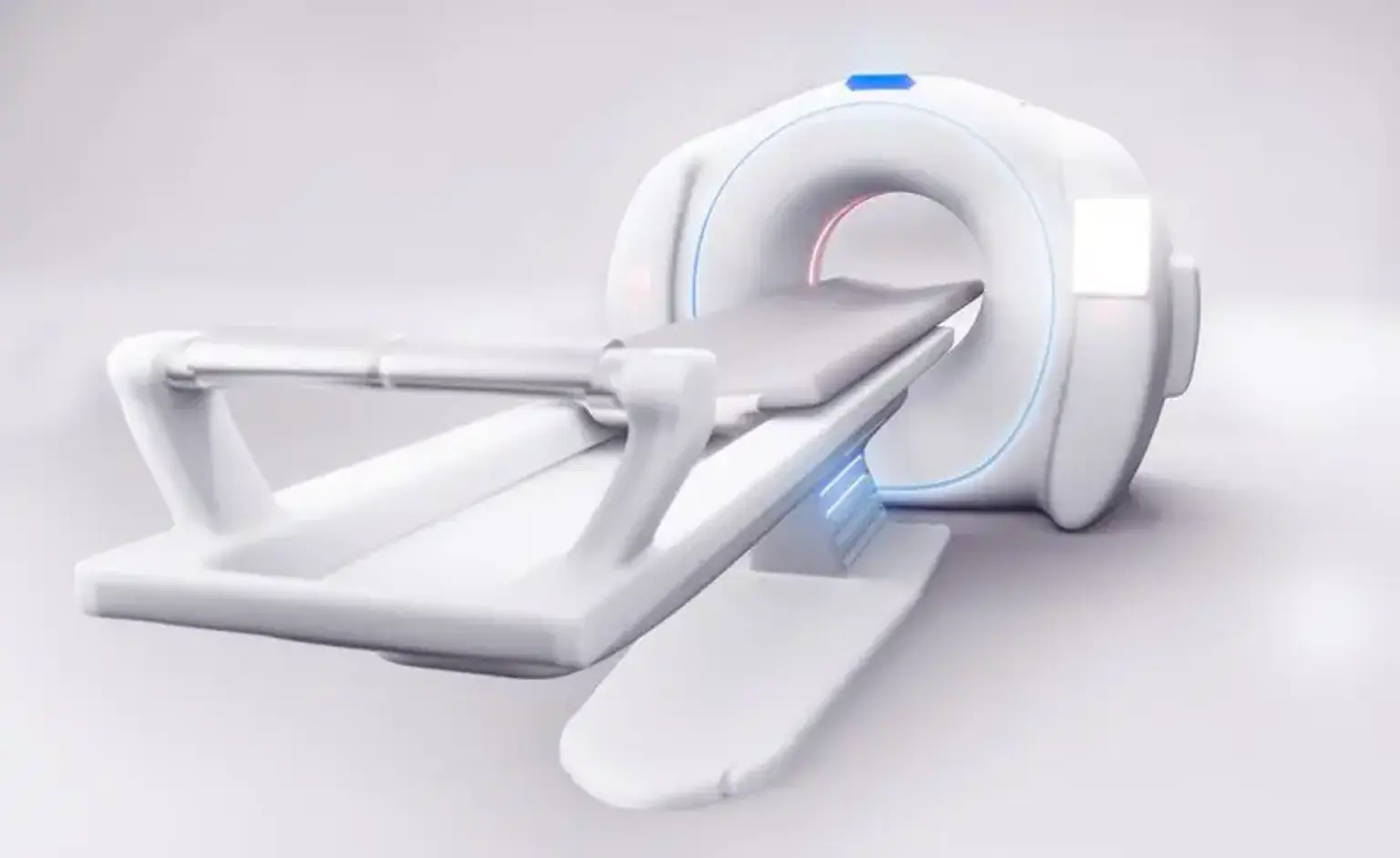
Multidetector computed tomography (MDCT) is a type of CT technology used for diagnostic imaging. A two-dimensional array of detector elements replaces the linear array of detector elements utilized in conventional and helical CT scanners in MDCT. The two-dimensional detector array enables CT scanners to capture many slices or sections at the same time, significantly increasing CT image acquisition speed.

To scan the chest, current CT technology employs multi-detector helical acquisition. Thin-section CT pictures with good spatial and temporal resolution are produced by multi-detector CT. The entire chest may be scanned in two or three seconds using the most recent detector arrays with 256 or 320 detector rows. Images obtained after intravenous contrast administration are tailored to examine pulmonary embolism, the aorta and its branches for congenital or acquired disease, and the coronary arteries for atherosclerotic disease.

The 1-2mm sections rebuilt from today's multi-detector CT scanners provide both high-resolution and thin-section pictures of the whole chest, with coronal and sagittal reconstructions having the same resolution as typical axial scans. Such imaging allows for a more accurate evaluation of the link between lesions and relevant anatomy, as well as an assessment of the distribution of lung or pleural illness in the cranio-caudal, medio-lateral, and antero-posterior planes.

When a series of images of the inner margins of the airways is viewed from an endoluminal perspective, as with fiberopt, three-dimensional rendering of intrathoracic structures that differ in attenuation from adjacent structures allows surface renderings of chest wall structures, enhancement of visualization of vessels such as the pulmonary arteries and aorta, and precise delineation of the outer and inner walls of the trachea and main bronchi (called "virtual bronchoscopy").


Why do people have MDCT?

MDCT Procedure

When other procedures, such as chest X-rays, electrocardiograms (ECG), echocardiograms (echocardiography), or stress tests, do not provide enough information about your heart, your doctor may request MDCT. Your physicians may obtain further information about:

  • Your heart’s structure and how well your heart pumps blood
  • Scarring of the heart muscle caused by a heart attack 
  • Fluid in the pericardial sac that covers the surface of the heart
  • The amount of plaque buildup and narrowing of your coronary arteries 
  • Any abnormalities in the large blood vessels leaving the heart
  • Your risk for a heart attack


Contraindications

Chest CT may be contraindicated due to two factors: radiation exposure and contrast delivery.

A standard chest CT exposes the patient to an effective dosage of 4-5 milliSieverts (mSv), whereas a low-dose scan exposes the patient to 1-2 mSv. High-resolution chest CT, which only samples around 10-15% of the chest, results in much lower effective radiation doses than typical volumetric multi-detector CT. The radiation dosage is of particular concern for younger patients, particularly young females, because ionizing radiation exposure is associated with a modest but discernible elevated risk of breast cancer.

Nephrotoxicity caused by iodinated contrast agents is a major problem in individuals with pre-existing renal failure, particularly those with diabetes-related chronic kidney disease. In most radiology departments, estimating the glomerular filtration rate (GFR) before to a study to estimate the risk of contrast delivery is standard practice. When such a risk is identified, prophylactic steps to limit the possibility of contrast-induced nephrotoxicity may be adopted, or an alternate imaging modality, such as sonography or MRI, may be considered.

A history of contrast allergy, a severe atopic history, or a previous anaphylactic reaction to intravenous contrast necessitates the prescription of corticosteroids and antihistamines at least six hours before contrast delivery. Another type of imaging should also be examined.


How do I prepare for MDCT?

Inquire with your doctor if contrast dye will be used during the exam. If this is the case, avoid eating for four to six hours before the test. If contrast dye will not be utilized, refrain from eating for two hours before the test.


What happens during MDCT?

 during multi-detector chest CT scans

Typically, multi-detector chest CT scans are conducted with the patient supine and arms lifted above the shoulders. A scanogram, often known as a "scout view," is generated to define the cranio-caudal extent of the scan and the reconstructed field of vision for diagnostic imaging.

The study's exposure parameters are determined by the patient's weight, with doses for smaller patients and minors decreased to the lowest levels necessary to provide diagnostic-quality pictures. For intravenous contrast imaging, an 18-20-gauge intravenous catheter placed in the antecubital fossa is preferable. When intravenous contrast is utilized, scans are scheduled to begin when a certain level of attenuation is identified by monitoring, and low-dose scans are performed through the pulmonary artery (for CT pulmonary angiography) or aorta (for evaluation of the aorta and great vessels).

All modern multi-detector CT scans are reconstructed in the axial, sagittal, and coronal planes and sent to PACS workstations for interpretation and system-wide access. Specialized CT technicians may build three-dimensional reconstructions from a thin-section data collection, which is especially valuable for displaying the intricate anatomy of aortic disorders and tracheobronchial pathologies. The three-dimensional, rebuilt pictures are displayed and saved on the PACS.


What happens after MDCT?

after Multi-detector computed tomography

  • Most people are able to resume their routine activities immediately.
  • The test findings will be sent to your doctor in writing. You should consult with that doctor about the results and following measures.

Other important steps include:

  • If you are overweight, your initial objective should be to lose 5 to 10 pounds. If you need to lose more weight, it is advised that you lose 1 to 2 pounds every week until you achieve a healthy weight.
  • Engage in some physical activity. Walk, ride a bike, or engage in other forms of moderate physical exercise for at least 150 minutes every week, ideally spaced out throughout the course of the week.
  • If you smoke, quit. Avoid secondhand smoke
  • If you drink alcohol, have no more than one drink a day if you’re a woman, two if you’re a man.


Interpretation of Results

Interpretation of Results of MDCT

Thoracic radiologists or radiologists with skills in cross-sectional imaging generally interpret chest CT tests. To give a full interpretation of scans, several radiologists have adopted a systematic analysis and reporting procedure. These elements are as follows:

  • Evaluation of the scan's technical sufficiency, including overall picture quality analysis, respiratory motion examination, and vascular contrast enhancement evaluation.
  • Support and monitoring devices, tubes, lines, and catheters must be identified, including their look and location.
  • An examination of the chest wall's bony and soft tissue components. Different window settings allow for bone (large window width) and soft tissue examination (narrower window widths).
  • The heart, pericardium, and major vessels are examined. The detection of both acute and chronic emboli is included in investigations to evaluate for pulmonary embolism.
  • Mediastinal examination, including lymph nodes, trachea and central bronchi, and esophagus.
  • Airway and lung evaluation, including assessment of localized lung disease (e.g., nodules, masses), air-space and interstitial disorders, cystic lung disease, and emphysema.
  • Pleural space evaluation, including pneumothorax monitoring and identification of pleural thickening, fluid, calcification, or lumps.
  • Search for free intraperitoneal air and observation of hepatic, splenic, gastric, duodenal, colonic, adrenal, pancreatic, upper abdominal aortic, and upper renal pathology.

MDCT

MDCT can detect blockages in your cardiac arteries if contrast dye (iodine) is administered during the scan. This is beneficial in patients with chest pain to determine whether the pain is caused by a shortage of blood flow to the heart muscle caused by clogged heart arteries (angina). If the heart arteries are normal, your doctor can investigate alternative reasons of chest discomfort that aren't connected to the heart with confidence.

MDCT can also be performed with contrast dye to see if coronary artery bypass grafts are still open, to look for congenital heart abnormalities (issues evident from birth), and to see how your ventricles are operating.

MDCT may be performed without contrast dye to determine the quantity of calcium in your heart arteries ("calcium score"). Your calcium score informs doctors about the amount of plaque in your heart arteries that hasn't yet produced difficulties. Your calcium score may help estimate your chance of having a heart attack and inform you and your doctor how active you should be in lowering your risk factors. This is especially beneficial if you are at "moderate" risk.

Calcium scoring is not suggested for regular screening of persons who do not have heart disease symptoms and are at low risk of having a heart attack. Calcium scoring will not give any further information if you have already had a heart attack, coronary bypass surgery, or a coronary stent. 


Can I have MDCT instead of a coronary angiogram?

coronary angiogram

MDCT is not a replacement for a coronary angiography (cardiac catheterization). Coronary angiography is the most reliable approach for detecting coronary artery blockages. It also provides detailed information about how your heart is operating.


Alternative and/or additional procedures to consider

Dual-energy subtraction (DES) radiography and tomosynthesis can be beneficial in identifying nodules in some individuals with suspected lung nodules at a much reduced cost and radiation dosage.

In certain patients with central lung cancer, MRI can be as effective as multi-detector CT in evaluating superior sulcus tumors and mediastinal masses, as well as assessing for cardiac or mediastinal invasion. In the assessment of heart masses, MRI outperforms CT. When patients with lung cancer are unable to obtain intravenous contrast, MRI might be used to evaluate them for liver or adrenal involvement.

CT-PET is more sensitive and somewhat more selective than MRI in detecting nodal involvement in lung cancer. In the identification of malignancy in solitary pulmonary nodules, CT-PET is more accurate than multi-detector CT.


What are the risks of MDCT?

risks of MDCT

Several significant difficulties of multi-detector CT scanning should be noted:

Contrast Extravasation – Intravenous contrast extravasation during CT power infusion occurs at a rate of less than 1%. There may be pain, swelling, tingling, redness, and warmth. The presence of clinically relevant sequelae is determined by the location of extravasation as well as the volume and osmolarity of the contrast agent utilized. Compartment syndrome is the most significant result of contrast extravasation; skin ulceration and tissue necrosis are less prevalent. The majority of contrast extravasation-related injuries heal spontaneously and without complications.

Patients who have contrast extravasation should be monitored for many hours to ensure that the physical results are stable. Hot or cold compresses, as well as elevation of the afflicted extremity, might help relieve discomfort. When there is a possibility of major harm, a surgical consultation should be sought. Progressive swelling or discomfort, skin ulceration or blistering, lack of capillary refill, or changes in sensation in the afflicted extremities should all require surgical evaluation.

For patients who have an acute contrast reaction, the following measures are taken:

  • Urticaria – No treatment needed.
  • Facial or laryngeal edema: Oxygen by mask; epinepherine (1:1000), 0.1 – 0.3 ml (0.1 – 0.3 mg) subcutaneously or intramuscularly, if hypotension develops, epinenephrine (1:10,000), 1 – 3 ml.
  • Bronchospasm: Oxygen by mask; beta-agonist inhaler; epinephrine, via route and at dose as for laryngeal edema.
  • Hypotension and tachycardia: Leg elevation; oxygen by mask; intravenous fluid.
  • Hypotension with bradycardia (vagal reaction): Leg elevation; intravenous fluid; atropine, 0.6 – 1.0 mg intravenously, up to a total of 2 – 3 mg.
  • Hypertension, severe: Nitroglycerin, sublingual, up to 3 doses; labetalol, 20 mg intravenously every 10 minutes, up to 300 mg.
  • Seizures: Diazepam, 5 mg, or midazolam, 1 mg, intravenously.
  • Pulmonary edema: Oxygen; furosemide, 20 – 40 mg intravenously; morphine, 1 – 3 mg intravenously.

The avoidance of contrast-induced nephrotoxicity (CIN) is the goal of treatment. Patients with pre-existing renal failure, diabetes, advanced age (>70 years), dehydration, hypertension, multiple myeloma, or hyperuricemia are at risk for CIN. The most essential preventative approach is to provide pre- and post-contrast hydration with intravenous fluids containing or not containing sodium bicarbonate. The use of preventive N-acetyl-cysteine in lowering the risk of CIN is still debatable.


Conclusion 

CT technology's revolution, including high scanning speed, wider coverage area, and high temporal and spatial resolution, is providing a credible, noninvasive diagnostic tool for a wide range of cardiovascular diseases and will likely replace catheter diagnostic angiography in most vascular beds in the near future. Cost-effectiveness studies and clear proof data on its clinical value, on the other hand, are still absent.