Myocardial disease

Last updated date: 18-Apr-2023

Originally Written in English

Myocardial disease

Overview

Myocardial diseases are prevalent and range from primary (cardiomyopathies) to secondary (hypertensive heart disease, alcoholic cardiomyopathy, Takotsubo cardiomyopathy, and more unusual types of secondary myocardial illness such as muscular dystrophy cardiomyopathy or peripartum cardiomyopathy.

Primary myocardial disorders are usually inherited, whereas secondary myocardial diseases are usually acquired. Subsequent forms may potentially have a genetic foundation that promotes secondary myocardial disease development. Inflammatory illnesses of the myocardium, such as myocarditis or viral cardiomyopathy, are examples of a subtype of cardiac disease.
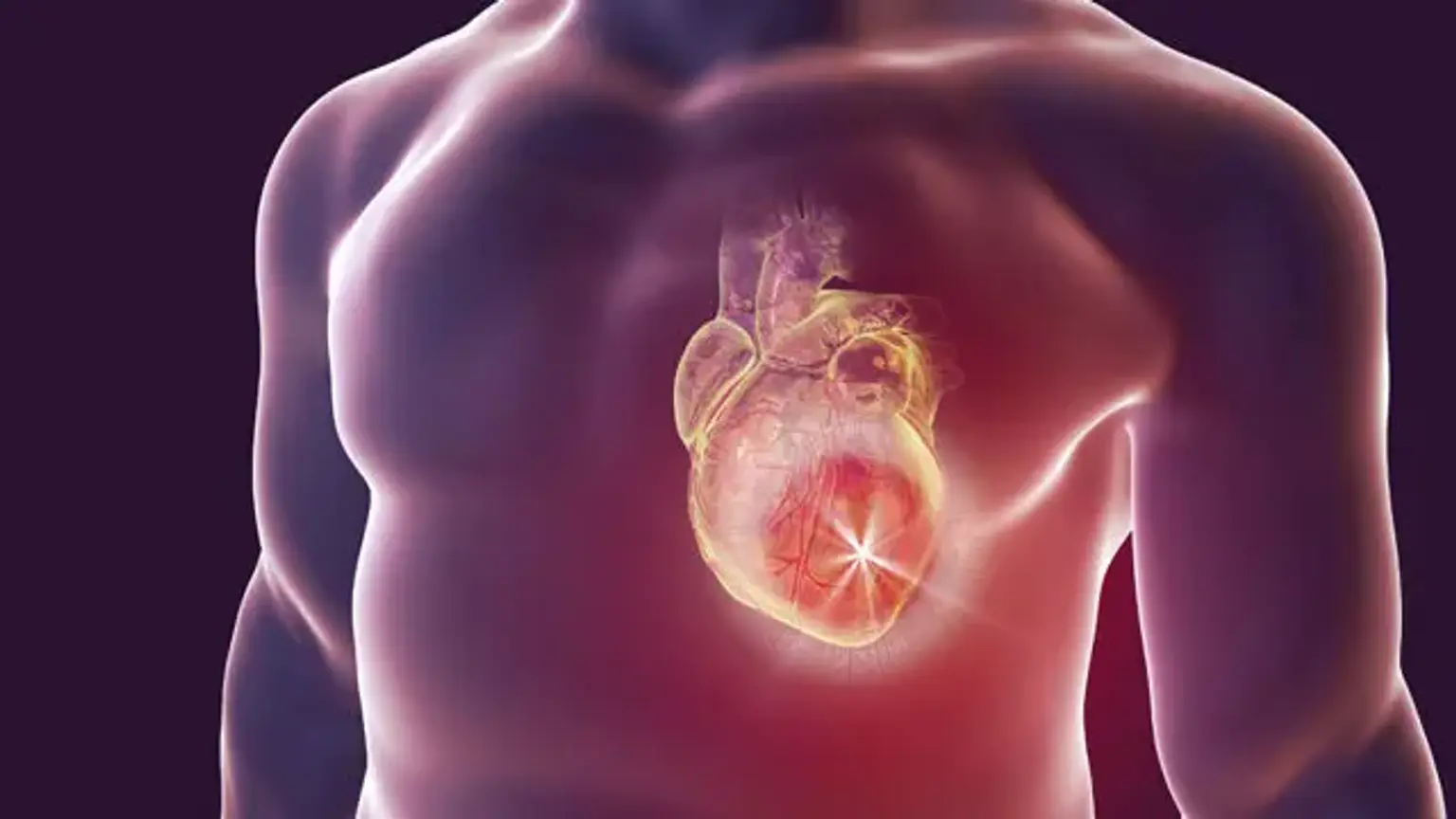
 

What is myocardial disease?

Primary and secondary Myocardial Disease 

There are five types of primary myocardial illness, which are classified as myocardium disorders with reduced heart function, commonly known as cardiomyopathies.

  • Hypertrophic cardiomyopathy (HCM)
  • Dilated cardiomyopathy (DCM)
  • Restrictive cardiomyopathy (RCM)
  • Arrythmic cardiomyopathy (ACM)
  • Unclassified cardiomyopathy (UCM)


Secondary myocardial illness is defined as cardiac disease that occurs after a recognized cause. Correction of the underlying condition in a timely manner may result in cardiomyopathy reversal. There are nine distinct etiologies that can be distinguished:

  • Hypertension
  • Ischemia
  • Valvular disease
  • Alcohol
  • Metabolic cardiomyopathy
  • Takotsubo cardiomyopathy
  • Peripartum cardiomyopathy
  • Tachycardia


Myocardial Infarction

Myocardial Infarction

A myocardial infarction (MI), sometimes known as a "heart attack," occurs when blood supply to a region of the myocardium is reduced or completely stopped. Myocardial infarction can be "quiet" and go unnoticed, or it can be a catastrophic occurrence that results in hemodynamic decline and untimely death.

The vast majority of myocardial infarctions are caused by underlying coronary artery disease, which is the leading cause of mortality in the United States. The myocardium is deprived of oxygen due to coronary artery blockage. Prolonged oxygen deprivation of the myocardium can result in cardiac cell death and necrosis.


Pathophysiology

Acute myocardial infarction can result from the obstruction of one or more big epicardial coronary arteries for more than 20 to 40 minutes. The blockage is often thrombotic and caused by the rupture of a plaque that has developed in the coronary arteries. Because of the blockage, there is a paucity of oxygen in the myocardium, which causes sarcolemmal rupture and myofibril relaxation.

These modifications are among the earliest ultrastructural changes that occur throughout the MI process, and they are followed by mitochondrial abnormalities. Prolonged ischemia eventually leads to liquefactive necrosis of cardiac tissue. The necrosis progresses from the subendocardium to the subepicardium. The subepicardium is thought to have higher collateral circulation, which causes it to die later.

The heart function is impaired depending on the region affected by the infarction. Due to the myocardium's limited regeneration ability, the infarcted region heals via scar formation, and the heart is frequently modified, characterized by dilatation, segmental hypertrophy of remaining viable tissue, and cardiac dysfunction.


Clinical presentation 

The mismatch between oxygen supply and demand causes myocardial ischemia and, in rare cases, myocardial infarction. The patient's history, electrocardiographic results, and increased serum biomarkers all contribute to the identification of ischemia symptoms. Myocardial ischemia can cause chest pain, upper extremity pain, mandibular or epigastric discomfort during exercise or at rest.

Myocardial ischemia can also manifest as dyspnea or weariness, both of which are ischemic equivalents. The pain in the chest is frequently retrosternal and is described as a feeling of pressure or weight. The discomfort frequently radiates to the left shoulder, neck, or arms with no evident cause, and it can be intermittent or continuous. The agony frequently lasts longer than 20 minutes.

It is typically unaffected by changes in posture or active movement of the area. Sweating, nausea, stomach discomfort, dyspnea, and syncope are some of the other symptoms that may be present. MI can sometimes develop atypically, with modest findings like palpitations or more catastrophic presentations like cardiac arrest. MI can occasionally appear with no symptoms.


Evaluation

Myocardial Disease Evaluation

Clinical characteristics, ECG abnormalities, and cardiac biomarkers are the three components of MI assessment.

  • ECG

The 12-lead resting ECG is the first-line diagnostic technique for acute coronary syndrome (ACS). It should be acquired within 10 minutes of the patient's arrival to the ER. Acute MI is frequently accompanied by dynamic alterations in the ECG waveform. If the initial EKG is non-diagnostic, serial ECG monitoring might offer vital clues to the diagnosis. Serial or continuous ECG recordings may aid in determining the state of reperfusion or re-occlusion. Reperfusion generally results in a significant and rapid decrease in ST-segment elevation. 


 Acute Management

All patients with symptoms of ischemia lasting fewer than 12 hours and persistent ST-segment elevation should get reperfusion treatment. If the treatment can be performed within 120 minutes after ECG diagnosis, primary percutaneous coronary intervention (PCI) is recommended over fibrinolysis. If PCI is not an urgent possibility (>120 minutes), fibrinolysis should begin within 10 minutes of STEMI once all contraindications have been ruled out.

If transfer to a PCI facility is achievable within 60 to 90 minutes following a fibrinolytic drug bolus and the patient fulfills reperfusion criteria, a normal PCI or a rescue PCI might be arranged. If fibrinolysis is intended, fibrin-specific drugs such as tenecteplase, alteplase, or reteplase should be used.


Myocarditis

Myocarditis

Myocarditis is an inflammation of the heart muscle's myocardium. Acute myocarditis is most usually caused by a viral infection, however it can also be caused by non-infectious etiologies. Myocarditis is classified as acute, subacute, or chronic, and it can affect a specific area of the heart or be widespread.

There are two types of causes of acute myocarditis: infectious and non-infectious. In half of the cases, no cause can be found; hence, myocarditis is usually idiopathic. In patients with a known cause, the most prevalent etiology is viral (similar to pericarditis), with enteroviruses, particularly Coxsackie B, being the most common. Human immunodeficiency virus (HIV), adenovirus, and hepatitis C are examples of other viral infections.

Other infectious causes include parasitic (e.g., Trypanosoma cruzi), bacterial (e.g., diphtheria or TB), helminths, and fungal infections. Granulomatous inflammatory illnesses (e.g., sarcoidosis or giant cell myocarditis), systemic lupus erythematosus, eosinophilic myocarditis, polymyositis and dermatomyositis, and collagen vascular disease are all non-infectious causes of myocarditis.  Cocaine-related cases have also been described in the literature.


Myocarditis symptoms 

Myocarditis symptoms

Acute myocarditis has a wide range of clinical manifestations, from asymptomatic to mild to cardiogenic shock and even rapid cardiac death. There are no pathognomonic clinical characteristics. The traditional presentation resembles that of heart failure, including symptoms such as dyspnea, orthopnea, and leg edema.
Palpitations and syncope are also possible. In one investigation of clinically suspected instances, around a quarter of patients showed decreased left ventricular ejection fraction (LVEF), prolonged ventricular arrhythmias, or symptoms of poor cardiac output.

When the pericardium is implicated, as in myopericarditis, chest discomfort with pericarditis-like symptoms develops. Because viral myocarditis is the most prevalent cause, the patient may describe a viral prodrome (fever, arthralgia, lethargy) 1 to 2 weeks before the development of heart failure symptoms.

Other signs of an infectious agent, such as dysphagia in Chagas disease (produced by Trypanosoma cruzi) or neurological symptoms in diphtheria patients, may also arise. Furthermore, individuals with a non-infectious etiology frequently show signs of their underlying systemic illness, such as skin or renal involvement in connective tissue disease patients.

Myocarditis is not related with any specific physical exam findings. Physical examination findings might range from normal to cardiac failure-like, including S3 gallop, jugular venous distention, peripheral edema, and tachycardia. Furthermore, individuals with ventricular dilatation may have a mitral regurgitation murmur, also known as an apical pansystolic murmur.

A pericardial friction rub may be welcomed, especially if pericarditis is present. In non-infectious etiologies, other results may point to an underlying condition. Eosinophilic myocarditis individuals will also have a pruritic maculopapular rash. When the pericardium is implicated, a friction rub on cardiac auscultation may be noted. 


Diagnosis of acute myocarditis

Acute myocarditis is difficult to diagnose since other clinical entities might mimic the diagnosis. The varying clinical appearance of acute myocarditis complicates matters even further. Acute myocarditis should be suspected in individuals with clinical signs and symptoms of the illness, regardless of workup, particularly in young patients (age 25-50) with no history of cardiac disease. Clues to the underlying etiology, such as a viral prodrome or evidence of connective tissue illness, may help with the diagnosis.

An EKG, echocardiography, serum troponin, and B-type natriuretic peptide should all be performed as part of the first examination for acute myocarditis (BNP). Troponin elevation, which is frequently severe, is found in more than half of the patients. In the correct clinical picture, BNP can be used to look for signs of heart failure and ventricular strain, which may indicate myocarditis. The EKG frequently reveals nonspecific ST alterations; however, it may also reveal sinus tachycardia or ventricular arrhythmias .

Changes similar with pericarditis, such as generalized ST elevation, may also be observed in patients with pericardial involvement. An echocardiography can assist determine the extent of heart failure and rule out other reasons, such as valvular disease.

A chest radiograph is neither sensitive nor specific for myocarditis, although it can reveal an enlarged heart, pulmonary vascular congestion, or pleural effusion. CT angiography may be recommended to rule out alternative causes of chest discomfort. In the correct circumstances, percutaneous coronary angiography is useful in individuals at high risk for coronary artery disease to help rule out an ischemic cause of heart dysfunction.

Cardiac magnetic resonance imaging is promising and may aid in distinguishing between ischemic and non-ischemic etiologies of dilated cardiomyopathy. A total blood count with eosinophilia in the differential may also indicate eosinophilic myocarditis. The erythrocyte sedimentation rate (ESR) and C-reactive protein (CRP) are commonly increased but nonspecifically. Viral antibody testing can be performed in the appropriate context; however, specificity is limited, which frequently results in a delay in diagnosis with little change in care.


Treatment of myocarditis 

Management is generally supportive, but it also involves the treatment of any discernible cause. Patients with heart failure should get routine care, which may include beta-blockers, angiotensin-converting enzyme inhibitors or angiotensin II receptor blockers, as well as diuretics if necessary. Immunosuppressive treatment has not been proved to be clinically beneficial and, as a result, should not be used frequently, except in patients with underlying systemic autoimmune or granulomatous inflammatory disorders.

Although viral etiology is the most usually recognized cause, antiviral medication's effectiveness is uncertain, and routine antiviral therapy is not indicated. Non-steroid anti-inflammatory medications should be avoided in the acute situation since they may hinder myocardium repair. Mechanical support devices, such as an intra-aortic pump or left ventricular assist devices, can be utilized in extreme situations, with heart transplants considered.

In these cases, it is critical to consider transferring to tertiary care hospitals for surgical assistance if necessary. Patients may develop dysrhythmias or a thrombus in the left atrial or ventricular chambers, necessitating anticoagulation. Finally, patients should be advised to minimize their physical activity and avoid alcohol, especially during the acute period, since it may boost viral replication. Long-term surveillance with serial echocardiography is advised.


Peripartum cardiomyopathy

Peripartum cardiomyopathy

Peripartum cardiomyopathy is defined as impaired left ventricular systolic function within one month of birth or within the first five months postpartum in the absence of pre-existing cardiac illness and in the absence of another identified reason for the cardiac dysfunction.

The typical presentation is with symptoms of left ventricular failure, albeit many of these characteristics are indistinguishable from normal changes in pregnancy, therefore minor variants may go unnoticed. In addition to malnutrition, viral infection, an aberrant immunological response, and hereditary susceptibility, an inflammatory component has been proposed. 

The condition recurs in successive pregnancies, making the previously indicated etiologies difficult to explain. Peripartum cardiomyopathy is most likely caused by a propensity to DCM, which is exacerbated by the increased circulatory load of pregnancy.

In peripartum cardiomyopathy, standard heart failure medication can be initiated, however the use of ACE inhibitors and angiotensin receptor antagonists is prohibited after the first trimester due to potential fetal harm. In severe circumstances, however, the possible hazards to the fetus should be evaluated against the crucial necessity to preserve ventricular function in order to provide both the mother and the fetus the greatest chance of a favorable clinical result.

 Pregnancy is also linked with a significant risk of thrombo-embolism since it is a hypercoagulable condition by definition, which is exacerbated by the presence of decreased ventricular function in the event of peripartum cardiomyopathy, and prophylaxis is therefore advised.

Despite optimum therapy, LV function may normalize in as few as 50% of patients and progress to end-stage heart failure in 15%. The possibility of recurrence in future pregnancies necessitates patient counseling in order to prevent additional bouts of symptomatic heart failure and advancement of ventricular dysfunction.


Hypertrophic cardiomyopathy

The hallmarks of hypertrophic cardiomyopathy (HCM) include unexplained left ventricular enlargement in the absence of underlying cardiac or systemic diseases. Myocyte disarray, familial incidence, and a relationship with abrupt cardiac death are further distinguishing markers (SCD).

Genetics

The discovery of HCM's genetic basis led to the concept that HCM is a disease of the sarcomere, the cell's contractile unit. First, mutations in the cardiac B-myosin heavy chain gene were discovered, and later, other sarcomeric proteins were discovered to have a role in HCM.

It was demonstrated that macroscopic hypertrophy of the myocardium is not required for diagnosis or prognosis, since mutations in troponin T, for example, might result in relatively modest or no hypertrophy while being related with a high incidence of SCD. HCM is now thought to be a genetic condition, inherited mostly autosomal dominantly with imperfect and age-related penetrance. 


Pathophysiology

The therapeutic significance of dividing HCM into obstructive and non-obstructive types is based on the existence or absence of an LV outflow tract gradient during rest and/or following provocation. When such a gradient exists, it usually causes a loud apical systolic ejection murmur. The blockage raises intraventricular pressures, which impairs LV function by raising myocardial wall stress and oxygen demand.

The blockage is induced by systolic anterior motion (SAM) of the mitral valve and mid-systolic contact with the ventricular septum and can be found either sub-aortic or mid-cavity. SAM is hypothesized to be caused by either an aberrant valvular apparatus that is free enough to allow movement or a hemodynamic force with an anterior component during systole.


Treatment of HCM

Medical treatment

Asymptomatic HCM patients should only be treated if diastolic dysfunction symptoms are observed. Verapamil is the preferred therapy, as it improves diastolic filling and ventricular relaxation while reducing diastolic filling pressures.

First-line medical therapy for symptomatic patients is a calcium-channel blocker or a betablocker. Verapamil is the preferred medication, but diltiazem can be used as an alternative. Second, when symptoms are severe, beta-blockers can be taken alone or in conjunction with a calcium-channel blocker. 

Diuretics may be given in extremely symptomatic individuals, but with extreme caution because a slight reduction in ventricular filling pressure can substantially impair stroke volume and cardiac output in HCM patients. A combination of calcium-channel blockers and beta-blockers is possible.

Amiodarone may be utilized in patients with ventricular tachyarrhythmia or supraventricular atrial fibrillation, and it may potentially improve symptoms and prognosis. Disopyramide has a negative inotropic effect, causing peripheral vasoconstriction, and may alleviate symptoms when diastolic dysfunction is severe.


Invasive treatment of obstructive HCM

When medicinal therapy is ineffective, invasive debulking of the myocardial septum may be explored in individuals with a significant outflow gradient. Percutaneous alcohol septal ablation or surgical septal myectomy are two treatment options.


Surgical myectomy 

Although surgical myectomy has shown excellent long-term results, 15-20% of patients may experience ventricular remodelling and dilatation of the left ventricle. Since the introduction of alcohol ablation, surgical myectomy has been reserved for patients with HCM who have concomitant disease that requires surgical correction on its own, such as coronary artery bypass grafting or valve repairs, and in whom surgical myectomy can be performed as part of the operation.


Septal ablation 

Septal ablation may be explored in individuals who have outflow tract gradients greater than 30 – 50 mmHg at rest or 60-100 mmHg following provocation. A minor myocardial infarction is caused by injecting 1-3 mL of pure alcohol into the first or second septal branch for 5 minutes. Furthermore, the alcohol causes septal hypokinesis, which contributes to a decrease in the outflow tract gradient. Although the gradient may disappear instantly, the procedure may take weeks or months. Patients can be treated a second time if the outflow tract blockage persists.


Infiltrative cardiomyopathy

Infiltrative cardiomyopathy

Amyloidosis

Amyloidosis is a condition caused by the deposition of fibrils with a specific secondary structure of a beta-pleated sheet configuration in the tissue, resulting in distinctive histological alterations. Amyloid depositions can develop in practically every organ, although until large depositions are present, they are typically clinically undiagnosed.


Types of amyloidosis

The AL (main) and AA (secondary) forms of amyloidosis are the most common. AL amyloidosis is a plasma cell dyscrasia that can arise independently or in conjunction with multiple myeloma. AA amyloidosis is a complication of chronic inflammatory disease states such as rheumatoid arthritis, with depositions consisting of fragments of serum amyloid A, an acute phase reactant.

Hereditary amyloidosis, which is caused by mutations in the thransthyretin gene, has become more well recognized in the previous decade. Some mutations are only found in the myocardium. Its prevalence increases with age, with a male preference, although its prognosis is better than that of the AL type. Senile systemic amyloidosis is caused by the buildup of normal wild-type transthyretin. This kind of amyloidosis is characterized clinically by an infiltrative cardiomyopathy, although development is gradual and the prognosis is better than in other acquired types.


Cardiac amyloidosis

Cardiac amyloidosis is a kind of infiltrative cardiomyopathy that worsens with time. The main variety is the most common, affecting around one-third to half of all patients, with deposits detectable even in the absence of clinical symptoms. Secondary amyloidosis is less commonly related with cardiac infiltration, occurring in roughly 5% of instances, and is less likely to be associated with ventricular dysfunction due to the smaller size and more advantageous placement of the depositions. 

In one-quarter of individuals, familial amyloidosis is linked with clinical symptoms of cardiac involvement, often manifesting beyond the age of 35 with a specific involvement of the cardiac conduction system. The degree of deposits in senile amyloidosis might range from isolated atrial involvement to widespread ventricular infiltration.


Clinical manifestations

Aside from the presence of known AL amyloidosis, connective tissue disease, or other chronic inflammatory disorders, cardiac amyloidosis should be considered in the following cases:

  • Restrictive cardiomyopathy of unknown origin
  • Left ventricular hypertrophy with a converse low-voltage ECG
  • Congestive heart failure of unknown origin, not responding to contemporary medical management.


Conclusion 

Myocardial disease is one of the most significant medical burdens that developed-world populations face. Diabetes and obesity are on the rise in both the developed and developing worlds, implying that this problem will worsen with time.

Cardiomyopathies are disorders that affect the myocardium. They are distinct in that they are not caused by hypertensive, congenital, valvular, or pericardial illnesses, and they are only infrequently caused by ischemic heart disease.

This type of cardiac disease is frequently distinguishable, both in terms of general symptoms and blood flow patterns, allowing a diagnosis to be made. Cardiomyopathy is a major source of morbidity and mortality, as evidenced by increased awareness of the disorder and improved diagnostic tools. It may responsible for up to 30% of all heart disease fatalities in various parts of the world.